Cook R.A., Stewart B. Colour Atlas of Anatomical Pathology
Подождите немного. Документ загружается.

BONES, JOINTS
AND
CONNECTIVE
TISSUE
Fig. 10.42
Fig. 10.38 Chondrosarcoma arising
in the
scapula.
F/35.
This
tumour
had
grown rapidly.
Its cut
surface shows
a
lobulated
pattern
with
the
white,
glistening
appearance
of
cartilage.
Fig. 10.39
Giant
cell
tumour
in the
lower
end of the
femur.
M/38. There
is a
haemorrhagic tumour expanding
the
lateral condyle
and
destroying
the
normal bone.
Fig. 10.40
Ewing's
tumour.
M/13.
A
large
tumour
in the
upper third
of the
tibia
has
eroded
the
cortical bone
and
extended beneath
the
periosteum. There
is a
pathological
fracture
through
the
tumour.
Fig. 10.41 Chordoma removed from
the
pelvis
anterior
to
the
sacrum.
M/58.
The
tumour
is
haemorrhagic
and has a
rather mucoid
cut
surface. Microscopic examination
was
required
to
make
a
definitive diagnosis.
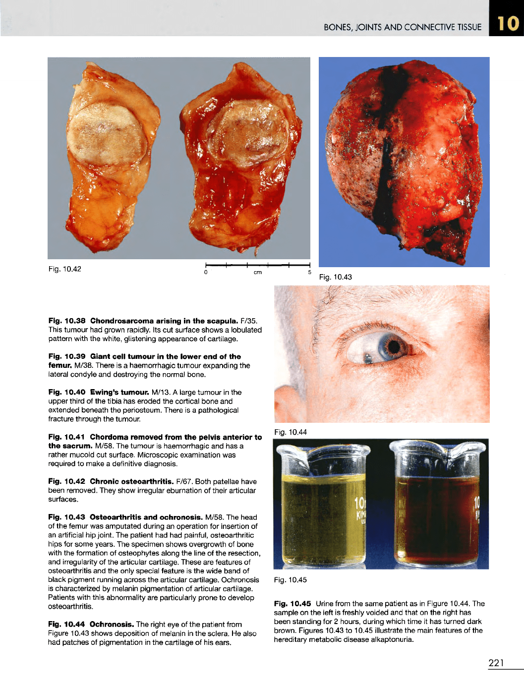
Fig. 10.42
Chronic
osteoarthritis.
F/67. Both patellae have
been
removed. They show irregular eburnation
of
their
articular
surfaces.
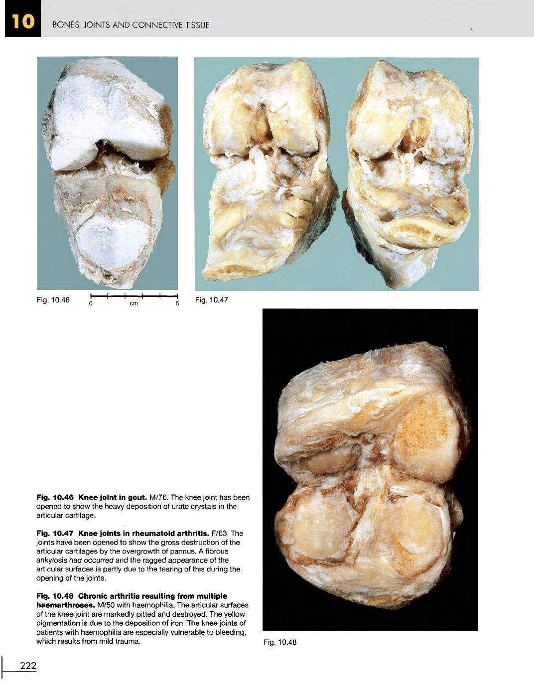
Fig. 10.43
Osteoarthritis
and
ochronosis. M/58.
The
head
of
the
femur
was
amputated during
an
operation
for
insertion
of
an
artificial
hip
joint.
The
patient
had had
painful, osteoarthritic
hips
for
some years.
The
specimen
shows overgrowth
of
bone
with
the
formation
of
osteophytes along
the
line
of the
resection,
and
irregularity
of the
articular cartilage. These
are
features
of
osteoarthritis
and the
only
special feature
is the
wide band
of
black pigment running across
the
articular cartilage. Ochronosis
is
characterized
by
melanin pigmentation
of
articular cartilage.
Patients with
this
abnormality
are
particularly prone
to
develop
osteoarthritis.
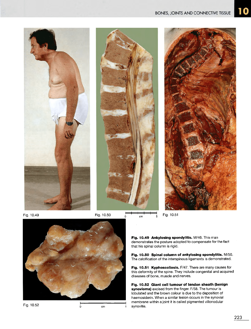
Fig. 10.44 Ochronosis.
The
right
eye of the
patient from
Figure
10.43 shows deposition
of
melanin
in the
sclera.
He
also
had
patches
of
pigmentation
in the
cartilage
of his
ears.
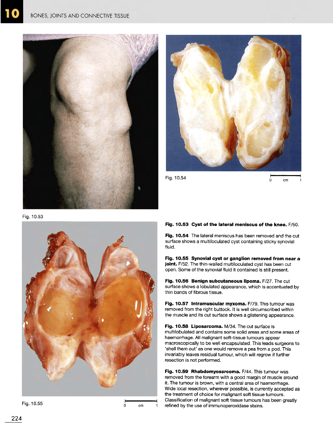
Fig. 10.45
Fig. 10.45 Urine from
the
same patient
as in
Figure 10.44.
The
sample
on the
left
is
freshly voided
and
that
on the
right
has
been standing
for 2
hours, during which time
it has
turned dark
brown. Figures 10.43
to
10.45 illustrate
the
main features
of the
hereditary metabolic disease alkaptonuria.
221
Fig. 10.43
Fig 10.44
BONES,
JOINTS
AND
CONNECTIVE
TISSUE
Fig. 10.46
Fig. 10.47
Fig. 10.46
Knee
joint
in
gout.
M/76.
The
knee joint
has
been
opened
to
show
the
heavy deposition
of
urate
crystals
in the
articular cartilage.
Fig. 10.47
Knee
joints
in
rheumatoid
arthritis.
F/63.
The
joints have been opened
to
show
the
gross destruction
of the
articular cartilages
by the
overgrowth
of
pannus.
A
fibrous
ankylosis
had
occurred
and the
ragged
appearance
of the
articular
surfaces
is
partly
due to the
tearing
of
this during
the
opening
of the
joints.
Fig. 10.48
Chronic
arthritis
resulting
from
multiple
haemarthroses.
M/50 with haemophilia.
The
articular surfaces
of
the
knee joint
are
markedly
pitted
and
destroyed.
The
yellow
pigmentation
is due to the
deposition
of
iron.
The
knee joints
of
patients with haemophilia
are
especially vulnerable
to
bleeding,
which results from mild trauma.
Fig. 10.48
222
BONES,
JOINTS
AND
CONNECTIVE
TISSUE
Fig.
10.49
Fig. 10.50
Fig.
10.52
Fig.
10.51
Fig. 10.49 Ankylosing
spondylitis.
M/46. This
man
demonstrates
the
posture adopted
to
compensate
for the
fact
that
his
spinal column
is
rigid.
Fig. 10.50
Spinal
column
of
ankylosing
spondylitis.
M/50.
The
calcification
of the
interspinous ligaments
is
demonstrated.
Fig. 10.51 Kyphoscoliosis. F/47. There
are
many causes
for
this deformity
of the
spine. They include congenital
and
acquired
diseases
of
bone, muscle
and
nerves.
Fig. 10.52
Giant
cell
tumour
of
tendon
sheath
(benign
synovioma) excised from
the
finger. F/58.
The
tumour
is
lobulated
and the
brown colour
is due to the
deposition
of
haemosiderin. When
a
similar lesion occurs
in the
synovial
membrane within
a
joint
it is
called pigmented villonodular
synovitis.
223
BONES,
JOINTS
AND
CONNECTIVE
TISSUE
Fig. 10.53
Fig. 10.55
Fig. 10.53
Cyst
of the
lateral
meniscus
of the
knee.
F/50.
Fig. 10.54
The
lateral meniscus
has
been removed
and the cut
surface
shows
a
multiloculated cyst containing sticky synovial
fluid.
Fig. 10.55 Synovial cyst
or
ganglion
removed from
near
a
joint.
F/52.
The
thin-walled multiloculated cyst
has
been
cut
open. Some
of the
synovial fluid
it
contained
is
still present.
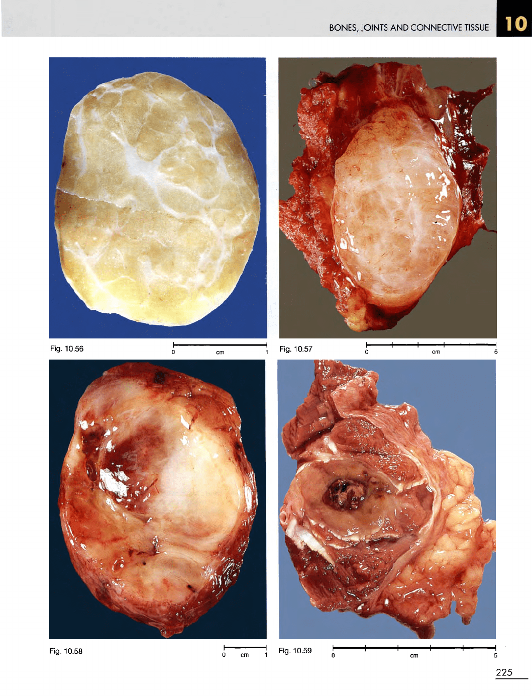
Fig. 10.56
Benign
subcutaneous
lipoma.
F/27.
The cut
surface
shows
a
lobulated appearance, which
is
accentuated
by
thin bands
of
fibrous tissue.
Fig. 10.57
Intramuscular
myxoma. F/79. This tumour
was
removed from
the
right buttock.
It is
well circumscribed within
the
muscle
and its cut
surface shows
a
glistening appearance.
Fig. 10.58 Liposarcoma. M/34.
The cut
surface
is
multilobulated
and
contains some solid areas
and
some areas
of
haemorrhage.
All
malignant soft-tissue tumours appear
macroscopically
to be
well
encapsulated.
This
leads
surgeons
to
'shell them out'
as one
would remove
a pea
from
a
pod. This
invariably
leaves residual tumour, which
will
regrow
if
further
resection
is not
performed.
Fig. 10.59 Rhabdomyosarcoma. F/44. This tumour
was
removed
from
the
forearm with
a
good
margin
of
muscle around
it. The
tumour
is
brown, with
a
central
area
of
haemorrhage.
Wide local resection, wherever
possible,
is
currently accepted
as
the
treatment
of
choice
for
malignant soft tissue tumours.
Classification
of
malignant soft tissue tumours
has
been greatly
refined
by the use of
immunoperoxidase stains.
224
Fig.
10.54
BONES, JOINTS
AND
CONNECTIVE
TISSUE
Fig.
10.58
225
Fig.
10.56
Fig.
10.57
Fig.
10.59
BONES,
JOINTS
AND
CONNECTIVE
TISSUE
Fig. 10.60
Fig. 10.63
Fig.
10.62
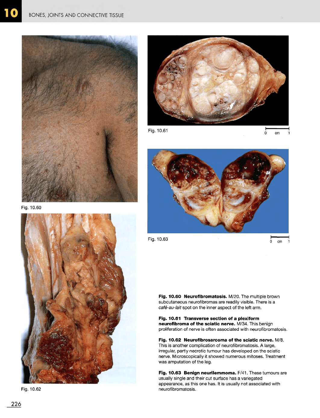
Fig. 10.60
Neurofibromatosis.
M/20.
The
multiple brown
subcutaneous neurofibromas
are
readily visible. There
is a
cafe-au-lait
spot
on the
inner aspect
of the
left arm.
Fig. 10.61 Transverse
section
of a
plexiform
neurofibroma
of the
sciatic
nerve.
M/34. This benign
proliferation
of
nerve
is
often associated with neurofibromatosis.
Fig. 10.62
Neurofibrosarcoma
of the
sciatic
nerve.
M/8.
This
is
another complication
of
neurofibromatosis.
A
large,
irregular,
partly necrotic tumour
has
developed
on the
sciatic
nerve.
Microscopically
it
showed numerous mitoses. Treatment
was
amputation
of the
leg.
Fig. 10.63
Benign
neurilemmoma.
F/41. These tumours
are
usually
single
and
their
cut
surface
has a
variegated
appearance,
as
this
one
has.
It is
usually
not
associated with
neurofibromatosis.
226
Fig. 10.61
BONES, JOINTS
AND
CONNECTIVE
TISSUE
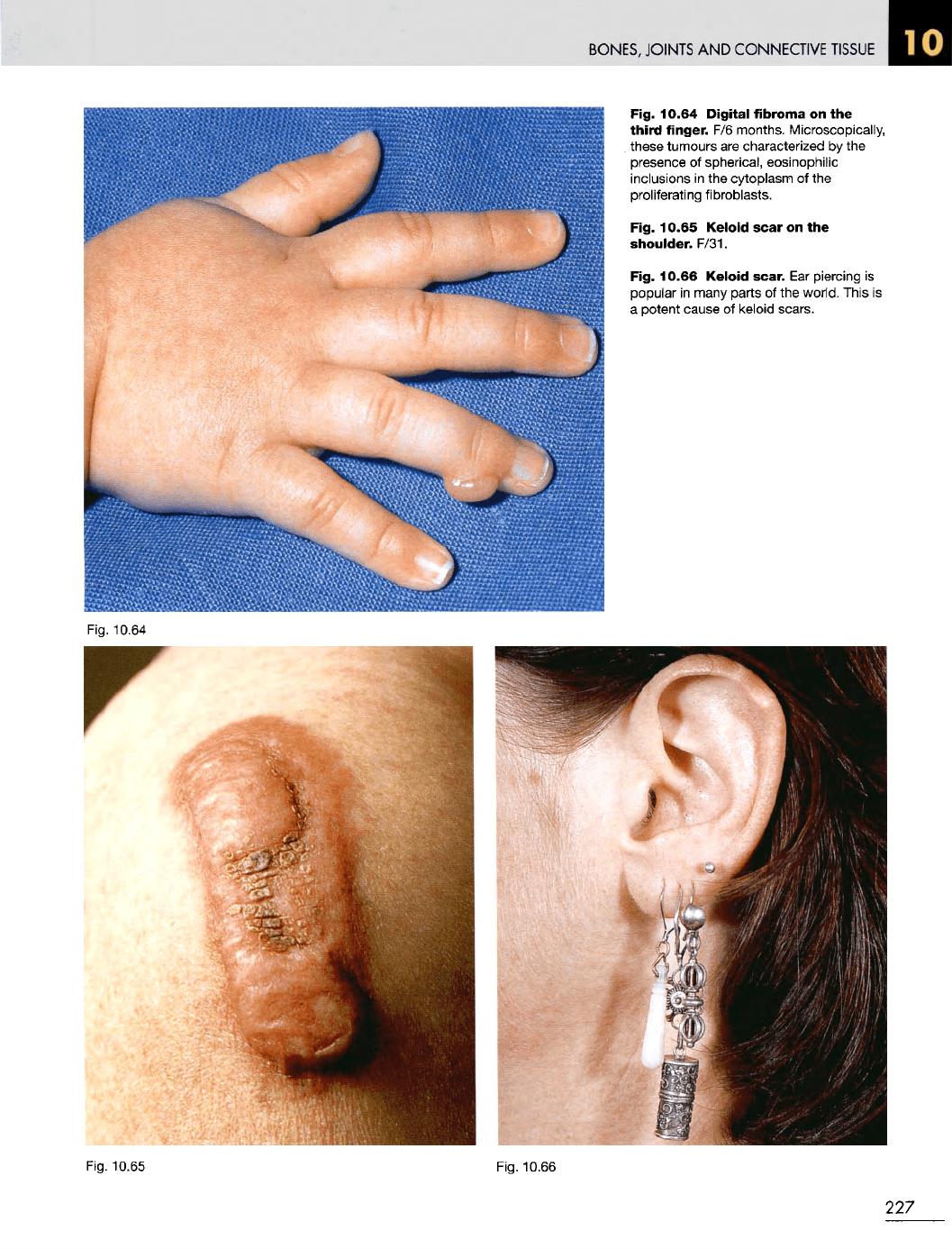
Fig. 10.64
Digital
fibroma
on the
third
finger.
F/6
months. Microscopically,
these tumours
are
characterized
by the
presence
of
spherical, eosinophilic
inclusions
in the
cytoplasm
of the
proliferating fibroblasts.
Fig. 10.65
Keloid
scar
on the
shoulder.
F/31.
Fig. 10.66
Keloid
scar.
Ear
piercing
is
popular
in
many parts
of the
world. This
is
a
potent cause
of
keloid scars.
Fig.
10.65
Fig. 10.66
227
Fig.
10.64
BONES,
JOINTS
AND
CONNECTIVE
TISSUE
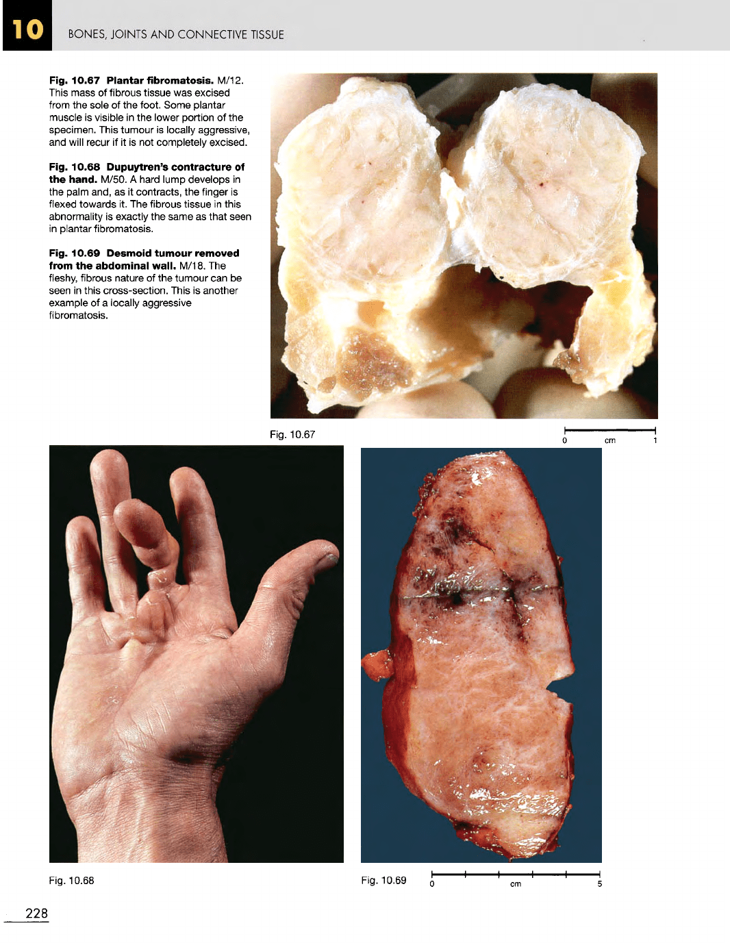
Fig. 10.67
Plantar
fibromatosis.
M/12.
This
mass
of
fibrous tissue
was
excised
from
the
sole
of the
foot. Some plantar
muscle
is
visible
in the
lower portion
of the
specimen. This tumour
is
locally aggressive,
and
will recur
if it is not
completely excised.
Fig. 10.68
Dupuytren's
contracture
of
the
hand.
M/50.
A
hard lump develops
in
the
palm and,
as it
contracts,
the
finger
is
flexed towards
it. The
fibrous tissue
in
this
abnormality
is
exactly
the
same
as
that seen
in
plantar fibromatosis.
Fig. 10.69
Desmoid
tumour
removed
from
the
abdominal
wall.
M/18.
The
fleshy,
fibrous nature
of the
tumour
can be
seen
in
this cross-section. This
is
another
example
of a
locally aggressive
fibromatosis.
Fig. 10.68
Fig. 10.69
228
Fig.
10.67

NERVOUS
SYSTEM
11
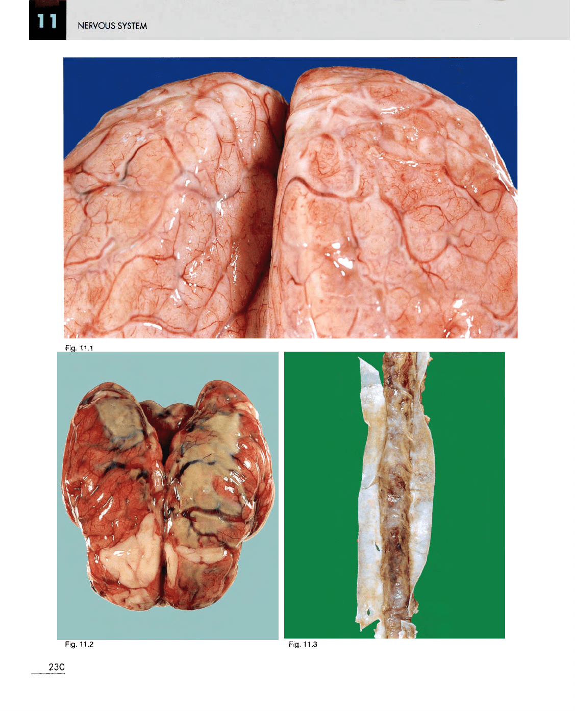
Fig.
11.2
Fig.
11.3
230
Fig.
11.1