Gladstone Geoffrey J., Black Evan H., Myint Sh. Oculoplastic Surgery Atlas
Подождите немного. Документ загружается.

Surgical Management 41
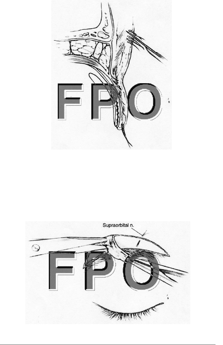
FIGURE 3-2. Incision is beveled away from the brow hairs to parallel the
direction of the follicles.
FIGURE 3-3. Skin and muscle flap are removed with scissors. In the region
of the supraorbital nerve, the dissection must be superficial to avoid damag-
ing the nerve.

POSTOPERATIVE CARE
Ophthalmic antibiotic ointment or a combination of antibiotic and
steroid is applied at the end of the case. Bandaging is generally not
necessary. The patient is instructed to use ice packs 15 minutes each
hour while awake for the next 2–3 days and to elevate the head with
several pillows at bedtime. Straining, lifting heavy objects, and bend-
ing should be avoided for 1 week. The sutures can be removed as soon
as 7 days after surgery to avoid epithelialization of the suture tracks.
Surgical adhesive strips may be used at this time if needed. To min-
imize the evidence of a scar the patient is instructed to reduce sun
exposure to the area by wearing a hat or applying lotions with a sun-
protection factor of 45 or more.
42 Direct Eyebrow Lift

4
R
HYTIDECTOMY SURGERY
A
variety of methods exist for facelift surgery (rhytidectomy). The
more complicated techniques involve extensive undermining of
the superficial musculoaponeurotic system (SMAS) or subpe-
riosteal dissection. The simpler techniques employ plication or im-
brication of the SMAS. Several of these techniques are presented here
so the surgeon can chose the technique with which he or she is most
comfortable and that most benefits the patient.
CLINICAL EVALUATION
The initial patient evaluation is of paramount importance. It is crit-
ical to understand the patient’s concerns. Unrealistic desires on the
part of patients can only lead to surgery that fails to please them.
Have patients hold a mirror and show you all the areas of their face
they would like to see improved. If they do not specifically point to
important areas ask them about their chin and neck, jowls, nasoju-
gal fold, and nasolabial fold. Make note of the quality of the skin, the
amount of redundant tissue, the presence or absence of platysmal
bands and the amount of submental fat. If significant platysmal bands
or deep nasolabial folds are present, they should be discussed preop-
eratively with the patient as they are difficult to correct fully.
Evaluation of the chin and neck directly affects the surgery per-
formed in this area. If minimal submental fat and skin laxity are
present, submental liposuction is performed and the skin dissection
in the neck does not extend into the central neck and submental area.
Conversely, if significant submental fat, platysmal bands, or skin lax-
43

ity is present, the dissection extends further into the neck (Fig. 4-1).
Direct lipectomy is performed through a submental incision, and
platysmal plication may be necessary.
In addition to evaluating surgically important facial features, it
is important to discuss smoking and medication usage with the pa-
tient. Smoking during the perioperative period can lead to flap necro-
sis secondary to vasoconstriction. It is imperative that the patient not
smoke for 2 weeks before and 2 weeks after surgery. Any medications
that inhibit platelets or coagulation must be stopped an appropriate
time prior to surgery. These include but are not limited to aspirin, many
other nonsteroidal antiinflammatory drugs, warfarin (Coumadin), and
clopidogrel (Plavix). These medications can increase the possibility of
the patient developing a hematoma underneath the facelift flaps,
which can lead to flap necrosis.
44 Rhytidectomy Surgery
Clinical Evaluation 45
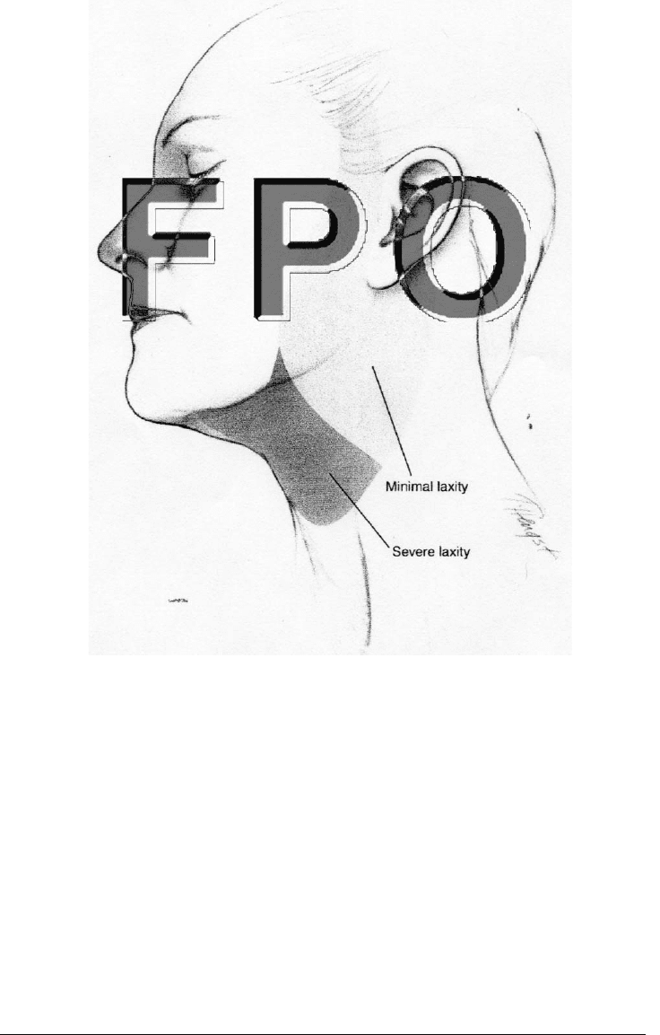
FIGURE 4-1. Extent of subcutaneous skin dissection for minimal and se-
vere laxity of neck skin.

SURGICAL MANAGEMENT
At the beginning of the procedure the facelift incision is marked in
front of the ear with a fine-tipped marker (Fig. 4-2). A heavier marker
is more appropriate in the temporal hairline and the retroauricular
area. Marking should be performed prior to administering the local
anesthetic. If platysmal bands, which represent the medial border of
the platysmal muscle, are present, they should be marked to facili-
tate finding the medial borders during the plication procedure. If sig-
nificant submental fat is present, the lateral extent of this compart-
ment should be marked to guide submental liposuction or direct
lipectomy.
The temporal incision is directed superiorly and temporally for
several centimeters. It parallels the temporal hairline and is placed
several centimeters posterior to it. The incision begins at the ante-
rior helix and extends superiorly and anteriorly. We typically utilize
a pretragal incision, which follows the anterior helix and the tragus.
This incision conforms to the natural contours of the ear and helps
avoid a straight-line scar, which tends to be more noticeable and cos-
metically objectionable. Many surgeons utilize a posttragal incision,
which must be used with care. If not performed carefully, the tragus
can be pulled forward during the healing process, causing an unnat-
urally open ear.
The incision continues under the lobule of the ear. In the area of
the lobule, a 1- to 2-mm rim of skin is left attached to the lobule. This
helps keep the lobule anchored in its normal position. If this rim of
tissue is too wide, it is noticeable postoperatively. The incision con-
tinues superiorly to the level of the external auditory canal several
millimeters onto the conchal cartilage. During the postoperative pe-
riod the incision migrates posteriorly into the fold. (If the incision is
initially placed in the fold, it migrates onto the mastoid area and is
visible.) The incision turns posteriorly and continues for several cen-
timeters into the occipital hairline. If a large skin excision is antici-
pated or the patient wears his or her hair quite short the incision can
be placed just anterior to the hair line. This prevents an unnatural
“step” in the hairline after skin excision.
An additional incision is placed in the submental area, permitting
submental liposuction, direct lipectomy, or modification of platysma
bands. The incision should be placed several millimeters posterior to
the naturally occurring submental crease. (An incision placed directly
on the crease can result in deepening of the crease, which is cosmet-
ically objectionable.)
46 Rhytidectomy Surgery
Various facelifting techniques require different degrees of skin un-
dermining. For SMAS plication or imbrication techniques a relatively
large skin flap (6–8 cm) is prepared in the preauricular area. For
techniques that develop a large SMAS flap, less skin undermining is
performed. In these cases a skin flap of 3 cm is typically raised.
Intravenous sedation or general anesthesia can be utilized de-
pending on the preference of the surgeon and the health and wishes
of the patient. An equal mixture of 1% lidocaine with epinephrine and
0.75% bupivacaine is injected along all incision lines. A small inci-
sion is made in the pre- and postauricular areas and in the submen-
tal area to allow introduction of a small cannula. Tumescent solution
is infiltrated through these incisions in a subcutaneous plane in all
areas to be undermined. A simple tumescent solution consisting of
1000 cc normal saline and 50 cc 2% lidocaine is employed. Hemosta-
sis is improved and anesthesia provided.
When direct lipectomy is planned or platysmal bands need to be
modified, a 2.5 cm incision is marked just posterior to the submen-
tal crease. If submental liposuction is planned, a much smaller (3
mm) incision is marked to allow introduction of a 3 mm liposuction
cannula. This incision must be kept small to prevent loss of suction
during the liposuction portion of the procedure.
Surgical Management 47
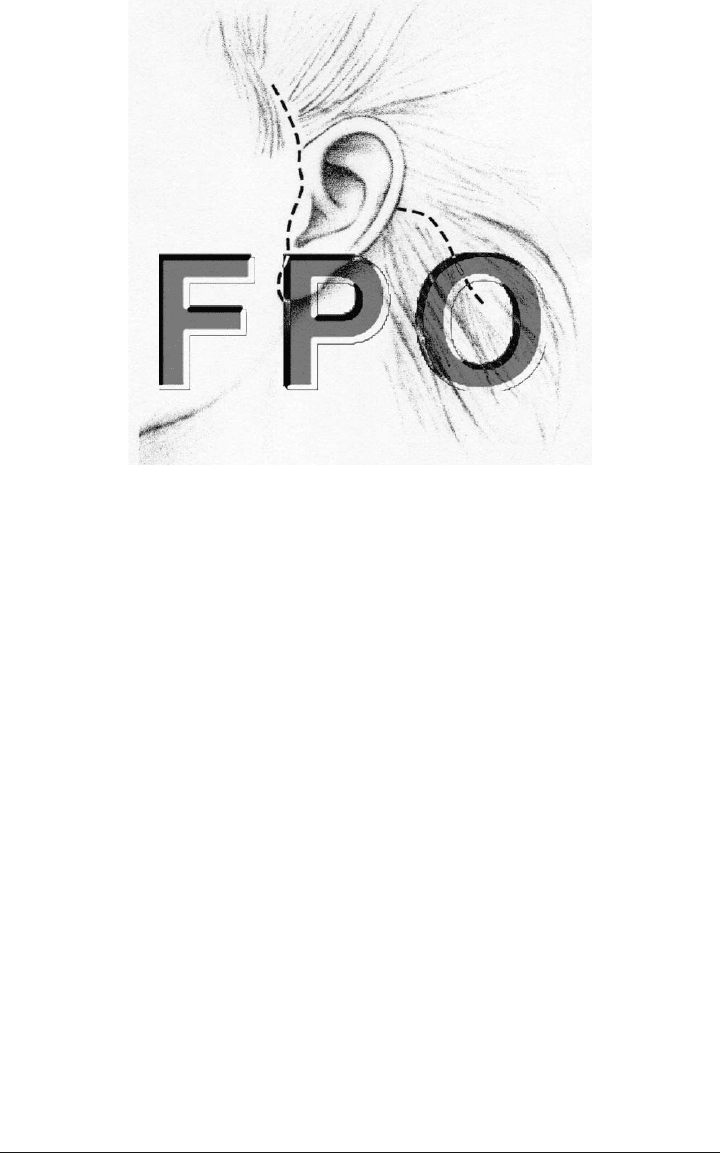
FIGURE 4-2. Typical incision for rhytidectomy.

Submental liposuction should be limited to the area bordered by
the mandible. The marginal mandibular nerve is superficial in the
area of the angle of the mandible, and liposuction should be avoided
in this region. If the submental fat has been properly marked preop-
eratively, liposuction need not extend outside this area.
When significant submental fat is present, direct lipectomy is indi-
cated. The 2.5 cm incision allows direct visualization of the fat and even-
tually the edges of the platysmal muscle. A subcutaneous dissection is
performed with facelift scissors in the previously marked submental area.
Approximately 5 mm of subcutaneous fat is left attached to the skin flap,
which helps prevent unusual adherence of the skin to the underlying tis-
sue during healing. A small (0.5 inch) lighted retractor aids in visuali-
zation and obtaining hemostasis. Preplatysmal fat is removed with the
scissors. The edges of the platysma muscle can be visualized and pli-
cated if significant platysmal bands are present (Fig. 4-3). Marking the
bands on the skin surface preoperatively helps locate the medial edges
of the muscle during dissection. The medial edges of the platysma are
plicated with multiple 4-0 permanent sutures. The skin in this area is
closed with a running 6-0 nylon suture. No skin excision is performed.
Once neck repair is complete, the facial incision can be performed.
This incision is made with a Bard-Parker No. 15 blade and is kept par-
allel with the hair follicles to avoid hair loss. In the temporal area the
dissection can be performed using the back of a blade handle. The han-
dle is pushed firmly in a plane just under the skin. Blunt dissection
avoids damaging the hair follicles, and it stays superficial to the frontal
branch of the facial nerve. The facial nerve is quite superficial in this
area, running within the superficial temporalis fascia. This subcutaneous
dissection terminates halfway between the ear and the lateral canthus.
Anterior to the ear the skin is tightly adherent to the underlying
tissue, making dissection difficult for the first several centimeters. A
No. 15 blade is useful for this initial portion of the dissection. A facelift
scissors is then used to complete an extremely superficial dissection.
Countertraction with a double skin hook simplifies the dissection and
leaves the underlying SMAS intact. Depending on the SMAS tech-
nique employed, this dissection creates a skin flap 3–8 cm in length.
The retroauricular portion of the incision is often started with the
No. 15 blade as well. The skin overlying the mastoid process is adher-
ent to the underlying tissue. Once the dissection becomes easier, a facelift
scissors can be employed. As the dissection proceeds inferiorly in the
neck, it is important to maintain a superficial dissection plane between
the subcutaneous tissue and the superficial musculature. The great au-
ricular nerve becomes extremely superficial as it crosses the body of the
48 Rhytidectomy Surgery
sternocleidomastoid muscle 6.5 cm below the external auditory canal.
Deep dissection in this area can sever the great auricular nerve.
Once the flaps have been raised and again prior to skin closure
meticulous hemostasis should be ensured. A wide (1.5 inch) lighted
retractor is invaluable for performing this task.
Various techniques can be used to treat the SMAS. The goal of
SMAS modification is to provide deep support and tightening for the
areas of the jowls, nasojugal fold, nasolabial fold, and neck. SMAS
modification can impart longevity to the procedure and allows less
Surgical Management 49
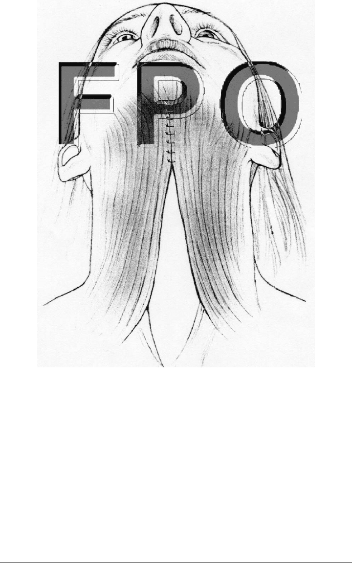
FIGURE 4-3. Platysma muscle plicated in the midline to reduce banding in
the neck.

tension to be applied directly to the skin, producing a more natural
appearance postoperatively. The techniques for SMAS plication,
SMAS imbrication, and deep SMAS dissection are presented.
SMAS Plication
SMAS plication is the simplest of the three techniques. Here, the
SMAS is sutured to itself in several locations without excising any
tissue. The platysma and the SMAS are easily seen after the skin
flaps are elevated. The SMAS has the appearance of muscle tissue
interspersed with lumpy fat deposits. This complex is mobile and can
be moved as a single unit. When the SMAS is grasped near the an-
terior mandibular ramus and the platysma is grasped in the neck,
the entire complex can be repositioned in a posterolateral direction.
The check and neck skin are observed to move in a similar direction,
tightening the neck, jowls, nasojugal fold, and nasolabial fold. A 4-0
nonabsorbable suture is used for the plication.
Three primary areas of the SMAS are tightened (Fig. 4-4). The
first and most superior suture tightens the nasolabial fold. The di-
rection of repositioning should be mostly posterior and slightly supe-
rior, perpendicular to the nasolabial fold. The next suture repositions
the jowls and nasojugal fold. This suture should exert a pull that is
mostly superior and slightly posterior. The final suture helps elevate
the neck. This suture, which should be directed superiorly and slightly
posteriorly, grasps the platysma below the ear and attaches it the
fascia overlying the mastoid process. Several interrupted or mattress
sutures are used in each location.
SMAS Imbrication
The technique for SMAS imbrication is similar to that for SMAS
plication. Instead of folding the SMAS on itself, however, an ellipse
of SMAS is excised, and the edges are sutured together. A relatively
safe area for SMAS excision lies between the zygomatic arch and the
angle of the mandible. The facial nerve is deep to the SMAS in this
area. Excision above the zygomatic arch would risk severing the
frontal branch of the facial nerve. The marginal mandibular nerve is
at risk if the excision extends over the angle of the mandible.
The SMAS excision starts just below the zygomatic arch and
1.5 cm anterior to the tragus. The excision extends inferiorly toward the
angle of the mandible: the greater the tissue laxity, the wider the exci-
sion should be. When the edges are sutured together, the degree of tis-
50 Rhytidectomy Surgery