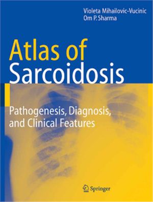
Springer Sience and Business Media, ISBN -1-85233-809-1, number of
pages- 119 with 300 illustrations, 111 in full color.
This Atlas of Sarcoidosis is designed to complement and provide a visual supplement to already existing excellent texts on pulmonary medicine and sarcoidosis. Medical students, postgraduate candidates, general practitioners, inteists, pulmonologists, dermatologists, and practitioners of many other disciplines of medicine who have to treat sarcoidosis patients will find the book useful and rewarding. In addition, paramedical personal such as nurses, physician’s assistants, and respiratory therapists will enjoy browsing the book.
Whatever the speciality of the reader, he or she must lea to obtain a careful history and perform a thorough physical examination. The next step is to link these observations with pertinent radiographic and laboratory information. If this amalgamation is not achieved the treatment of the patient will remain incomplete and ineffective.
The atlas aspires to facilitate this process. Sarcoidosis is a complex multisytem disease. The
lungs are the most commonly involved organs by Sarcoidosis, but no structure of the body is immune to its ravages. Each organ involvement is dealt in a brief and easy to comprehend manner. Various radiographic and laboratory abnormalities are then linked to the clinical
features in order to encourage a smooth and easy integration at the bedside. Finally it is worth remembering that the Atlas is not a repository of all that is known about Sarcoidosis. Its goal is to provide the reader a tantalizing visual interpretation of a fascinating and mysterious illness.

