Becker W. Advanced Time-Correlated Single Photon Counting Techniques
Подождите немного. Документ загружается.


154 5 Application of Modern TCSPC Techniques
acceptor fluorescence decay should show an excimer-like behaviour (see Fig. 5.3,
page 64), with a negative-coefficient decay component with the same decay time
as the quenched donor molecules. An
a/b image of the acceptor decay should
display the ratio of the acceptor emission excited via FRET and directly. Unfortu-
nately, in the CFP-YFP system, the acceptor decay cannot be observed directly
because of the strong overlap of the donor fluorescence with the acceptor fluores-
cence spectrum. An attempt was made in [39] to subtract the donor bleedthrough
from the acceptor decay and to build up an
a/b image.
A general characterisation of TCSPC-FLIM FRET for monitoring protein inter-
actions is given in [62, 93, 94, 405, 468]. Applications to protein interaction re-
lated to Alzheimer’s disease are described in [15, 16, 45, 46, 47]. Interactions
between the PCK and NKNB signalling pathways have been investigated in [372].
FRET between GFP and RFP and FRET cascades from GFP via Cy3 into Cy5 are
demonstrated in [406] and [7]. The agglutination of red blood cells by monoclonal
antibodies was studied using FRET between Alexa 488 and DiI [433]. Interaction
of the neuronal PDZ protein PSD95 with the potassium channels and SHP1-
target interaction were studied in [61, 62]. It has also been shown that FRET can
be used to monitor conformational changes of proteins in cells by FLIM-FRET
[80, 331].
A detailed description of a TCSPC-FLIM-FRET system is given in [147]. The
system is used for FRET between ECPF-EYFP and FM143 - FM464 in cul-
tured neurones. FRET between ECFP and EYFP in plant cells was demonstrated
in [68]. FRET measurements in plant cells are difficult because of the strong auto-
fluorescence of the plant tissue. It is possible to show that two-photon excitation
can be used to keep the autofluorescence signal at a tolerable level.
5.7.7 Technical Details of TCSPC Laser Scanning Microscopy
Laser Blocking in Two-Photon Microscopes
The laser intensity in two-photon imaging is several orders of magnitude higher
than in one-photon imaging. Good blocking of the excitation wavelength is there-
fore essential. In fact, complete failure to obtain two-photon FLIM images by
TCSPC results in most cases from insufficient laser blocking. The laser is
scattered not only in the sample, but also at the edge of the microscope lens, at the
dichroic mirror, in the AOM, and at various glass surfaces in the light path. Scat-
tering inside the microscope is so strong that often not even a reflection image of
the sample can be recorded. Moreover, some microscopes use IR LEDs to control
filter wheels. Leakage from these LEDs can add a substantial amount of continu-
ous background.
Moderate leakage of excitation light is often not recognised in steady state im-
ages. In TCSPC images the same amount of leakage can be disastrous. Due to
different path lengths of the individual reflections, false pulses can appear at any
time in the laser pulse period. The typical appearance of moderate leakage in time-
resolved images is shown in Fig. 5.90.
5.7 TCSPC Laser Scanning Microscopy 155
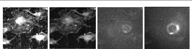
Fig. 5.90 Effect of insufficient laser blocking in two-photon imaging. TCSPC images in
different time windows in the laser period, left to right 0 ns, 4 ns, 8 ns, 10 ns. The laser
pulse period is 12 ns
The image at the maximum of excitation (0 ns) shows speckles due to scatter-
ing in the sample. The image at 4 ns is almost free of scattered laser light. Images
at 8 ns and 10 ns show a reflection in the microscope optics and a large amount of
diffusely scattered light caused by the next laser pulse on its way through the mi-
croscope. Of course, such data are entirely useless for fluorescence lifetime de-
termination.
Reliable blocking in the wavelength range of the Ti:Sapphire laser can be ob-
tained by BG39 filter glasses. In most cases 1 mm BG39 glass is required for
bialkali-detectors, and 3 mm for multialkali detectors, see Fig. 5.91.
The drawback of the BG39 filter is the relatively soft slope that results in a poor
transmission above 540 nm. Interference filters have a steeper slope than glass
filters but often do not achieve a sufficient blocking factor. Stacking of interfer-
ence filters should be avoided because the blocked light is reflected and bounces
between the filters. Good blocking with a relatively steep filter slope can, how-
ever, be obtained by combining a thin BG39 glass filter with an interference filter.
If two interference filters are used, the BG39 should be placed between them.
Choosing the Detectors
Detectors should be chosen based on the IRF width, the quantum efficiency versus
wavelength, the background count rate, and the size of the active detector area.
The fluorescence lifetime of the fluorophores commonly used in microscopy is
between 1 and 5 ns. Single-exponential lifetimes in this range can reliably be
measured with a detector IRF width of 200 to 400 ps. Medium speed detectors,
such as the Hamamatsu H742240 or the H5773 or H5783 modules are sufficient
for these applications. Side-window PMTs are not recommended. The IRF of
these detectors depends on the illuminated area of the photocathode and can be
between 350 ps and about 1 ns.
However, in the more sophisticated FLIM applications, the decay functions
cannot be considered single-exponential. Multiexponential decay analysis requires
either the IRF to be accurately known, or an IRF considerably shorter than the
lifetime components to be resolved. Medium-speed detectors, such as the H5773
or H5783 are actually fast enough to resolve double-exponential decay compo-
nents down to 100 ps. However, the IRF of these detectors has secondary bumps.
If the shape of the IRF is not accurately known, these bumps can mimic additional
decay components. Unfortunately it is difficult to obtain an accurate IRF in a mi-
156 5 Application of Modern TCSPC Techniques
croscope, especially a multiphoton microscope (see below). The R3809U MCP
PMTs with their clean and ultrafast IRF are a better choice.
The H5773 or H5783 and the R3809U detectors come in different cathode ver-
sions (see also Fig. 6.16, page 230, and Fig. 6.39, page 250). Figure 5.91, left,
shows the relative sensitivity (counts per input power) versus wavelength of the
commonly used bialkali and multialkali cathodes. The multialkali cathode is
clearly more sensitive above 500 nm.
However, in two-photon microscopes the cathode sensitivity must be consid-
ered in combination with the laser blocking filter. The standard filter is the Schott
BG 39. Normally, 1 mm BG39 are necessary to block the laser for a bialkali PMT,
and 3 mm for a multialkali PMT. The transmission of the BG39 filter and curves
of the relative radiant sensitivity for the cathodes detecting through the filters are
shown in Fig. 5.91, right. With the filters, there is little difference between the
bialkali and the multialkali cathode. It can therefore be worthwhile to sacrifice
some sensitivity at the red end of the spectral range and benefit from the lower
dark count rate of a bialkali cathode.
300
400
500 600 700 nm
1
0.1
0.01
0.001
10
Relative Sensitivity
Multialkali
Bialkali
Wavelength
300
400
500 600 700 nm
1
0.1
0.01
0.001
10
Transmission or Relative Sensitivity
1mm BG39
3mm BG39
Bialkali + 1mm BG39
Multialkali + 3mm BG39
Wavelength
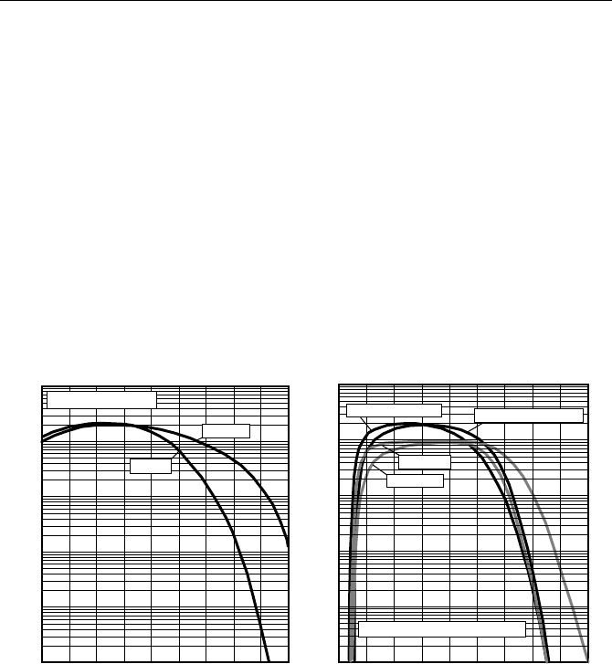
Fig. 5.91 Left: Relative radiant sensitivity (relative counts per incident power) of the bial-
kali and the multialkali cathode. Right: Transmission of a BG39 NIR blocking filter, and
relative sensitivity of the cathodes with the filter. Curves calculated from Hamamatsu sensi-
tivity curves and Schott filter data
Exceptionally high sensitivity can be achieved with GaAsP photocathodes. A
practical solution is the H7422P40 module of Hamamatsu. Between 500 and
600 nm, the GaAsP cathode has 2 to 4 times the sensitivity of a multialkali or
bialkali cathode (see Fig. 6.16, page 230 and Fig. 6.33, page 246). Unfortunately
the GaAsP cathode is intrinsically slow, so that the H742240 has an IRF width of
200 to 350 ps. The detector is currently the best compromise for combined
FCS / lifetime measurement.
Single photon avalanche photodiodes (SPADs) achieve the highest radiant sen-
sitivity of all detectors in the NIR. Currently available APD detectors have ex-
5.7 TCSPC Laser Scanning Microscopy 157
tremely small detector areas and are therefore not applicable to nondescanned
detection. Moreover, the count-rate-dependent timing shift found in many SPAD
modules makes them less useful for FLIM applications.
Transferring the Light to the Detectors
The fluorescence light collected by the microscope objective lens must be trans-
ferred to the detector. In descanned (confocal) systems this is relatively easy. The
light emerging from a small pinhole can be transferred efficiently to the detector
by a lens or an optical fibre, or simply by placing the detector close to the pinhole.
If a fibre is used, a large-diameter fibre should be preferred. The pulse dispersion
in a fibre depends on the effective NA, but not on the fibre diameter. A large-
diameter fibre can be used at a smaller NA, and it is easier to obtain a good cou-
pling efficiency (see Sect. 7.2.5, page 282).
If an MCP PMT is used the light should be spread over the entire cathode area.
This not only prevents premature degradation of the microchannels but also yields
a better timing stability at high count rates (see Sect. 7.2.13, page 296). For con-
ventional PMTs the IRF can be optimised by focusing the light on a small area.
However, focusing makes sense only if the location on the photocathode can be
adjusted.
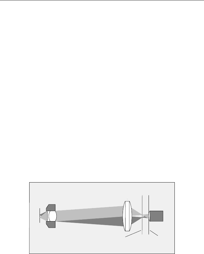
For nondescanned detectors it is important to use a field lens in front of the de-
tector. The general optical principle is shown in Fig. 5.92. In this figure it was
assumed that the fluorescence light is separated from the excitation before it
passes the tube lens; the lens diameters are exaggerated.
Sample
Microscope
Lens
Field
Lens
plane of sample
Conjugate image
Plane of image
of microscope
lens
Detector
Fig. 5.92 Field lens for nondescanned detection
The fluorescent spot in the sample moves with the scanning. Consequently, the
bundle of the fluorescence light wobbles. If a detector is placed in some distance
from the microscope lens, vignetting of the image is almost inevitable. A correctly
designed field lens projects a stationary image of the microscope objective lens
onto the detector. The field lens should be made as large as possible to collect
scattered light from deep sample layers. It must be at least large enough to collect
the light at the maximum scanning amplitude.
If the detector is not in the plane of the image of the microscope lens, either in-
tensity may be lost or the image may be vignetted, or both. By no means should
158 5 Application of Modern TCSPC Techniques
the detector be placed in the conjugate sample plane. This plane is closer to the
field lens than the image of the microscope lens. Placing the detector there results
in scanning the fluorescent spot over the detector. The result is that the detector
structure prints through into the image. An example for an H5773 detector is
shown in Fig. 5.93.
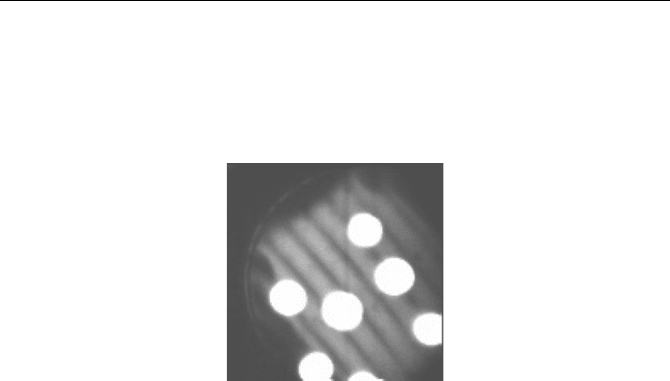
Fig. 5.93 Result of placing the detector in a conjugate sample plane. The dynode structure,
in this case of a H5773, appears in the image. The bright circles are fluorescing beads in the
sample
If a tube lens is in the fluorescence detection path, the beam configuration may
be slightly different than that shown in Fig. 5.90. The microscope may also have
additional lenses in the beam path to project an image on a camera, or to increase
the light-collection area of direct detection. In any case, there is a simple way to
find the image of the microscope lens behind the field lens: Turn on the micro-
scope lamp in the transmission beam path, so that the condenser lens fully illumi-
nates the aperture of the microscope lens. The image of the microscope lens can
then easily be found by holding a sheet of paper behind the field lens.
The Problem of IRF Recording
Recording the IRF in a one-photon microscope may appear simple at first glance.
The sample would be replaced with a scattering medium or a mirror, and a TCSPC
image or a single waveform would be recorded at the wavelength of the excitation
laser. In practice, recording an accurate IRF in a microscope can be very difficult.
The laser is scattered and reflected at many places in the microscope itself. A
recording taken at the laser wavelength therefore does not represent the true exci-
tation profile of the sample. Moreover, it can be difficult to remove the laser
blocking filter, or to find a sample that has appropriate scattering and no fluores-
cence. Reflecting the laser light back from a mirror requires the mirror to be
placed accurately in the focus of the microscope lens and perpendicular to the
optical axis. Therefore, some scepticism is recommended if an IRF is recorded this
way.
For a two-photon microscope the situation is even more complicated. Even if
the NIR blocking filter is removable, a detector with a bialkali or GaAsP cathode
is insensitive at the laser wavelength. Of course, by increasing the laser power
something is detected in any PMT. However, in the NIR a photocathode for the

5.7 TCSPC Laser Scanning Microscopy 159
visible is almost transparent, and a large fraction of the photoelectrons may be
emitted not from the cathode but from the first dynode. Therefore an IRF recorded
this way is not the true excitation profile of the sample.
The logical way to record an IRF in a two-photon microscope is to use second-
harmonic generation (SHG). SHG in a crystal is not very useful because the SHG
is emitted in the direction of the laser radiation. Returning it to the microscope
lens with the right NA is difficult. The best way to record an IRF in a microscope
is SHG by hyper Rayleigh scattering in a suspension of gold nanoparticles [206,
375].
If the IRF cannot be recorded, it is often derived from the data themselves. The
IRF is approximated by a gaussian function, and the width is adjusted to give the
best fit to the rising edge of the decay functions. The method gives acceptable
results for lifetimes down to the FWHM of the IRF. It does, however, not take into
account the low-amplitude bumps present in the IRF of many PMTs. In multiex-
ponential decay analysis the simplification of the IRF can result in false lifetime
components of low amplitude.
Count Rate of FLIM Experiments
Compared to steady-state imaging, the count rates typically obtained in FLIM
experiments are relatively low. An exact comparison is difficult because the re-
corded photon rates for steady-state imaging are not explicitly known. However,
an approximate estimation can be based on the signal-to-noise ratio of the ac-
quired intensity images. Video-rate imaging systems record images at rates of
more than 100 frames per second. The individual images of the video sequence are
noisy, but nevertheless show the spatial structure of the sample. That means that
the images contain 1 to 10 photons per pixel. For 10
5
pixels and 10 ms acquisition
time per image, the count rate must be of the order of 10
7
to 10
8
photons per sec-
ond. Steady-state images in confocal laser scanning microscopes are often ob-
tained by recording a single frame of about 1 s. Assuming a signal-to-noise ratio
of 10, i.e. 100 photons per pixel, the count rate amounts to about 10
7
photons per
second.
The count rates of FLIM in laser scanning microscopes are substantially lower.
The CFP-YFP FRET images shown above were recorded at 5010
3
s
-1
. CFP-YFP
FRET in
Caenorhabditis Elegans [73] was recorded at < 10
5
s
-1
. Two-photon
autofluorescence of skin delivers about 6010
3
s
-1
. These count rates are by factor
of 10 to 100 lower than the maximum count rate of the TCSPC devices used. It
should be expected that much higher count rates are obtained from stained tissue.
Imaging of the pH in skin tissue by BCECF was performed at an average rate of
only 210
6
s
-1
[216], although the frequency-domain technique used is capable of
processing much higher rates. A count rate of 1410
6
s
-1
was used to record the
image shown in Fig. 5.84. This count rate comes close to the rates used in steady-
state imaging. However, it caused severe photobleaching and lifetime changes (see
Fig. 5.85).
The low count rates of FLIM experiments require an explanation.
Tolerable photobleaching. If steady-state images are used to investigate the
spatial structure of a specimen, a large amount of photobleaching can be tolerated.

160 5 Application of Modern TCSPC Techniques
Photobleaching may even remain unnoticed if the image is recorded in a single
scan. Z stacks recorded by two-photon imaging may entirely bleach the imaged
plane and still reveal the spatial structure of the specimen. In FLIM recordings,
photobleaching must be kept at a much lower level. In most cases the photo-
bleaching rate is higher for types of molecules with longer lifetimes. Photobleach-
ing can therefore change the lifetime distribution considerably. The change of the
decay function is real and should not be confused with photobleaching-related
artefacts in techniques with sequential recording of several images.
Photobleaching rate. The photodamage and photobleaching rates are different
for one-photon and two-photon excitation. Although this is not commonly ac-
cepted, photobleaching seems to be faster for two-photon excitation [140]. More-
over, with increasing excitation intensity the photobleaching rate increases more
rapidly than the fluorescence intensity [239, 396]. However, two-photon
photobleaching is confined to the scanned image plane. Photobleaching for one-
photon excitation is usually considered to vary linearly with the excitation dose.
However, recent experiments have shown that nonlinear effects can also be pre-
sent for one-photon excitation [52]. Consequently, keeping the photobleaching
low for a given number of emitted photons means keeping the excitation power
low and acquiring photons over a longer time period.
Concentration of fluorophores. The fluorophore concentration can vary over a
wide range. Stained beads can contain almost any dye concentration, and a cell
can contain a highly concentrated fluorophore in the entire cytoplasm. However,
such samples are rarely interesting for FLIM experiments. In samples investigated
by FLIM, either specific targets in the cells are labelled or the cells are transfected
to express a fluorophore in highly specific subunits. The total amount of fluoro-
phore in these cases can be 100 times lower than in the first case. Autofluores-
cence in unstained samples is particularly weak because both the concentration
and the quantum efficiency of the fluorophores are low.
Excitation and detection geometry. The sample volume from which the fluores-
cence is detected can differ considerably. In two-photon imaging the excited vol-
ume is of the order of 0.1 µm
3
. Confocal imaging with a wide pinhole detects from
a considerably larger sample volume. Consequently, the fluorescence comes from
a larger number of molecules, and a correspondingly higher intensity is available.
The majority of FLIM experiments are performed in two-photon systems with a
small focal volume and low intensity.
Figure of Merit and Counting Efficiency of TCSPC FLIM
An ideal lifetime technique would record all photons detected within the fluores-
cence decay function, over a time interval much longer than the fluorescence de-
cay time, in a large number of time channels, and with an infinitely short temporal
instrument response function. The standard deviation,
V
W
of the fluorescence life-
time,
W
, for a number of recorded photons, N, would be
N/
WV
W
(5.16)

5.7 TCSPC Laser Scanning Microscopy 161
and the signal-to-noise-ratio
NSNR
W
VW
/ (5.17)
In other words, a single-exponential fluorescence lifetime can ideally be derived
from a given number of photons per pixel with the same relative uncertainty as the
intensity [187, 274]. The efficiency of a lifetime technique is often characterised
by the „Figure of Merit“ [19, 84, 274, 409]. The figure of merit,
F, compares the
SNR (signal-to-noise ratio) of an ideal recording device with the SNR of the tech-
nique under consideration:
real
ideal
SNR
SNR
F
(5.18)
The loss of SNR in a real technique can also be expressed by the counting effi-
ciency. The counting efficiency,
E, is the ratio of the number of photons ideally
needed to the number needed by the considered technique:
2
/1 FE (5.19)
As long as the signal processing time (the „dead time“) for the photons is small
compared to the average time between the photons, the TCSPC technique yields a
near-perfect counting efficiency and a maximum signal-to-noise ratio for a given
acquisition time. For higher count rates an increasing number of photons is lost in
the dead time, and the efficiency decreases (see Sect. 7.9.2, page 338). The count-
ing efficiency, E, is
d
tr
E
det
1
1
(5.20)
and the figure of merit
d
trF
det
1 (5.21)
with
r
det
= detector count rate, t
d
= dead time.
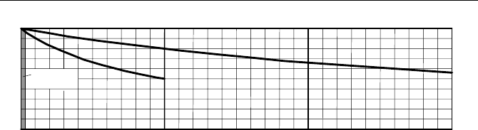
The efficiency versus the count rate of a single TCSPC channel and a four-
module TCSPC system is shown in Fig. 5.94. The efficiency of the single-channel
system remains better than 0.9 and the figure of merit better than 1.05 for count
rates up to 1 MHz detector count rate. This is better than for any other lifetime
imaging technique. For a detector count rate of 10 MHz, the values are 0.5 and
1.4, respectively. Higher count rates not only result in a substantial loss in effi-
ciency but also increase lifetime errors by pile-up-effect (see Sect. 7.9.1, page
332). For detector count rates above 10 MHz the solution is multimodule systems;
see Sect. 5.7.5, page 146.
162 5 Application of Modern TCSPC Techniques
12345678910 MHz
1.0
0.9
0.8
0.7
0.6
0.5
0.4
0.3
0.2
0.1
Detector Count Rate
Efficienc
y
Typical Rates
20 MHz 30 MHz
2p Excitation
a
b
Fig. 5.94 Efficiency versus count rate for a single TCSPC channel (a) and a system of four
parallel TCSPC channels (b). Dead time 100 ns
A system with four parallel TCSPC channels can be used up to 40 MHz detec-
tor count rate. When this count rate is compared to the count rates of other time-
resolved detection techniques, the high efficiency of TCSPC must be taken into
account. Consider a gated image intensifier that is operated at a gate width of 100
to 200 ps, i.e. at a time resolution equivalent to a mediocre TCSPC system. The
short gate width then results in an efficiency of 0.05 to 0.1. A four-channel
TCSPC system operated at 40 MHz has an efficiency of 50%. The 40 MHz detec-
tor count rate of the TCSPC system therefore corresponds to an input count rate of
200 to 400 MHz in the image intensifier.
It should be noted that in practice the values of
F and E also depend on the
width of the IRF, the detector background rate, the width of the used TAC win-
dow, and the numerical stability of the lifetime analysis algorithm. However, the
impact of these parameters can be kept small by using a fast detector and appro-
priate system setting. Moreover,
F was originally defined for a single-detector
device and single-exponential decay. The definition of
F is therefore not directly
applicable to multiwavelength TCSPC and multiexponential decay analysis.
Acquisition Time of FLIM
Acquisition times for TCSPC FLIM measurements can vary widely. In vivo life-
time measurements of the human ocular fundus in conjunction with an ophthalmic
scanner delivered single exponential lifetimes for an array of 128 u 128 pixels
within a few seconds [451, 452, 454]. High-quality double exponential lifetime
images of microscopic samples were obtained within 10 seconds by a four-module
TCSPC system (see Fig. 5.84) [39]. On the other hand, for the double exponential
decay data of FRET measurements in live cells (see Fig. 5.87), acquisition times
ranged from 5 to 30 minutes [32, 37]. In practice the acquisition time depends on
the size and the photostability of the sample and the requirements for accuracy
rather than on the counting capability of the TCSPC device.
Figure 5.95 shows the acquisition time as a function of the product of the num-
ber of pixels and the number of wavelength channels. The left diagram is for a
count rate of 10
6
/s. Count rates of this order require highly fluorescent samples of
good photostability. The right diagram is for a count rate of 10
4
/s. Count rates this
low are typical for autofluorescence of cells and tissue and for samples of poor
photostability. The number of photons per pixel ranges from 100 for rough single
exponential decay mapping to 10
5
for precision multiexponential decay analysis.

5.8 Other TCSPC Microscopy Techniques 163
Figure 5.95 shows that relatively long acquisition times must be used, espe-
cially for large numbers of pixels and multiexponential decay measurements of
samples of low photostability. The only way to obtain high-accuracy lifetime data
for such samples is often to reduce the effective number of pixels. This is possible
without sacrificing spatial resolution by binning only the decay curves. The image
is scanned with high resolution, and the intensity is calculated from the photon
numbers of the individual pixels. Then the fluorescence decay curves of several
adjacent pixels are binned, and the decay analysis is performed on the binned data.
10000s
1000s
100s
10s
1s
100ms
10ms
1ms
0.1ms
1 10 100 1000 10,000 100,000
Acquisition
Time
Pixels x Wavelength Channels
100
1000
10,000
100,000
Photons/
Pixel
Count Rate
10 /s
6
10000s
1000s
100s
10s
1s
100ms
10ms
1ms
0.1ms
1 10 100 1000 10,000 100,000
Acquisition
Time
Pixels x Wavelength Channels
100
1000
10,000
100,000
Photons/
Pixel
Count Rate
10 /s
4
Fig. 5.95 Acquisition times for a count rate of 10
6
/s (left) and 10
4
/s (right) for various
numbers of photons per pixel.
It should be pointed out that long acquisition times are not a specific feature of
TCSPC imaging. The long acquisition times result from the higher quality stan-
dards usually expected with TCSPC data and from the fact that TCSPC can work
at intensities so low that other techniques fail.
5.8 Other TCSPC Microscopy Techniques
5.8.1 TCSPC Lifetime Imaging by Scan Stages
Commercial confocal and two-photon laser scanning microscopes scan the laser
beam by fast galvano-driven mirrors. Typical pixel dwell times are of the order of
a few microseconds. Depending on the number of pixels, a complete frame is
scanned within 25 ms to several seconds.
Images can also be obtained by scanning the sample by a piezo-driven scan
stage. The principle is the same as in the laser scanning microscope, but the opti-
cal beam scanner is replaced with a scan stage that moves the sample. The sample
scanning technique is shown in Fig. 5.96.
From the vantage point of TCSPC, imaging by a piezo stage is a „slow scan“
procedure. That means a full fluorescence decay curve is recorded in the current
pixel before the scanner moves to the next one. Lifetime images can be therefore
acquired by almost any TCSPC module and operation mode. Applications of one-
dimensional TCSPC with slow scanning are described in [76] and [74]. These