Berg J.M., Tymoczko J.L., Stryer L. Biochemistry
Подождите немного. Документ загружается.

I. The Molecular Design of Life 3. Protein Structure and Function 3.2. Primary Structure: Amino Acids Are Linked by Peptide Bonds to Form Polypeptide Chains
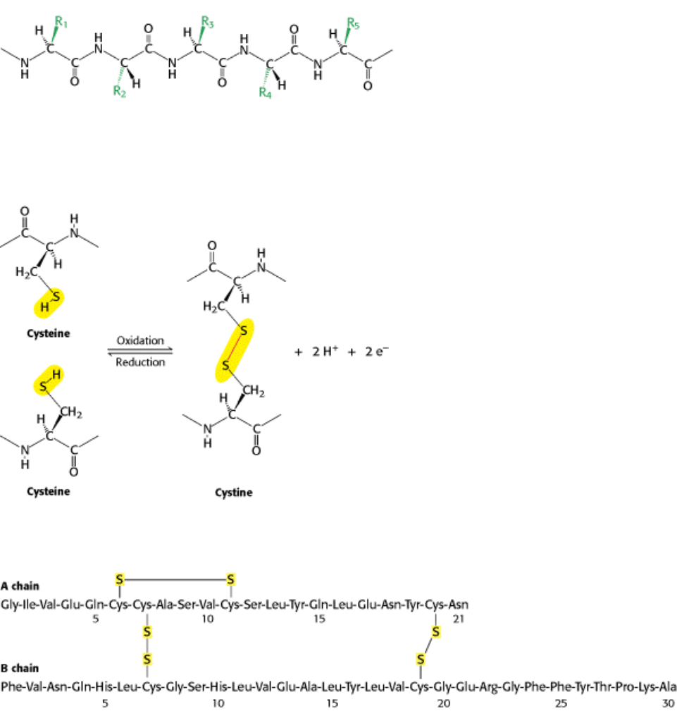
Figure 3.20. Components of a Polypeptide Chain. A polypeptide chain consists of a constant backbone (shown in
black) and variable side chains (shown in green).
I. The Molecular Design of Life 3. Protein Structure and Function 3.2. Primary Structure: Amino Acids Are Linked by Peptide Bonds to Form Polypeptide Chains
Figure 3.21. Cross-Links. The formation of a disulfide bond from two cysteine residues is an oxidation reaction.
I. The Molecular Design of Life 3. Protein Structure and Function 3.2. Primary Structure: Amino Acids Are Linked by Peptide Bonds to Form Polypeptide Chains
Figure 3.22. Amino Acid Sequence of Bovine Insulin.
I. The Molecular Design of Life 3. Protein Structure and Function 3.2. Primary Structure: Amino Acids Are Linked by Peptide Bonds to Form Polypeptide Chains
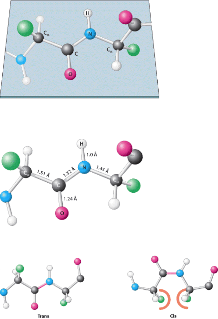
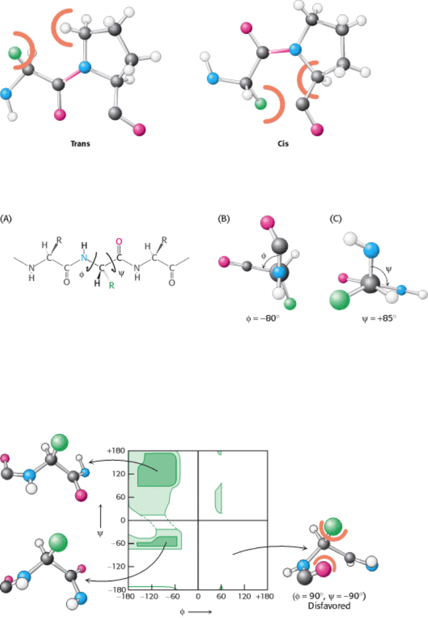
Figure 3.23. Peptide Bonds Are Planar. In a pair of linked amino acids, six atoms (C
α
, C, O, N, H, and C
α
) lie in a
plane. Side chains are shown as green balls.
I. The Molecular Design of Life 3. Protein Structure and Function 3.2. Primary Structure: Amino Acids Are Linked by Peptide Bonds to Form Polypeptide Chains
Figure 3.24. Typical Bond Lengths Within a Peptide Unit. The peptide unit is shown in the trans configuration.
I. The Molecular Design of Life 3. Protein Structure and Function 3.2. Primary Structure: Amino Acids Are Linked by Peptide Bonds to Form Polypeptide Chains
Figure 3.25. Trans and Cis Peptide Bonds. The trans form is strongly favored because of steric clashes that occur in
the cis form.
I. The Molecular Design of Life 3. Protein Structure and Function 3.2. Primary Structure: Amino Acids Are Linked by Peptide Bonds to Form Polypeptide Chains
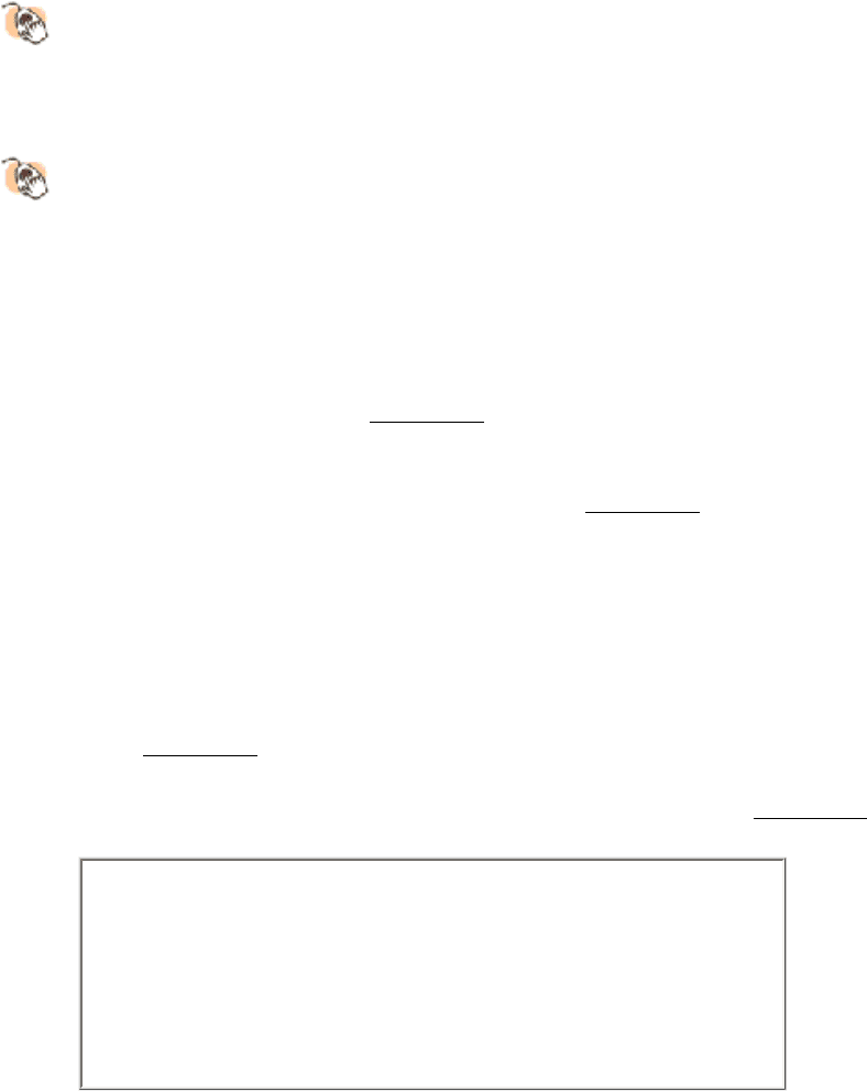
Figure 3.26. Trans and Cis X-Pro Bonds. The energies of these forms are relatively balanced because steric clashes
occur in both forms.
I. The Molecular Design of Life 3. Protein Structure and Function 3.2. Primary Structure: Amino Acids Are Linked by Peptide Bonds to Form Polypeptide Chains
Figure 3.27. Rotation About Bonds in a Polypeptide. The structure of each amino acid in a polypeptide can be
adjusted by rotation about two single bonds. (A) Phi (φ) is the angle of rotation about the bond between the nitrogen and
the α-carbon atoms, whereas psi (ψ) is the angle of rotation about the bond between the α-carbon and the carbonyl
carbon atoms. (B) A view down the bond between the nitrogen and the α-carbon atoms, showing how φ is measured. (C)
A view down the bond between the α-carbon and the carbonyl carbon atoms, showing how ψ is measured.
I. The Molecular Design of Life 3. Protein Structure and Function 3.2. Primary Structure: Amino Acids Are Linked by Peptide Bonds to Form Polypeptide Chains
Figure 3.28. A Ramachandran Diagram Showing the Values of φ and ψ. Not all φ and ψ values are possible without
collisions between atoms. The most favorable regions are shown in dark green; borderline regions are shown in light
green. The structure on the right is disfavored because of steric clashes.
I. The Molecular Design of Life 3. Protein Structure and Function
3.3. Secondary Structure: Polypeptide Chains Can Fold Into Regular Structures Such
as the Alpha Helix, the Beta Sheet, and Turns and Loops
Can a polypeptide chain fold into a regularly repeating structure? In 1951, Linus Pauling and Robert Corey proposed two
periodic structures called the α helix (alpha helix) and the β pleated sheet (beta pleated sheet). Subsequently, other
structures such as the β turn and omega ( Ω ) loop were identified. Although not periodic, these common turn or loop
structures are well defined and contribute with α helices and β sheets to form the final protein structure.
Structural Insights, appearing throughout the book, are molecular modeling-
based tutorials that enable you to review structure and learn what the latest
research tells us about the workings of the molecule. To access, go to the Web
site: www.whfreeman.com/biochem5, and select the chapter, Structural
Insights, and the title.
Structural Insights, Elements of Protein Structure provides interactive
representations of some of the important elements of protein architecture
described in this chapter, including a summary of secondary structure motifs.
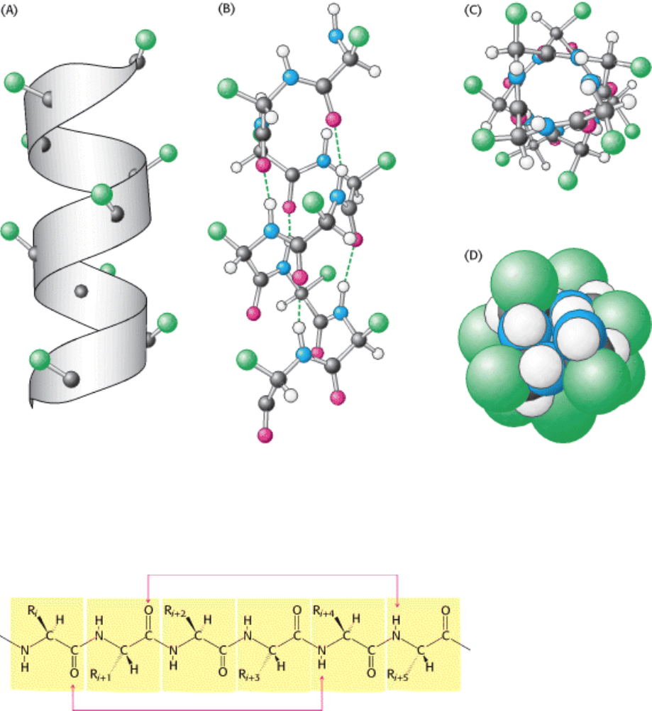
3.3.1. The Alpha Helix Is a Coiled Structure Stabilized by Intrachain Hydrogen Bonds
In evaluating potential structures, Pauling and Corey considered which conformations of peptides were sterically allowed
and which most fully exploited the hydrogen-bonding capacity of the backbone NH and CO groups. The first of their
proposed structures, the α helix, is a rodlike structure (Figure 3.29). A tightly coiled backbone forms the inner part of the
rod and the side chains extend outward in a helical array. The α helix is stabilized by hydrogen bonds between the NH
and CO groups of the main chain. In particular, the CO group of each amino acid forms a hydrogen bond with the NH
group of the amino acid that is situated four residues ahead in the sequence (Figure 3.30). Thus, except for amino acids
near the ends of an α helix, all the main-chain CO and NH groups are hydrogen bonded. Each residue is related to the
next one by a rise of 1.5 Å along the helix axis and a rotation of 100 degrees, which gives 3.6 amino acid residues per
turn of helix. Thus, amino acids spaced three and four apart in the sequence are spatially quite close to one another in an
α helix. In contrast, amino acids two apart in the sequence are situated on opposite sides of the helix and so are unlikely
to make contact. The pitch of the α helix, which is equal to the product of the translation (1.5 Å) and the number of
residues per turn (3.6), is 5.4 Å. The screw sense of a helix can be right-handed (clockwise) or left-handed
(counterclockwise). The Ramachandran diagram reveals that both the right-handed and the left-handed helices are
among allowed conformations (Figure 3.31). However, right-handed helices are energetically more favorable because
there is less steric clash between the side chains and the backbone. Essentially all α helices found in proteins are right-
handed. In schematic diagrams of proteins, α helices are depicted as twisted ribbons or rods (Figure 3.32).
Screw sense
Describes the direction in which a helical structure rotates with
respect to its axis. If, viewed down the axis of a helix, the chain turns
in a clockwise direction, it has a right-handed screw sense. If the
turning is counterclockwise, the screw sense is left-handed.
Pauling and Corey predicted the structure of the α helix 6 years before it was actually seen in the x-ray reconstruction of
the structure of myoglobin. The elucidation of the structure of the α helix is a landmark in biochemistry because it
demonstrated that the conformation of a polypeptide chain can be predicted if the properties of its components are

rigorously and precisely known.
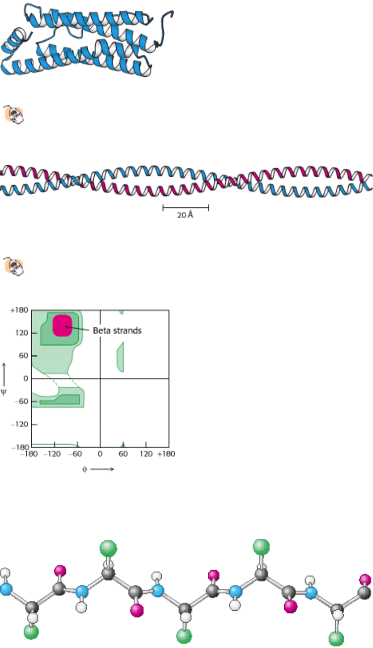
The α-helical content of proteins ranges widely, from nearly none to almost 100%. For example, about 75% of the
residues in ferritin, a protein that helps store iron, are in α helices (Figure 3.33). Single α helices are usually less than 45
Å long. However, two or more α helices can entwine to form a very stable structure, which can have a length of 1000 Å
(100 nm, or 0.1 µ m) or more (Figure 3.34). Such α-helical coiled coils are found in myosin and tropomyosin in muscle,
in fibrin in blood clots, and in keratin in hair. The helical cables in these proteins serve a mechanical role in forming stiff
bundles of fibers, as in porcupine quills. The cytoskeleton (internal scaffolding) of cells is rich in so-called intermediate
filaments, which also are two-stranded α-helical coiled coils. Many proteins that span biological membranes also contain
α helices.
3.3.2. Beta Sheets Are Stabilized by Hydrogen Bonding Between Polypeptide Strands
Pauling and Corey discovered another periodic structural motif, which they named the β pleated sheet (β because it was
the second structure that they elucidated, the α helix having been the first). The β pleated sheet (or, more simply, the β
sheet) differs markedly from the rodlike α helix. A polypeptide chain, called a β strand, in a β sheet is almost fully
extended rather than being tightly coiled as in the α helix. A range of extended structures are sterically allowed (Figure
3.35).
The distance between adjacent amino acids along a β strand is approximately 3.5 Å, in contrast with a distance of 1.5 Å
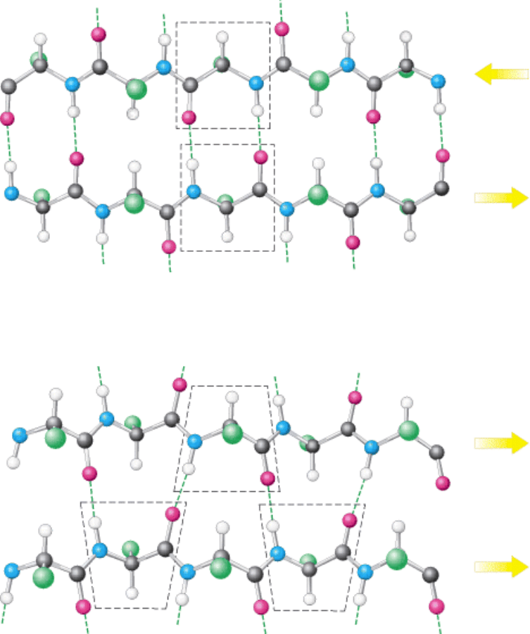
along an α helix. The side chains of adjacent amino acids point in opposite directions (Figure 3.36). A β sheet is formed
by linking two or more β strands by hydrogen bonds. Adjacent chains in a β sheet can run in opposite directions
(antiparallel β sheet) or in the same direction (parallel β sheet). In the antiparallel arrangement, the NH group and the
CO group of each amino acid are respectively hydrogen bonded to the CO group and the NH group of a partner on the
adjacent chain (Figure 3.37). In the parallel arrangement, the hydrogen-bonding scheme is slightly more complicated.
For each amino acid, the NH group is hydrogen bonded to the CO group of one amino acid on the adjacent strand,
whereas the CO group is hydrogen bonded to the NH group on the amino acid two residues farther along the chain
(Figure 3.38). Many strands, typically 4 or 5 but as many as 10 or more, can come together in β sheets. Such β sheets can
be purely antiparallel, purely parallel, or mixed (Figure 3.39).
In schematic diagrams, β strands are usually depicted by broad arrows pointing in the direction of the carboxyl-terminal
end to indicate the type of β sheet formed
parallel or antiparallel. More structurally diverse than α helices, β sheets can
be relatively flat but most adopt a somewhat twisted shape (Figure 3.40). The β sheet is an important structural element
in many proteins. For example, fatty acid-binding proteins, important for lipid metabolism, are built almost entirely from
β sheets (Figure 3.41).
3.3.3. Polypeptide Chains Can Change Direction by Making Reverse Turns and Loops
Most proteins have compact, globular shapes, requiring reversals in the direction of their polypeptide chains. Many of
these reversals are accomplished by a common structural element called the reverse turn (also known as the β turn or
hairpin bend), illustrated in Figure 3.42. In many reverse turns, the CO group of residue i of a polypeptide is hydrogen
bonded to the NH group of residue i + 3. This interaction stabilizes abrupt changes in direction of the polypeptide chain.
In other cases, more elaborate structures are responsible for chain reversals. These structures are called loops or
sometimes Ω loops (omega loops) to suggest their overall shape. Unlike α helices and β strands, loops do not have
regular, periodic structures. Nonetheless, loop structures are often rigid and well defined (Figure 3.43). Turns and loops
invariably lie on the surfaces of proteins and thus often participate in interactions between proteins and other molecules.
The distribution of α helices, β strands, and turns along a protein chain is often referred to as its secondary structure.
I. The Molecular Design of Life 3. Protein Structure and Function 3.3. Secondary Structure: Polypeptide Chains Can Fold Into Regular Structures Such as the Alpha Helix, the Beta Sheet, and Turns and Loops
Figure 3.29. Structure of the α Helix. (A) A ribbon depiction with the α-carbon atoms and side chains (green) shown.
(B) A side view of a ball-and-stick version depicts the hydrogen bonds (dashed lines) between NH and CO groups. (C)
An end view shows the coiled backbone as the inside of the helix and the side chains (green) projecting outward. (D) A
space-filling view of part C shows the tightly packed interior core of the helix.
I. The Molecular Design of Life 3. Protein Structure and Function 3.3. Secondary Structure: Polypeptide Chains Can Fold Into Regular Structures Such as the Alpha Helix, the Beta Sheet, and Turns and Loops
Figure 3.30. Hydrogen-Bonding Scheme For an α helix. In the α helix, the CO group of residue n forms a hydrogen
bond with the NH group of residue n+ 4.
I. The Molecular Design of Life 3. Protein Structure and Function 3.3. Secondary Structure: Polypeptide Chains Can Fold Into Regular Structures Such as the Alpha Helix, the Beta Sheet, and Turns and Loops
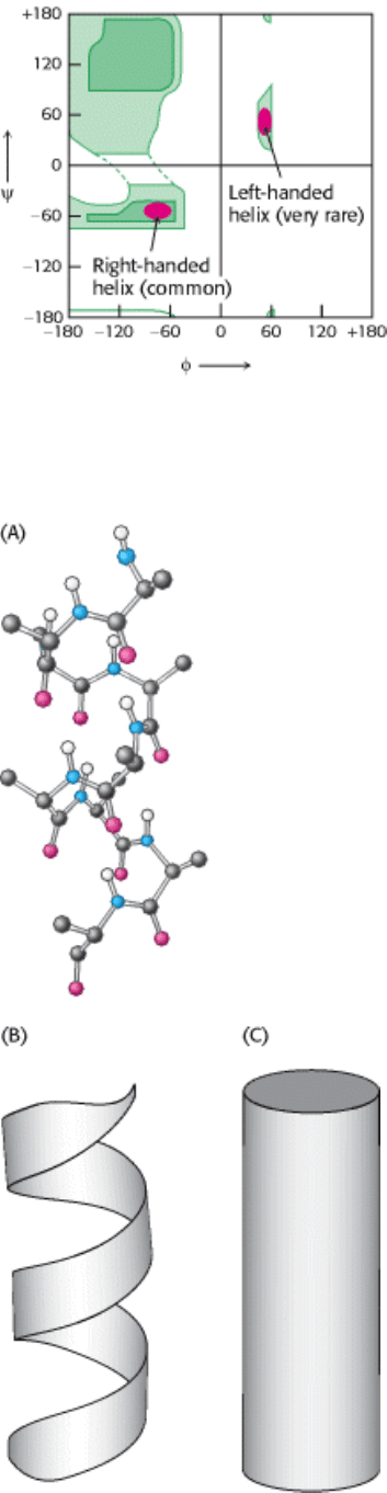
Figure 3.31. Ramachandran Diagram for Helices. Both right- and left-handed helices lie in regions of allowed
conformations in the Ramachandran diagram. However, essentially all α helices in proteins are right-handed.
I. The Molecular Design of Life 3. Protein Structure and Function 3.3. Secondary Structure: Polypeptide Chains Can Fold Into Regular Structures Such as the Alpha Helix, the Beta Sheet, and Turns and Loops
Figure 3.32. Schematic Views OF α Helices. (A) A ball-and-stick model. (B) A ribbon depiction. (C) A cylindrical
depiction.
I. The Molecular Design of Life 3. Protein Structure and Function 3.3. Secondary Structure: Polypeptide Chains Can Fold Into Regular Structures Such as the Alpha Helix, the Beta Sheet, and Turns and Loops
Figure 3.33. A Largely α Helical Protein. Ferritin, an iron-storage protein, is built from a bundle of α helices.
I. The Molecular Design of Life 3. Protein Structure and Function 3.3. Secondary Structure: Polypeptide Chains Can Fold Into Regular Structures Such as the Alpha Helix, the Beta Sheet, and Turns and Loops
Figure 3.34. An α -Helical Coiled Coil.
The two helices wind around one another to form a superhelix. Such structures
are found in many proteins including keratin in hair, quills, claws, and horns.
I. The Molecular Design of Life 3. Protein Structure and Function 3.3. Secondary Structure: Polypeptide Chains Can Fold Into Regular Structures Such as the Alpha Helix, the Beta Sheet, and Turns and Loops
Figure 3.35. Ramachandran Diagram For β Strands. The red area shows the sterically allowed conformations of
extended, β-strand-like structures.
I. The Molecular Design of Life 3. Protein Structure and Function 3.3. Secondary Structure: Polypeptide Chains Can Fold Into Regular Structures Such as the Alpha Helix, the Beta Sheet, and Turns and Loops
Figure 3.36. Structure of a β Strand. The side chains (green) are alternately above and below the plane of the strand.
I. The Molecular Design of Life 3. Protein Structure and Function 3.3. Secondary Structure: Polypeptide Chains Can Fold Into Regular Structures Such as the Alpha Helix, the Beta Sheet, and Turns and Loops
Figure 3.37. An Antiparallel β Sheet. Adjacent β strands run in opposite directions. Hydrogen bonds between NH and
CO groups connect each amino acid to a single amino acid on an adjacent strand, stabilizing the structure.
I. The Molecular Design of Life 3. Protein Structure and Function 3.3. Secondary Structure: Polypeptide Chains Can Fold Into Regular Structures Such as the Alpha Helix, the Beta Sheet, and Turns and Loops
Figure 3.38. A Parallel β Sheet. Adjacent β strands run in the same direction. Hydrogen bonds connect each amino acid
on one strand with two different amino acids on the adjacent strand.
I. The Molecular Design of Life 3. Protein Structure and Function 3.3. Secondary Structure: Polypeptide Chains Can Fold Into Regular Structures Such as the Alpha Helix, the Beta Sheet, and Turns and Loops
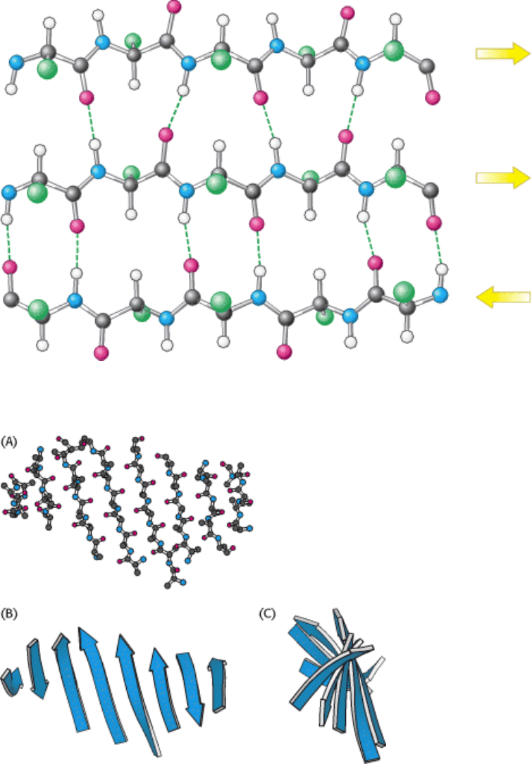
Figure 3.39. Structure of a Mixed β Sheet.
I. The Molecular Design of Life 3. Protein Structure and Function 3.3. Secondary Structure: Polypeptide Chains Can Fold Into Regular Structures Such as the Alpha Helix, the Beta Sheet, and Turns and Loops
Figure 3.40. A Twisted β Sheet. (A) A ball-and-stick model. (B) A schematic model. (C) The schematic view rotated
by 90 degrees to illustrate the twist more clearly.