Chen C.C. (ed.) Selected Topics in DNA Repair
Подождите немного. Документ загружается.

12
The Nuclear Compartmentation of Glutathione:
Effect on Cell Cycle Progression
Jelena Markovic, Nancy Mora, Amparo Gimeno, Consuelo Burguete,
José Luis García-Gimenez and Federico V. Pallardó
University of Valencia School of Medicine,
Spain
1. Introduction
The oxidizing environment, shared by all aerobic organisms and crucial for their survival,
poses a continuous threat to cellular structures all the living beings are made of. Structural
proteins and lipids, and cellular membranes they compose, nucleic acids and enzymes that
govern vital cellular processes are all susceptible to oxidative damage. Dealing with this
inevitable and constant danger is one of the greatest challenges the living being with aerobic
metabolism has to meet.
2. The implication of cellular redox balance in cell proliferation
2.1 Reactive oxidative species in cell proliferation
Classical work of Kelvin Davies (Davies, 1999), more than ten years ago, showed that the
cells show a whole range of responses to oxidative stress that depends on the intensity of the
stress. Low level of hydrogen peroxide induced mitogenic responses and stimulation of
proliferation; this observation was firstly reported by Oberly (Oberly et al., 1981) who have
described that oxidative stimuli, such as superoxide and hydrogen peroxide, could activate
signalling pathways that lead to proliferation. Davies et al. further assert that considerable
increase in the oxidant concentrations caused temporary growth arrest which became
permanent with a progressive increase. When high H
2
O
2
concentrations were used,
apoptosis took place and at very high oxidant levels the cells were killed by necrosis. A year
later, (Pani et al., 2000) demonstrate a causal link between redox changes and growth control
by cell density: they show that low level of oxygen species in the environment of
proliferating cells was not only stimulating but necessary for the correct mitogenic
signaling. This study was immediately followed by the work of Menon et al., 2003 who
suggested that an oxidation event early in G1 phase may be a critical regulatory step in the
progression of the cells into S phase. This lead to the development of the model of the
“redox cycle within a cell cycle” proposed by the same group several years later (Menon &
Goswami 2007).
According to this model, the transient change in ROS could modify the redox state of cell
cycle regulatory proteins, at their critical cysteine residues, and thus determine progression
or arrest in the proliferation. Antioxidant mechanism could scavenge ROS and reverse the
process. In accordance to these reports, Barry Halliwell (Halliwell, 2007) draw a complete

Selected Topics in DNA Repair
272
view of the present knowledge of the role of oxidative stress in promoting cancer, its
damaging effects to DNA, and its action on cell proliferation and apoptosis. Malignant cells
produce more radical species and, although antioxidant defence could also be induced in
these cells, they display a pro-oxidant state. However, apparently the oxidative stress
generated in these high proliferative cells does not exceed the level where oxidative damage
becomes so severe that cell function is impaired. This finding is in line with previously cited
reports and many others that support the role of reactive oxidative species mediated
signalling in the promotion of cell growth.
2.2 The bridge between the oxidative stress and cell proliferation - Glutathione
Glutathione (GSH) is the most abundant non-protein thiol in
mammalian cells (Meister &
Anderson, 1983). It is considered essential for survival in mammalian cells (Viña, 1990) and
yeast Meister & Anderson, 1983; Viña et al., 1978), but not in prokaryotic cells. The exact
nature of this important difference has not been elucidated. Glutathione was discovered in
1888 by Rey Pailhade as “organic hydrogenate of sulphur” (Rey Paihade, 1988) and
“rediscovered” and fully described by Sir Frederic Gowland Hopkins in the 1920s (Hopkins,
1929) and quoted by Sies several years later (Sies, 1999).
Glutathione has attracted the scientific interest with variable intensity along the century
since its discovery and many important cellular functions of this tripeptide were revealed
along the years. Glutathione shows a widespread localization within cells and considerably
high concentration in cells and tissues (up to 10 mM) (Tateishi et al., 1974). Examples of
normal physiological functions of glutathione known for a long time include regulation of
the transport of certain amino acids (Viña & Viña, 1983) control of cytoskeleton assembly
(Burchil et al. 1978) and regulation of enzymatic activity (Ernst et al., 1978; Ziegler, 1985).
During 1960s, GSH was demonstrated to be a co-substrate for a number of important
enzymatic reactions: GSH-S-transferase was described (Booth, 1961) and its role in a first–
line defence against electrophilic insult, obviously dependent on glutathione, was suggested
(Boyland, 1969). These pioneer works became the bases for many studies that lead to the
development of concepts such as drug and foreign compound detoxification, and multidrug
resistance (Smith, 1977) of crucial importance in the modern cancer therapy. Glutathione, as
it lacks toxicity linked to cysteine (Viña et al., 1983), is considered perfect as a cellular thiol
“redox buffer” with a purpose to maintain a given thiol/disulfide redox potential (Sies,
1999). Therefore, the redox properties and abundance that characterize this molecule grant it
a major role in protecting the cell against oxidants and electrophiles, and during 1980s this
particular role of glutathione is central in many research efforts.
Association of redox regulation with toxicity events lead to the introduction of the concept
of “oxidative stress” at biochemical and cellular level (Sies & Cadenas, 1985). Oxidative
stress is generally defined as an imbalance between prooxidants and antioxidants with a
considerable effect on other cellular components, including redox sensitive functional
groups of proteins. Nowadays, with the increasing awareness of the importance of ROS and
glutathione in cellular signalling, and the cellular redox environment in fundamental
physiological processes, a new definition of oxidative stress is proposed. According to Jones,
2006, oxidative stress may be better defined as a disruption of redox signaling and control.
Interestingly, more than 10 years ago, searching for a molecular link between oxidative
stress and cell proliferation, Cotgrave IA and Gerdes RG recommended similar term:
“oxidant mediated regulation” (Cortgreave and Gerdes, 1998).

The Nuclear Compartmentation of Glutathione: Effect on Cell Cycle Progression
273
2.3 Glutathione in cell proliferation
Several studies from more than 20 years ago have suggested that changes in low molecular
weight thiols (LMWT) are associated with regulation of cell growth. Harris and Patt
published (Harris & Pat, 1969) that nonproliferating mouse tumour cells contained LMWT
than proliferating cells and in early eighties various authors report similar results: human
lung and ovarian tumour cells during the exponential growth demonstrate higher GSH
levels than during nondividing state (Harris & Pat, 1969; Post, 1983). In accordance to these
findings, Kosower and Kosower (Kosower & Kosower, 1978) have demonstrated that
decrease of GSH biosynthesis in vivo inhibits tumour growth rate. Moreover, it was
suggested that cellular GSH may have to reach certain critical levels before proliferation can
be initiated and that variations in the protein sulphydryl redox status may directly relate to
regulation of cell growth (Atzori et al. 1990).
Defining the intrinsic cellular redox environment by estimation of glutathione
(GSH)/glutathione disulfide (GSSG) redox state, the group of Dean P. Jones (Nkabyo et al.
2002) concluded that each phase in the life of the cell is characterized by the certain redox
state. Proliferating cells are in the most reduced state, with the values of Eh between -260mV
and -230mV (Schafer & Buettner, 2001). Upon a growth arrest caused by differentiation
(Nkabyo et al. 2002) or contact inhibition (Schafer & Buettner, 2001) cells are 40 mV more
oxidised (-220mV to -190mV) while the apoptotic process is accompanied by further
oxidation up to -165mV (Sun & Oberley, 1996).
Therefore, while the cell progresses from proliferation, through contact inhibition,
differentiation, and finally apoptosis, there is an intrinsic and natural progression from more
reduced to more oxidised cellular redox environment. The universality of this model which
applies to various cells from different organisms (reviewed in Schafer & Buettner, 2001)
inspired a daring hypothesis of Schafer and Buettner on the implication and function of
thiols and disulfides as nano-switches. The GSSG/2GSH couple is imagined as a
switchboard that move the cell from proliferation through differentiation towards
programmed cell death, if the redox environment could not be maintained, or necrosis when
the oxidative insult is to severe.
2.4 Glutathiolation of regulatory proteins as a link between a stimulating oxidative
event and reduced cell environment in cell proliferation
During the last two decades the increasing body of evidence reveals that several
transcription factors undergo oxidant modification necessary for their activation. For
instance, the property of binding DNA and thus regulate gene expression of AP1, NfkB, p53,
and SP1 depends on the redox status of cysteinyl thiols in their structures (Sun & Oberley,
1996). Thus, the idea of protein glutathiolation as a regulatory mechanism of importance in
cell proliferation came into sight.
Glutathiolation is a protein modification which consists in the covalent union of the
tripeptide glutathione to the SH group of the cysteine residue. For a long time this reaction
was considered to be a consequence of the equilibrium between protein thiols and GSSG
inevitably related to oxidative stress. From this point of view, glutathiolation fulfills two
important functions. Firstly, its reversibility enables the preservation of glutathione in the
cell and serves as a buffer for the reduction potential; otherwise, GSSG efflux would cause
the loss of GSH from the cell, decreasing the reducing capacity which could be recovered
only by the synthesis of new GSH (Schafer & Buettner, 2001). Secondly, it provides

Selected Topics in DNA Repair
274
protection for protein-SH against irreversible modifications and protein damage in response
to higher levels of oxidative stress (Dalle-Donne et al., 2007). Interestingly, it was
demonstrated that glutathiolation as a posttranslational modification occurs not only during
oxidative stress, but also under basal conditions and is involved in regulating distinct
transcription factors, such as NF-kB (Pineda-Molina et al., 2001), its inhibitor factor IKK
(Reynaert et al., 2006) and c-Jun (Canela et al. 2007). Apparently, the binding capacity of
these proteins to DNA or other proteins is modulated by glutathiolation. This relatively
recent focus on the implication of glutathiolation modulatory effects on protein function
yielded important breakthrough in elucidation of the implication of this modification in
various physiological and pathological situations (Giustarini et al. 2004) and raises
interesting questions about its possible implications in cell proliferation.
2.4.1 The role of glutathione in DNA synthesis
Among the important roles that GSH plays in cellular physiology, and among the first to be
described, was its role in DNA synthesis. The pentose phosphate pathway is a cellular
source of NADPH that is involved in reductive biosynthesis. In this process, ribose-5-
phosphate is formed and subsequently used for the synthesis of RNA, DNA and nucleotide
coenzymes. Apart from the synthesis of nucleotides, NADPH is also required for the
formation of amino acids, fatty acids, cholesterol, neurotransmitters and nitric oxide (NO).
Furthermore, NADPH is the source of electrons in the process of reduction of
ribonucleotides to deoxyribonucleotides catalyzed by the ribonucleotide reductase
(Thelander & Reichard, 1979).
The route is initialized by two distinct but complemented systems, the thioredoxin system
and the glutaredoxin system. Thioredoxin operates by transferring the electrons to
ribonucleotide reductase, and they are supplied by the thioredoxin reductase and NADPH.
The glutaredoxin system is initialized by the glutathione reductase, which reduces the GSSG
to GSH using the NADPH as source of electrons. GSH is used by the glutaredoxin to
provide the reducing power to the ribonucleotide reductase (Zahedi et al., 2009).
The crucial role of glutathione in DNA synthesis has been extensively documented
(Thelander & Reichard, 1979; Holmgren, 1976). For instance, Dethlefsen and co-workers
(Dethlefsen et al. 1988) showed that glutathione depletion inhibits DNA synthesis in
mammary carcinoma cells. In addition, the vital importance of GSH in this process has also
been demonstrated in human T lymphocytes (Suthanthiran et al., 1990). As mentioned
above GSH is an indispensable requirement in eukaryotes. In contrast, it does not
demonstrate the same importance in prokariotes. It has been shown that E. Coli lacking
gshA, the rate limiting enzyme in the synthesis of GSH, can grow without GSH
supplementation (Greenberg & Demple, 1986; Miranda-Vizuete et al., 1996). On the
contrary, in yeast, the depletion of GSH does affect cell proliferation on the level different
from DNA synthesis: mutants deficient in GCS after GSH withdrawal arrest cells in G1,
whereas a strain with a defect in ribonucleotide reduction arrest cells in S phase (Wang et
al., 1997). Since other possible explanations, such as protection against oxidative stress or
protection against non-native protein disulfides, have been discarded (Spector el al., 2001), it
appears that the essential function of GSH in yeast is related to the redox properties of its
thiol group. Consequently, glutathione can be replaced by dithiothreitol, but not with a GSH
analog where a thiol group has been substituted by a methyl group (Grant et al., 1996). Since
GSH is the reductant for glutaredoxin as explained previously, and glutaredoxin is also

The Nuclear Compartmentation of Glutathione: Effect on Cell Cycle Progression
275
essential (Rodriguez-Manzaneque et al., 1999) presumably their vital importance may be
interdependent. Then GSH seems to be important in S phase. During the process of DNA
replication, errors, such as double-strand breaks (DSBs) that arise from stalled replication
forks, require attention by the DNA damage response proteins. Thus, the correct control of
DNA synthesis and probably essential molecules, such as GSH, are necessary for the correct
DNA processing.
One of the most important proteins involved in DNA damage signaling pathway is the
ataxia-telangiectasa mutated protein (ATM). This central signaling protein, mainly for DSBs,
is involved in the repairing DNA process necessary after replication stress. Thus cells
lacking ATM fail to execute many of the cellular responses to DNA damage (Zhou &
Elledge, 2000). In addition, control of ATM responses after DNA replication may be
necessary for the correct cell cycle control. In that way, ATM is a central component in the
cell cycle regulation. Therefore, patients with ataxia telangiectasia have reduction in DNA
synthesis (Painter & Young, 1980). Furthermore, a recent work published by Guo Z. and
coworkers describes using a series of elegant experiments how ATM sense the redox
changes to modulate their activity (Guo et al, 2010). Interestingly, these authors propose that
ATM may regulate global cellular responses to oxidative stress, remarking the essential link
between redox control and DNA interacting, remodeling or repairing proteins. In Fanconi
anemia for instance, Castillo and coworkers have shown that ATM dependent
phosphorylation of FANCD2, one of the main proteins in the Fanconi anemia pathway of
DNA repair, is necessary for normal S-phase checkpoint activation after oxidative stress
(Castillo et al, 2011).
2.4.2 Regulation of telomerase activity by glutathione
The eukaryotic chromosomes are capped by telomeres, which consist of TTAGGG DNA
sequences repeated in tandem, associated with several proteins, which protect the final
regions of chromosomes. These structures play an important role in the stability and the
complete replication of the chromosomes. Conventional DNA polymerases cannot fully
replicate the 3’-end of the lagging strand of linear molecules, and therefore in every cell
division telomeric sequences are lost (Komberg, 1969). Telomerase is an important enzyme
that ensures the maintenance of normal telomere length. This activity is high in human
cancers (Kim et al., 1994), but virtually absent in normal human tissues, except germinal
cells (Harley et al., 1990). Telomerase regulation is not completely understood, but its
changes are related to both cancer and aging (Sharpless & Depinho, 2004). Studies carried
out by Jady et al. show that human telomeres are more accessible during the S-phase (Jady
et al., 2006) and that the telomerase assembly with telomeres takes place at this specific
moment of the cell cycle (Jady et al., 2006; Tomlison et al., 2006). Telomerase plays a key role
in cellular homeostasis, because it maintains the length of the telomeres. This especially
important in germinal cells in which it is necessary to keep a normal telomeric length after
many cellular divisions. Important contributions about the epigenetic control of telomeres
have been reported recently (Koziel et al., 2011). In that way, Maria Blasco has suggested
that telomeres are under epigenetic control (García-Cao et al., 2004). Mammalian telomeres
and subtelomeric regions are enriched in epigenetic marks that are characteristic of
heterochromatin. In addition, histone deacetylase enzymes, such as Sirt6, regulate the
telomeric chromatin conformation in order to allow the interaction of WRN protein with
these chromosomal regions (Michishita et al., 2008).
Selected Topics in DNA Repair
276
There are evidence that point to a role of redox environment in a short term regulation of
the activity of this important enzyme. Minamino et al., 2001, using vascular smooth muscle
cells, reported that hypoxia up-regulates telomerase activity. Hypoxia is known to lower
oxidative stress and thus to increase levels of glutathione. A specific inhibitor of telomerase,
2-[3-(trifluoromethyl) phenyl]isothiazolin-3-one, reacts with a key cysteine residue, which is
essential for telomerase activity and must be kept reduced. Consequently, it has been
reported that dithiothreitol reverses this inhibition (Hayakawa et al., 1999). Furthermore,
antioxidants have been shown to inhibit nuclear export of telomerase reverse transcriptase
and thus delay replicative senescence of endothelial cells (Haendeler et al., 2004). In
conclusion, a critical cysteine residue must be kept reduced in order to maintain full
telomerase activity. It is likely that the glutathione redox potential may be important in this
process.
Previous findings of our group showed that telomerase is regulated by the shift in
glutathione redox potential within values similar to those found in vivo and alterations in
telomerase activity are coordinated with changes in critical cell cycle proteins, particularly
Id2 and E2F4 (Borras et al., 2004).
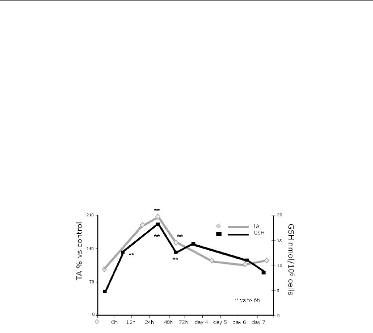
Fig. 1. Reduced glutathione regulates telomerase activity in 3T3 fibroblasts.
Thus, physiological variations in glutathione level induce changes in telomerase activities
that are in concordance with changes in cell cycle regulatory proteins. A number of reports
have shown similar results. Brown et al., 2007 demonstrated for the first time in vivo that
high hepatic glutathione levels correlate with increased telomerase activity. Also, the
importance of glutathione regulation in telomerase activity has been proved in endothelial
progenitor cells (EPC): impairment of antioxidant defences in EPC promoted oxidant
mediated apoptosis and telomerase inactivation which subsequently lead to development
and/or progression of atherothrombosis (Fujii et al., 2006).
Recent data suggest that telomerase activity is regulated and ordered by telomere structure
and telomerase assembly. Experimental evidence suggests that the telomere structure may
change in a cell cycle-dependent manner to restrict telomerase activity to S phase (Hug &
Ligner, 2006). In addition, telomere structure and specially the telomeric G-overhangs
generation are strictly regulated during S phase and prolonged to other cell cycle phases
depending on whether are telomeres from the lagging or the leading telomere (Dai et al.,
2010). For this to happen, the precise control of the changes not only in telomerase
conformation, but in chromatin structure (i.e. in its compactation level) as well, is of vital

The Nuclear Compartmentation of Glutathione: Effect on Cell Cycle Progression
277
importance. This finding that telomere length, and, therefore, telomere structure, is tightly
regulated in telomerase proficient cells invokes a connection between cell cycle, telomerase
and telomere structure (Blasco, 2002). In other words, the mechanism that lies beneath
telomerase regulation might be related with the mechanism that control cell proliferation.
This opens a highly significant area for exploratory study and the diversity of processes and
control mechanisms that could be involved in this phenomenon remain to be elucidated.
3. Compartmentalization of glutathione
3.1 The physiological importance of compartmentalization of glutathione
Pioneer work from Meister, (Meister & Anderson, 1983) correlated GSH synthesis and its
degradation throughout the so-called -glutamyl cycle, and defined it as a cytosolic
processes. The importance of cellular compartmentalization of GSH is two fold, first because
it plays an important role in fighting against radical oxygen species (ROS). It is well known
that these molecules have a very short half life and exert their action close to the place they
were produced. Thus, the presence or absence of GSH could determine the development of
localized oxidative damage for the cell structure or metabolic function developed in the
vicinity. Secondly, GSH compartmentalization is of vital importance because of its role as a
cellular detoxifying agent; it is known that tumours that have high glutathione levels are
more resistant to chemotherapy, and the importance of nuclear (Voheringer et al., 1998) and
mitochondrial (Benlloch et al. 2005) compartmentalization of GSH has been pointed out.
The overview of compartmentalization of glutathione in mammal cells is a complicated
matter. This is due to the presence in the literature of a number of contradictory reports. The
reason for the controversy is mainly methodological. Until very recently most reports were
mainly based on cell-fractionation techniques. Those techniques appear to be reliable for
mitochondrial studies; however their usefulness in nuclear or even endoplasmic reticulum
measurements is at least controversial.
3.1.1 Nucleus
Although the role of nuclear GSH in the synthesis of DNA (Thelander & Reichard, 1979) and
in protection against oxidative damage or ionizing radiation (Biaglow et al., 1983) is well
established, little is known about the concentration
of GSH in the nucleus and its regulation.
This is due to two
main factors. The first is methodological: it is impossible
to determine the
nuclear concentration of GSH using standard
cell fractionation and analytical approaches
(for a review see
Söderdahl et al., 2003). The second factor is that most, if not all, of the
reports share
the common view of nuclear GSH distribution in a static situation.
Cells are
usually studied under steady state conditions i.e.
when they are confluent (G
0
/G
1
phase of
the cell cycle). The
nuclear membrane dissolves during mitosis and is formed again around
newly replicated DNA packed in chromosomes; this spectacular change involves a variety
of regulatory mechanisms. Therefore, if the nuclear GSH distribution is studied, the cell
cycle physiology should be carefully considered.
The role of GSH in cell cycle regulation has been addressed mainly from the point of view of
its overall cellular content. This is surprising since it is in the nucleus where most cell cycle
progression events take place. The nucleus changes dramatically during the different phases
of cell cycle, and failing to consider the corresponding changes in its redox environment
could confer an important disadvantage in elucidating the actual importance of glutathione
in the control of cell proliferation.
Selected Topics in DNA Repair
278
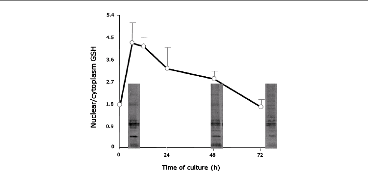
Fig. 2. Modifications of nuclear proteins along the cell cycle.
Our results, in general, are in accordance with the work of Hutter at al, (Hutter et al., 1997),
who have studied the redox potential (E) in the normal and malignant cells along the cell
cycle. The E of normal cells fluctuates during the cell cycle; for the proliferation to start it has
to decrease at least 30mV comparing to its level in the G
0
phase (where the cells are 100%
confluent). On the contrary, in cancer cells, the E remains low throughout the complete cell
cycle, even at a high cell density (Hutter et al., 1997). Our evaluation of the GSH levels along
the cell cycle in different models indirectly confirms these findings.
The work of Hutter et al. was further developed and completed by Hoffman et al. (Hoffman
et al., 2008), who proposed recently a novel redox model of cell proliferation. They postulate
the existence of a redox switch that helps regulate the proliferation within normal cells; its
absence in cancer cells enables the bypass of the restriction point and leads to the loss of the
control of cell cycle. The authors offer this model as a base to understand the aberrant
cellular proliferation that leads to malignant transformation.
According to “redox model of cell proliferation”, in normal cells exist a threshold value (θ)
of E≤ 207±11mV which initiates the phosphorylation of different regulatory proteins
associated with different phases of the cell cycle and, consequently, cell proliferation. When
E>θ cell enters G1/G0 phase. However, when cancer cells are concerned, Hoffman does not
take into account the fluctuations in the E level. In our hands, the increase in GSH level
along the cell cycle (before the onset of the proliferation comparing to the GSH level at the
final time point) is, indeed, less striking than in 3T3 fibroblasts; two-fold comparing to four-
fold, respectively. The author hypothesizes, though, the existence of a reductive limit of -
260mV which normal cell could not survive, but would not jeopardise cancer cell.
It was proposed previously that the proliferation occurs within the range of ROS levels,
concentrations above or below this range could lead to growth arrest or cell death; hence
ROS could act like a dual-edged sword (Davies, 1999).
3.2 Glutathione controls the cell cycle regulatory proteins
3.2.1 Id2 as a redox sensitive protein
The study of the expression of the Id2 along the cell cycle gave further support to this
premise. The family of helix-loop-helix proteins denominated Id (inhibitor of DNA binding

The Nuclear Compartmentation of Glutathione: Effect on Cell Cycle Progression
279
or inhibitor of differentiation) was demonstrated to be of considerable importance in the
regulation of cell growth, differentiation and cancer in many mammalian tissues (Norton,
2000; Yokota & Mori, 2002). Id2, in particular, was shown to disrupt antiproliferative effects
of tumour suppressor proteins of the Rb family, thus allowing cell cycle progression
(Lasorella et al., 1996). Indeed, the pattern of the Id2 expression detected by Western
blotting, confirmed the distribution of the phases of the cell proliferation detected by flow
cytometry. This observation was previously published by our group suggesting a redox
regulation of this protein (Borras et al., 2004). In addition, the studies of liver regeneration,
process that involves DNA synthesis and cell proliferation, gave further support to our
findings.
It was demonstrated that when the increase of GSH after partial hepatectomy was
prevented, the liver regeneration was delayed and the total liver amount of the DNA was
lower than in the control group (Huang et al., 2001). Furthermore, an early increase in Id2
gene has been demonstrated as well as the contribution of Id2 in the control of hepatocyte
priming through modulation of c-myc expression (Rodriguez et al., 2006). All these support
our notion that Id2 could be an excellent candidate as a protein marker of the redox
regulation of cell proliferation in our models.
3.2.2 PCNA as a possible redox sensor in the onset of DNA synthesis
PCNA, a proliferating cell nuclear antigen, is a central protein in both DNA replication and
repair. It’s a “sliding clamp” that localizes proteins, such as DNA polymerase, to DNA and
thus enables the correct DNA replication.
Replication of mammalian genome starts at thousands of origins, called replication foci,
which contain PCNA and are activated at different times during S phase. The dynamics of
replication foci is still a matter of debate; there are contradictory reports on the organization
of the DNA replication sites in diverse cell types attributable to the differences in the
technical approach (Dimitrova & Gilbert, 2000; Kennedy et al., 2000). According to
Dimitrova, SD and Berezney, R (Dimitrova & Berezney, 2002) there is no fundamental
difference in the spatiotemporal organization of the DNA replication in primary,
immortalized and malignant mammalian cells. On the contrary, Kennedy’s group (Kennedy
et al, 2000), observed different patterns of replication foci in primary versus immortalized
cell lines, as well as their perinuclear localization in the contact-inhibited cells prior to cell
cycle exit (Barbie et al., 2004). Another fundamental question was weather the replication
foci are moving along the DNA in the process of the replication, or the DNA is spooling
through fixed replication factories. It seems that the important body of evidence is
accumulating supporting the fixed-replication-site model (Dimitrova & Gilbert, 2000;
Leonhard et al., 2000). The replication machinary bind to DNA, but they are also tethered to
an underlying framework called nuclear matrix or skeleton (Leonhardt et al., 2000).
Regardless of the discrepancies in their findings, all the authors call attention to the
importance of the preserving nuclear architecture in order to guarantee the correct
development of the process of DNA replication (Dimitrova & Gilbert, 2000; Barbie et al.,
2004; Leonhardt et al., 2000). In addition, it has been shown that chromosome territory
organization depends on association with the nuclear skeleton (Leonhardt et al., 2000). More
than 20 years ago, Dijkwell et al. (Dijkwell & Wenink, 1986) postulated that the maintenance
of the nuclear matrix, especially nuclear lamina, by preserving disulphide bonds depended
on the level of nuclear thiols. In accordance to this work, Oleinick et al. (Oleinik et al., 1987)

Selected Topics in DNA Repair
280
reported that DNA-protein cross links, present at basal level as normal associations of
chromosomal loops with the nuclear matrix proteins, can be increased by ionising radiation
and removal of intracellular glutathione, and decreased by hydroxyl radical scavengers. The
importance of the GSH in cell proliferation could be extrapolated to the safeguarding of the
nuclear architecture, providing in that way a proper milieu for the DNA replication. Our
results could provide support to the hypothesis of Bellomo et al (Bellomo et al., 1997) that
reduced nuclear glutathione may modulate the structural organization of chromatin. It is
tempting to speculate that the high nuclear GSH level we observed before (late G1 phase)
and at the onset of cell proliferation (S phase) could provide the redox environment that
stimulates chromatin decompaction by reducing disulfide bonds.
3.3 Nuclear compartmentalization of glutathione as an important feature of
proliferating cell: reduce to replicate
The functional compartmentalization is an obvious characteristic of eukaryotic cell. The
organelles, visible by light microscopy, are surrounded by membranes, which, although
permitting communication, provide unique and defined environment in each one, which
guarantee its accurate function. Probably the most remarkable examples of
compartmentalization are oxidative phosphorylation in mitochondria, protein folding in
endoplasmic reticulum and, for the purpose of this study the most interesting of all, DNA
synthesis. It is interesting to note that, when the first two organelles are concerned, the
dependence of their function on the correct GSH level has been thoroughly studied. The
high intramitochondrial concentration of GSH is maintained by an active multicomponent
transport system “pumping” glutathione from the cytosol into the matrix (Matensson et al.,
1990). On the contrary, for the correct folding of proteins into a native structure by disulfide
bond formation, the GSH level and the ratio GSH/GSSG in the endoplasmic reticulum is
maintained at extremely low level by the limited permeability of the vesicle membrane to
GSH (Hwang et al., 1992). However, in the case of nuclear compartmentalization of
glutathione the reports were scarce and contradictory over the years. This could be
attributed to two main factors: methodological difficulties in measuring nuclear glutathione
content and the focus of the research generally limited to confluent cells.
3.4 Modifications of nuclear proteins along the cell cycle
Various studies have demonstrated that the nucleus is more reduced than the cytosol
(15mM GSH vs. 11 mM, respectively) (Bellomo et al., 1997; Schafer & Buettner, 2001; Soboll
et al., 1995). An important number of nuclear proteins, including transcription
factors,
require a reduced environment to bind to DNA. More
than 62 proteins are involved directly
in transcription, nucleotide
metabolism, (de)phosphorylation, or (de)ubiquitinylation, which
are all essential processes for cell cycle progression (Connour et al., 2004). For instance, it
appears that, at the onset of cell proliferation in the early G1 phase, an increase of ROS in the
cytoplasm is necessary for the initiation of the phosphorylation cascade mediated by
epidermal growth factor (EGF) that, subsequently, activates DNA replication and the cell
division (Carpentes & Cohen, 1990). According to Jang and Surh (Jang & Surh, 2003) nuclear
GSH may act as a transcriptional
regulator of NF- B, AP-1, and p53 by altering their nuclear
redox
state. The transcription factor NF-κB is an example of distinct redox-sensitive
activation and DNA binding (Hansen et al., 2006); it is activated by various physiological
stimuli known to produce ROS; on the contrary, to permit DNA binding, similar to Fos, Jun,