George Paxinos, Charles Watson. The Rat Brain in Stereotaxis Coordinates (Мозг крысы в стереотаксических координатах)
Подождите немного. Документ загружается.

-1
0
1
2
3
4
5
6
7
8
10
9
10
9
8
7
6
5
4
3
2
0
1
11
Olfactory
Olfactory
Bulb
Cerebral Cortex
Fornix
Corpus Callosum
3V
Septum
Preoptic
Hypothalamus
Optic Chiasm
Thalamus
Midbrain
Superior
Pineal
4V
4V
Pituitary
Pons
Medulla Oblongata
Spinal
Cord
1
2
3
4
5
6
6
6
7
8
9
9
10
Cerebellum
Lateral 0.40 mm
Interaural
Bregma
Area
Tubercle
Anterior
Commissure
Posterior
Commissure
Colliculus
Inferior
Colliculus
Hippocampus
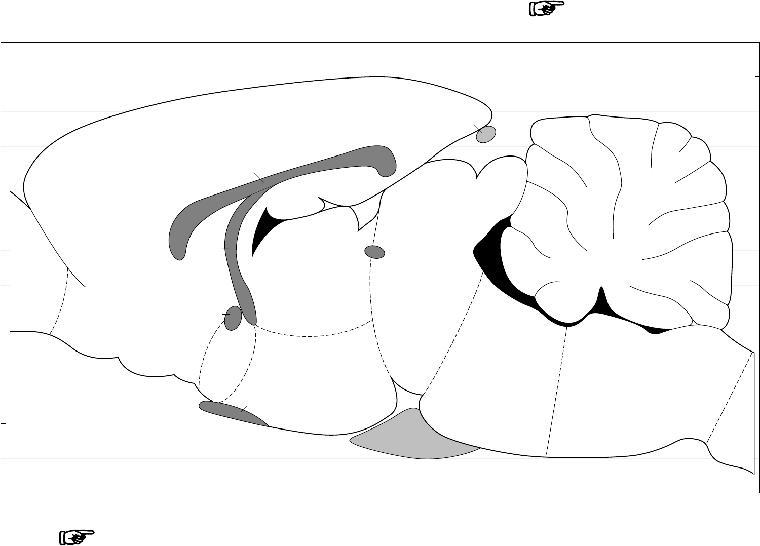
Click on sagittal diagram to go the corresponding horizontal diagram
Go to START PAGE
PREFACE: FOURTH EDITION
The Compact Third Edition of
The Rat Brain in
Stereotaxic
Coordinates
presented revised drawings of 76 coronal sections of
the brain. The Fourth Edition includes these coronal diagrams,
but in addition presents the fully revised sagittal and horizontal
figures and the photographs on which all the diagrams are based.
We are brain cartographers, and, like our geographer colleagues,
our central aim is to provide precise coordinates and accurate
identification of features of interest. In this fourth edition of our
atlas, we have used the proven stereotaxic reference system that
we constructed for the first edition (Paxinos and Watson, 1982).
This reference system is now universally recognized as the optimum
grid for stereotaxic work in the rat brain. We have completely
revised the identification of brain structures in order to produce
an atlas that will be useful beyond the end of this century.
Features of the Fourth Edition
• The most accurate stereotaxic reference system available
displayed in three planes
• Completely revised diagrams of 10 sagittal and 27 horizontal
sections that previously appeared in the second edition of this
atlas (Paxinos and Watson, 1986)
• Includes the 1997 revisions of the 76 coronal diagrams
(Paxinos and Watson, 1997) augmented by two new drawings of
coronal sections at the level of the anterior diencephalon
• Each diagram is accompanied by a matching photograph of
the section on which it is based
• Delineations assisted by consideration of separate sets of brain
sections stained for parvalbumin, calbindin, calretinin, SMI-32,
tyrosine hydroxylase, and NADPH diaphorase (Paxinos et al., in
press [a,b]), the rat nervous system textbook (Paxinos, 1995),
(Paxinos and Watson 1997), and other recent neuroanatomical
literature
• Spinal cord drawings from the atlas of Molander and Grant
(1995)
• Diagrams available on CD-ROM for printing at different
magnifications
Reproduction of Atlas Figures
in Other Publications
As authors, we are happy for our atlas figures to be reproduced in
other publications, but we expect our work to be suitably
acknowledged. If coordinates from the atlas are used, they should
be acknowledged as in the following example: “Bregma 1.60 mm
(Paxinos and Watson, 1998).” Permission to reproduce the figures
contained in the atlas can be obtained from the publisher:
Academic Press
Permissions Department
6277 Sea Harbor Drive
Orlando, FL
USA 32887
Telephone: (407) 345-3990
Fax: (407) 352-8860.
When requesting permission, please identify all the figures you
wish to reproduce. Allow four weeks for your request to be
processed. We recommend that you use the nomenclature and
abbreviation scheme that we developed for this book. This scheme
has been developed on a systematic basis (see below) and is now
widely recognized.
ACKNOWLEDGMENTS
We are indebted to Lewis Tsalis for outstanding accuracy and speed
in entering our drawings into the computer using Adobe Illustrator.
We thank Paul Halasz for his excellent work in designing and
assembling the CD-Rom version of this atlas.
We thank Hong-Qin Wang for preparation of excellent quality
photographic plates.
We thank David Kopf for providing us with his extremely accurate
stereotaxic instrument.
We thank Kodak Australia for assistance with the provision of
photographic paper.
We thank Fine Science Tools Inc (Vancouver, Fax: 415-349-3729)
for providing us with precision forceps used for the preparation of
specimens. We thank Faulding Imaging for providing us with an
Olympus Provis Research Microscope system at a reduced price
We thank Professor Kevin McConkey, Head of the School of

TABLE 1 Craniometric and stereotaxic data (means ± S.D.) for rats of different sex, strain, and weight
Mean AP AP DV AP AP
AP DV
weight I – B I – L I – B I – Acb B – ac
I – 7n I – incisor bar
Subject (g)* (mm) (mm) (mm) (mm)** (mm)**
(mm)** (mm)
‘Atlas’ Wistar 290 9.1 ± 0.3 0.3 ± 0.3 10.0 ± 0.2 11.7 0.0
–1.3 –3.3 ± 0.4
Coronal plates 300 9.2 0.2 10.1
Sagittal plates 270 8.9 0.0 10.0
Horizontal plates 290 9.1 0.2 10.1
Female Wistar 282 9.3 ± 0.2 0.5 ± 0.3 10.0 ± 0.1 11.6 0.1
–1.2 –3.2 ± 0.5
Hooded290 9.4 ± 0.4 0.3 ± 0.6 9.8 ± 0.2 11.9 0.0 –1.2
–3.9 ± 0.6
Sprague 299 9.0 ± 0.2 0.7 ± 0.2 10.1 ± 0.1 11.7 0.1
–1.2 –3.9 ± 0.5
Juvenile Wistar 180 7.7 ± 0.4 –0.4 ± 0.3 9.9 ± 0.2 10.2 –0.1
–1.6 –2.0 ± 0.4
Mature Wistar 436 9.7 ± 0.3 0.6 ± 0.3 10.7 ± 0.4 12.4 –0.1
–0.8 –2.7 ± 0.3
* S.D.s. ≤ 20 g.
** S.D.s. ≤ 0.4 mm.
ac, anterior commissure; Acb, accumbens nucleus; AP, anterior-posterior; B, bregma; DV, dorsal-ventral; 7n, facial nerve; I, interaural line; L, lambda.
Reprinted with permission from J. Neuroscience Methods. 13 (1985) 139-143.
Psychology at the University of New South Wales, and Professor
Gerard Sutton, Vice Chancellor of the University of Wollongong,
for their generous support. We thank Professor David Tracey for
allowing us to use facilities in the School of Anatomy. We thank
Apple Australia for providing equipment to assist with digitizing
the drawings for this atlas.
G.P. Thanks Elly for direct help as well as tolerance during
construction of this and previous editions of this atlas.
INTRODUCTION
There are many reasons why the rat is the most commonly selected
subject for research in mammalian neuroscience. First, rats are
the right size: neither too small for accurate stereotaxic localization
of discrete brain areas nor too large for cost-effective laboratory
management. Second, rats are generally hardy animals and are
resistant to infections. Third, a number of inbred strains are
available commercially; so, animals of consistent size can be used
for stereotaxic procedures.
When the first edition of
The Rat Brain in Stereotaxic Coordinates
was published in 1982, it was the first atlas to be based on the flat-
skull position. It offered a choice of bregma, lambda, or the
midpoint of the interaural line as the reference point. Although
the coordinates were developed from study of adult male Wistar
rats with weights ranging from 270 to 310 g, the atlas can be
successfully used with male or female rats, with weights ranging
from 250 to 350 g (Paxinos et al., 1985).
The present atlas presents 78 photographs and accompanying
diagrams of coronal sections of the brain, at intervals that average
0.25 mm. The sections were cut from unfixed brains that were
frozen. The diagrams in this atlas were originally based on the
study of Nissl- (cresyl violet) and acetylcholinesterase- (AChE)
stained sections. The mapping of the sections was assisted by the
use of our sections showing the distribution of a number of
antibody-based and enzyme-based stains (parvalbumin, calbindin,
calretinin, SMI-32, tyrosine hydroxylase, and NADPH diaphorase).
A comprehensive atlas based on chemical markers will be published
by Academic Press (Paxinos et al., in press [a,b]).
It also presents photographs and fully revised diagrams of 10
sagittal and 27 horizontal sections.
Stereotaxic Surgery
In establishing the stereotaxic coordinate system for this atlas we
studied sections from over 100 rats. To prepare these sections, we
positioned the skull in a standard way (the flat-skull position) and
marked the vertical and horizontal planes with needle tracks. We
placed anesthetized rats in a Kopf small-animal stereotaxic
instrument, and the incisor bar was adjusted until the heights of
lambda and bregma were equal. This flat-skull position was
achieved when the incisor bar was lowered 3.3 ± 0.4 mm below
horizontal zero (Table 1).
Because the point of intersection of the lambdoid and sagittal
sutures is variable, we have chosen to define lambda as the midpoint
of the curve of best fit along the lambdoid suture (see skull
diagram). This redefined reference point is considerably more
reliable than the true lambda (the point of intersection of the
sagittal and lambdoid sutures), and it is located 0.3 ± 0.3 mm
anterior to the interaural line. We defined bregma as the point of
intersection of the sagittal suture with the curve of best fit along
the coronal suture. When the two sides of the coronal suture meet
the sagittal suture at different points, bregma usually falls midway
between the two junctions. The anteroposterior position of bregma
was 9.1 ± 0.3 mm anterior to the coronal plane passing through
the interaural line, but for the brain represented in this atlas bregma
is deemed to lie at 9.0 mm. The top of the skull at bregma and
lambda was 10.0 ± 0.2 mm dorsal to the interaural zero plane.
To confirm the stereotaxic orientation of sections in the brain
used for this atlas, reference needle tracks were made perpendicular
to the horizontal and coronal planes. For brains sectioned in the
coronal plane, vertical needle insertions were made at 2.0 mm
intervals through the brain, except for the penetrations at 0.7 mm
anterior to the interaural line, which was chosen to avoid rupture
of a venous sinus. Ten such needle tracks appear on coronal plates
of this atlas. Three horizontal needle insertions perpendicular to
the coronal plane were made from the posterior of the brain at
1.0, 3.0, and 5.0 mm above the interaural line and approximately
1.0 mm lateral to the midline. The reference tracks from the
horizontal needles appear as small holes in coronal sections.
For brains sectioned in the sagittal plane, vertical needles were
inserted in both hemispheres at 3.0 mm posterior to the interaural
line and at 1.0 and 2.0 mm lateral to the midline. A second pair
was inserted 11 mm anterior to the interaural line at 1.0 and 2.0
mm lateral to the midline. Horizontal needle tracks perpendicular
to the coronal plane were made at 5.0 mm and 6.0 mm dorsal to
the interaural line and 2.0 mm lateral to the midline. Horizontal
needle tracks perpendicular to the sagittal plane were made at 5.0
mm dorsal to the interaural line and 2.0 mm and 8.0 mm anterior
to it.
For brains sectioned in the horizontal plane, two vertical reference
tracks were made by inserting a needle at 2.0 and 9.0 mm anterior
to the interaural line and approximately 1.4 mm lateral to the
midline. These reference tracks appear as pinholes in the horizontal
sections. Five horizontal needle tracks perpendicular to the coronal
plane were made by inserting needles at 1.0, 3.0, 5.0, 7.0, and 8.0
mm above the interaural lines.
Histology
After surgery, the animals were deeply anesthetized and
decapitated, and their brains were frozen on dry ice within 3 min
of decapitation. For the coronal plane, brains were divided into
two blocks at a plane 3.0 mm anterior to the interaural line prior
to placement on the freezing microtome stage. At the level of the
blocking some sections were lost and it was necessary to insert
three plates (Figs. 42–44) from another brain.
Brains remove from the skull for sectioning on the horizontal or
sagittal planes initially presented a problem in that they assumed
the shape of the stage on which they were positioned. To avoid
this distortion, the brains were frozen in the skull, and the skull
bones were then prized off the frozen brains.
Spinal cord segments were obtained from 9 rats and were fresh
frozen in propane cooled with liquid nitrogen. Representative
sections from the major regions of the spinal cord are included in
the atlas (Plates 117a,b). None of these plates are accompanied by
a drawing, but their approximate location in the Mollander and
Grant (1995) spinal cord atlas is given and the diagrams from the
Mollander and Grant atlas are reproduced with permission of the
authors.
Frozen brains were sectioned on an American Optical Cryocut
microtome at 40 µm. Sections were obtained parallel to the
stereotaxic planes by adjusting the angle of cutting until the needle
tracks encountered were judged to be parallel to the plane of
section. Sections were taken directly from the cryotome knife on
uncoated slides with the help of an anti-roll device. At each 0.5
mm interval, three sections were taken for staining with cresyl
violet or for the demonstration of AChE. Staining was carried out
on the same day as cutting. The two stains were used in an
alternating fashion in relation to the section intervals. One of these
sections at each interval was presented in the first edition of the
atlas. In the second edition we interpolated an additional section
between the 0.5 mm interval sections in most cases. In the present
atlas, the interpolated sections commence after Fig. 11. An attempt
was made to select interpolated sections close to the midpoint of
the interval of the plates in the first edition of the atlas, but such
sections often were not available, and the closest suitable section
was selected. Because of this process of selection of interpolated
sections, the regular alternation of Nissl- and AChE-stained sections
was broken.
Cresyl Violet Staining
Slides were immersed for 5 min in each of the following: xylene,
xylene, 100% alcohol, 100% alcohol, 95% alcohol, and 70% alcohol.
They were dipped in distilled water and stained in 0.5% cresyl
violet for 15–30 min. They were differentiated in water for 3–5
min and then dehydrated through 70% alcohol, 95% alcohol, 100%
alcohol, and 100% alcohol. They were then put in xylene and cover-
slipped.
To make 500 mL of 0.5% cresyl violet of about pH 3.9, mix 2.5 g
of cresylecht violet (Chroma Gesellschaft, Postfach 11 10, D-73257,
Kongen, Germany, Fax number: 49-7024-82660), 300 mL of water,
30 mL of 1.0 M sodium acetate (13.6 g of granular sodium acetate
in 92 mL of water), and 170 mL of 1.0 M acetic acid (29 mL of
glacial acetic acid added to 471 mL of water). Mix this solution for
at least 7 days on a magnetic stirrer, then filter.
AChE Histochemistry
The method for the demonstration of AChE followed the procedures
of Koelle and Friedenwald (1949) and Lewis (1961). Slides were
incubated for 15 h in 100 mL of stock solution (see below) to which
had been added 116 mg of S-acetylthiocholine iodide and 3.0 mg
ethopropazine (May & Baker). The slides were rinsed with tap water
and developed for 10 min in 1% sodium sulphide (1.0 g in 100 mL
of water) at pH 7.5. They were then rinsed with water and immersed
in 4% paraformaldehyde in phosphate buffer for
8 h, and then allowed to dry. Subsequently, they were dehydrated
for 5 min in 100% alcohol, then immersed in xylene and cover-
slipped with Permount.
The stock solution was a 50 mM sodium acetate buffer at pH 5.0
which was made 4.0 mM with respect to copper sulphate and 16
mM with respect to glycine. This was done by
adding 6.8 g of sodium acetate, 1.0 g of copper sulphate crystals,
and 1.2 g of glycine to 1.0 L of water and lowering the pH to 5.0
with HCl.
We found that fresh, unfixed tissue from the frozen brains showed
a substantially stronger reaction for both stains than tissue fixed
with formalin, paraformaldehyde, glutaraldehyde, or alcohol.
A detailed protocol of the staining procedures may be obtained
from George Paxinos (http://www.psy.unsw.edu.au/~ paxinos).
Photography
The drawings of the brain were traced from photographs of stained
brain sections which were taken with a Nikon Multiplot
macrophotographic apparatus on 4 × 5 inch Kodax Plus X film.
High contrast (grade 4) Ilfospeed paper was used for the Nissl
sections, whereas lower contrast (grade 2) paper was used for the
AChE sections.
Drawings
As far as possible, we have attempted to make the drawings
accurately represent the photographs. Small adjustments have been
made to some sections in which the midline or cerebral cortex
were distorted, but otherwise the drawings depict the asymmetries
present in the sections. When part of an atlas section was missing
or severely distorted, the missing part was drawn in after
consideration of sections obtained from other brains.
Fiber tracts in the drawings are outlined by solid lines, and nuclei
and cell groups are outlined by broken lines. In general, each
abbreviation is placed in the center of the structure to which it
relates; where this is not possible, the abbreviation is placed
alongside the structure and a leader line is used. The abbreviations
for fiber tracts and fissures are almost always positioned on the
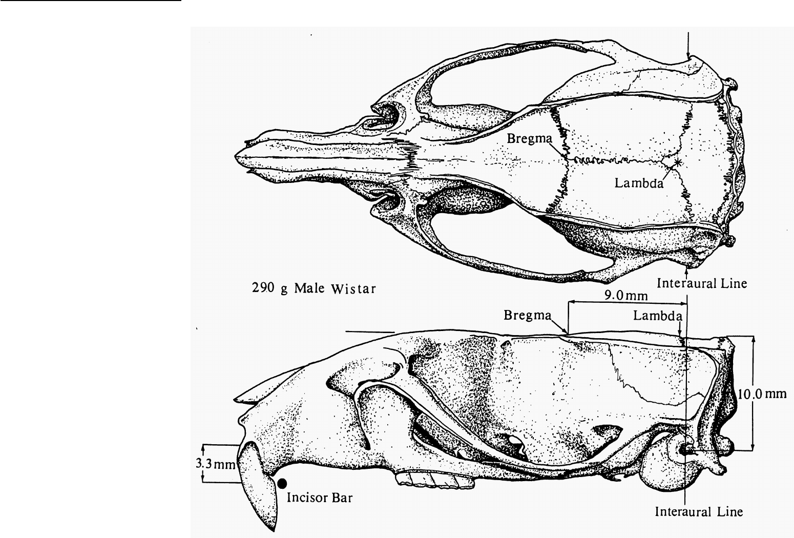
SKULL DIAGRAM
Dorsal and lateral views of
the skull of a 290 g Wistar rat.
The positions of bregma,
lambda, and the plane of the
interaural line are shown above
the lateral view. The distance
between the horizontal plane
passing through the interaural
line is shown on the right of the
lateral view. The distance
between the incisor bar and the
horizontal plane passing
through the interaural line is
shown on the left of the lateral
view. Lambda (midpoint of the
curve of best fit along the
lambdoid suture) is 0.3 mm
anterior to the coronal plane
passing through the interaural
line.

TABLE 2 Coordinates of the bed nucleus of the anterior
commissure and the trochlear nucleus obtained from the
three planes
Bed nucleus of the ant. comm. Trochlear nucleus
Plane A-P D-V Lat A-P D-V Lat
Coronal 8.2 3.4 0.9 1.7 3.4 0.4
Sagittal 8.0 3.4 0.9 1.8 3.5 0.4
Horizontal 8.1 3.4 0.9 1.8 3.4 0.4
left side of the figure, and the abbreviations for nuclei and other
cell groups are generally positioned on the right side. The outlines
of the ventricles and aqueduct are filled in with solid black. The
drawings were entered into a Power Mac using Adobe Illustrator 6.
Stereotaxic Reference System
Two coronal and two horizontal zero-reference planes are used in
these drawings; one coronal plane and one horizontal plane are
related to the interaural line and the other two are related to
bregma. Lambda is located 0.3 ± 0.3 mm anterior to the interaural
line, and it can be used as an alternative reference point in
conjunction with the dorsoventral coordinate of bregma. The
position of the stereotaxic reference points and planes are indicated
on the skull diagram. The stereotaxic reference grid shows 0.2 mm
intervals.
Coronal Drawings
In each of the coronal drawings, the large number at the bottom
shows the anteroposterior distance of the corresponding plate from
the vertical plane passing through the interaural line. The large
number at bottom right shows the anteroposterior distance of the
plate from bregma. The small numbers on the left margin show
the dorsoventral distance from the horizontal plane passing
through the interaural line. The numbers on the right margin show
the dorsoventral distance from the horizontal plane passing
through bregma and lambda on the surface of the skull. The
numbers on the top and bottom margins show the distance of
structures from the midline.
Sagittal Drawings
The large number at the bottom left of each drawing shows the
distance of the corresponding plate from the midline. The numbers
on the left margin show the dorsoventral
distance from the horizontal plane passing through the interaural
line. The numbers on the right margin show the dorsoventral
distance from the horizontal plane passing through bregma and
lambda on the surface of the skull. The numbers on the bottom
margin show the anteroposterior distance from the coronal plane
passing through the interaural line. The numbers on the top margin
show the anteroposterior distance from the coronal plane passing
through bregma.
Horizontal Drawings
The large number at the bottom left of each drawing shows the
dorsoventral distance of the corresponding plate from the
horizontal plane passing through the interaural line. The large
number at the top right shows the dorsoventral distance of the
plate from the horizontal plane passing through the bregma and
lambda on the surface of the skull. The numbers on the bottom
margin show the anteroposterior distance from the coronal plane
passing through the interaural line. The numbers on the top margin
show the anteroposterior distance from the coronal plane passing
through bregma. The numbers on the left and right margins show
the distance of structures from the midline.
Values ventral to the interaural horizontal zero or posterior to
either the interaural line or bregma are preceded by a minus sign.
An Example of Use of the Stereotaxic
Reference System
In this example, we will consider the insertion of an electrode into
the basolateral amygdaloid nucleus. Figure 31 shows that the center
of the basolateral amygdaloid nucleus is 6.2 mm anterior to the
interaural line, 1.5 mm dorsal to it, and 5.0 mm lateral to the
midline. The nucleus is 2.8 mm posterior to bregma, 8.5 mm ventral
to it, and 5.0 mm lateral to the midline.
Stereotaxic Accuracy
In most cases, the position of a structure is represented to an
accuracy of less than 0.5 mm. Although we used medium-sized
(average 290 g) male Wistar rats in the construction of this atlas,
we recognize that researchers often use animals of different sex,
strain, and weight. Because of this, we have estimated the error
that will occur if this atlas is used with female Wistar rats, male
hooded (Long Evans) rats, male Sprague Dawley rats of 290-g
weight, juvenile (180 g) Wistar rats, and mature (436 g) Wistar
rats. The results of these estimations are shown in Table 1
(reproduced from Paxinos et al., 1985). It is evident from these
studies that no substantial stereotaxic error will occur when rats
of different sex and strain are chosen, provided that the rats are of
similar weight to those on which the atlas is based (290 g).
For example, for rats of different sex and strain but of similar
weight, the anteroposterior distance between the interaural line
and bregma is between 9.0 and 9.4 mm. Similarly, the dorsoventral
distance between the interaural line and the surface of the skull at
bregma and lambda is very stable (9.8–10.1 mm). By contrast,
craniometric data for juvenile (180 g) and mature (436 g) Wistar
rats differ substantially from those of other groups. The
anteroposterior distance between the interaural line and bregma
is 7.7 mm in the juvenile and 9.7 mm in the mature rats (9.0 mm
in 290-g male rats). Lambda is 0.4 mm posterior to the interaural
line in the juvenile rats and 0.6 mm anterior to this line in the
mature rats (0.3 mm anterior in 290-g rats). Unexpectedly, the
dorsoventral distance between the interaural line and bregma for
juvenile rats (9.9 mm) was almost the same as that of 290-g rats
(10.0 mm). In the mature rats, the interaural line to bregma vertical
distance was 10.7 mm.
In female rats, as well as in hooded, juvenile (180 g), mature
(436 g) and 290-g Wistar rats, bregma was found to be above the
most forward crossing fibers of the anterior commissure. This is
the point at which the posterior limbs of the anterior commissure
appear. These data confirm the observation of Whishaw et al. (1977)
that bregma is more stable than the interaural line for positioning
of electrodes in brain structures close to, or anterior to, bregma.
However, data from insertion of needles aimed at the level where
the facial nerve leaves the facial genu (Fig. 60 in this atlas) show
that the interaural reference point is more stable than bregma for
localization of such posterior structures. Therefore, if juvenile or
mature rats are used, greater accuracy can be achieved if bregma
is used as the reference point for work with rostral structures and
the interaural line for work with caudal structures. A further
improvement in accuracy can be obtained by taking into account
the actual location of the accumbens nucleus (anterior part of Acb
shown on Fig. 9) and the genu of the facial nerve (Fig. 60). In
agreement with Slotnick and Brown (1980), we noticed that
coordinates of structures were closer to target if the coordinates
given by the interaural and bregma reference systems were
averaged.
In studying the drawings of coronal sections in this atlas, it should
be noted that the sections in Figs. 1–3, 42-44, and 69–78 were
taken from rat brains different from the one pictured in the
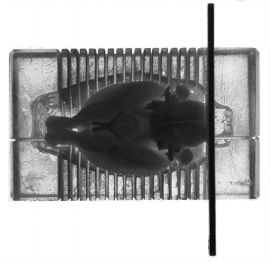
The Brain Blocker ␣ ␣ Designed to reproduce the plane of section of The Rat Brain in
Stereotaxic Coordinates (PA 001; David Kopf Instruments, P.O. Box 636, Tujunga, CA
91043).
remaining coronal drawings. This supplementation was necessary
because of distortion of the olfactory bulbs and caudal medulla in
the brain from which most of the sections were taken, and because
of the loss of a number of sections at the level at which the brain
was blocked.
Use of the David Kopf Stereotaxic Instrument
and Brain Blocker
In our work on this and other stereotaxic atlases we found the
David Kopf Small Animal Stereotaxic instrument of high precision.
However, no atlas or stereotaxic instrument will compensate for
using bregma and lambdoid points inappropriately. These
reference skull marks are the midpoints of the curve of best fit
along the coronal and the lambdoid suture, respectively. They are
not necessarily the points of intersection of these sutures with the
midline suture.
Researchers usually wish to block the brain at the same coronal
(or sagittal) plane as the atlas plane (flat skull position) so that
they can determine most readily the location of their electrode
placements and identify structures according to the atlas. The brain
blocker depicted in the accompanying photograph allows the
blocking of the brain at the stereotaxic plane or the slicing of the
brain at 1-mm intervals. After blocking, the brain needs to be placed
on a chuck with surface parallel to the cryotome knife. For
cryotomes without zero-tilt position, this can be achieved by
freezing some mounting medium directly onto the chuck and
cutting through the mounting medium to create an aligned surface
on which to position the blocked tissue.
Nomenclature
There is a critical need for a stable neuroanatomical nomenclature
to accurately and efficiently convey information between
neuroscientists. However, many terms are still used to describe a
single structure, and, in some cases, the same term is used for
completely different structures. We urge you to consider the merits
of our system of nomenclature because it is systematic and derived
after extensive consultations with neuroanatomy experts.
In considering the merit of a particular term over synonyms, we
have chosen terms that have been ratified by modern usage,
particularly usage by experts in that field. We have used anglicized
versions of terms rather than older latinized versions wherever
possible, and we have in all but a handful of cases avoided the use
of eponyms.
Principles of Construction of Abbreviations
Neuroscience communities concerned with different systems have
developed identical abbreviations for completely different
structures; for example SO stands for both supraoptic nucleus and
superior olive, SC for suprachiasmatic nucleus and superior
colliculus, and IC for inferior colliculus and internal capsule. In
dealing with the entire nervous system (as increasingly more
researchers do) these parochial abbreviation schemes become
impossible to implement. An additional complication arises when
homologous structures are nonetheless named or abbreviated
differently in different species. We have made an effort to establish
homologies and are using the same abbreviations for homologous
structures in atlases of the rat (Paxinos and Watson, 1986), mouse
(Franklin and Paxinos, 1996), monkey (Paxinos et al., in press), and
human (Paxinos et al., 1990; Mai et al., 1997). Atlas users may wish
to reflect on the construction of the abbreviations for
acetylcholinesterase (AChE) and magnesium (Mg) to get a good
idea of how our list was produced. The abbreviations used in the
present and in our other work were developed using the following
principles:
1. The abbreviations represent the order of words as spoken in English (e.g.,
DLG = dorsal lateral geniculate nucleus).
2. Capital letters represent nuclei, and lower case letters represent fiber
tracts. Thus, the letter “N” has not been used to denote nuclei, and the
letter “t” has not been used to denote fiber tracts.
3. The general principle used in the abbreviations of the names of elements
in the periodic table was followed: the capital letter representing the first
letter of a word in a nucleus is followed by the lower case letter most
characteristic of that word (not necessarily the second letter; e.g., Mg =
magnesium; Rt = reticular thalamic nucleus).
4. Compound names of nuclei have a capital letter for each part (e.g., LPGi
= lateral paragigantocellular nucleus.
5. If a word occurs in the names of a number of structures, it is usually
given the same abbreviation (e.g., Rt = reticular thalamic nucleus; RtTg =
reticulotegmental nucleus of the pons). Exceptions to this rule are made
for well-established abbreviations such as VTA.
6. Abbreviations of brain regions are omitted where the identity of the
region in question is clear from its position (CMn = centromedian
thalamic nucleus; not CMnTh).
7. Arabic numerals are used instead of Roman numerals in identifying (a)
cranial nerves and nuclei (as in the Berman, 1968, atlas), and (b) layers
of the spinal cord. While the spoken meaning is the same, the detection
threshold is lower, ambiguity is reduced, and they are easier to position in
small spaces available on diagrams.
The Basis of Delineation of Structures
in This Atlas
For the third edition, we completely reviewed our delineations of
all areas of the brain. Our primary guide was an extensive collection
of histochemically stained sections (monoclonal antibodies and
enzyme-based stains; Paxinos et al., in press [a,b]). Our work has
been made easier by the availability of an excellent rat brain atlas
published by Swanson (1992). We have also made extensive use of
other publications from our laboratory (Paxinos, 1995; Paxinos
and Huang, 1995; Paxinos et al., 1994) as well as of authoritative
studies published in the Journal of Comparative Neurology and
other journals.
We present below a brief account of the basis of delineation of
structures. We have not repeated here the rationale for the
delineation of structures presented in the second edition (Paxinos
and Watson, 1996). Readers may wish to refer to that book or
consult the work of Swanson (1992) and Kruger et al. (1995) for the
history of identification of many structures in the rat brain.
Olfactory System
Refer to Shipley et al. (1995) for a general description of the olfactory
system. We based our delineations in part on the work of de Olmos
et al. (1978).
The existence of the semilunar nucleus was established on the
basis of NADPH-diaphorase histochemistry (Paxinos et al., in press
[a]). We acknowledge assistance of R. Harlan and P.-Y. Wang in the
identification of this structure (Ahima and Harlan, 1990; Wang
and Zhang, 1995).
Basal Ganglia and Basal Forebrain
Refer to Heimer et al. (1995) and Fallon and Loughlin (1995) for a
general description of the basal ganglia and to Alheid et al. (1995)
for a discussion of the substantia innominata and extended
amygdala.
Immunoreactivity for parvalbumin and the neurofilament protein
SMI-32 identifies the ventral pallidum (Paxinos et al., in press [a]).
We retained the term substantia innominata and identified dorsal,
ventral (as in Grove, 1988), and basal components with the
assistance of G. Alheid. The basal component is marked by some
positivity in tyrosine hydroxylase but is negative for SMI-32
(although surrounding areas are positive).
The concept of ventral pallidum, first proposed by Heimer and
his associates, has been the guiding principle for structure/function
relations of the basal forebrain (Barragan and Ferreyra-Moyano,
1995; Heimer et al., 1997). The researchers at the University of
Virginia and Universidad Nacional de Cordoba have recently carved
out of the substantia inominata another big territory, the
sublenticular extended amygdala (Alheid et al., 1995). Franklin and
Paxinos (1997) adopted the new scheme for their mouse brain atlas.
We retained the name substantia inominata (for the part remaining
after the removal of the ventral pallidum) in keeping with earlier
editions of this atlas. The dorsal substantia inominata roughly
corresponds to the sublenticular extended amygdala, central part,
while the ventral substantia inominata corresponds to the
sublenticular extended amygdala, medial part.
The area previously called fundus striati resembles the striatum
proper in some respects and the accumbens shell in others. Given
that the use of the term fundus striati creates problems with primate
homologues, we followed the advice of G. Alheid and called it the
lateral accumbens shell. The remaining accumbens is delineated
in accordance with Zaborszky et al. (1985) and Heimer et al. (1991).
We followed Alheid et al. (1995) in the identification of the
interstitial nucleus of the posterior limb of the anterior
commissure (IPAC).
The reticular part of the substantia nigra can be divided into a
ventrolateral and a dorsomedial component on the basis of
parvalbumin and calbindin distribution (Paxinos et al., in press
[b]). The remainder of the substantia nigra and the ventral
tegmental area were delineated according to the work of
McRitchie et al. (1996).
Septum, Hypothalamus, and
Neurosecretory Nuclei
Refer to Simerly (1995), Armstrong (1995), Jakab and Leranth
(1995), and Oldfield and McKinley (1995) for a general description
of these structures.
Jutting ventrolaterally from the anterodorsal preoptic nucleus
is a strip which is negative for parvalbumin which we have called
the
alar nucleus
. The alar nucleus displays substance P positive
cell bodies but little reactivity in its neuropil (Larsen, 1992).
In the preoptic area we followed Simerly (1995) and Simerly
et al. (1984) except for the identification of the ventromedial
and ventrolateral preoptic nuclei, for which we followed Elmquist
et al. (1996) and Sherin et al. (1996).
The compact part of the medial preoptic nucleus is negative for
substance P (see Fig. 7 of Halliday et al., 1995, in which this structure
is visible but not labeled).
In the lateral hypothalamus we identified a ventrolateral
hypothalamus nucleus on the basis of NADPH-diaphorase reactivity