Hawkes P.W., Spence J.C.H. (Eds.) Science of Microscopy. V.1 and 2
Подождите немного. Документ загружается.

Chapter 6 In Situ Transmission Electron Microscopy 475
refl ection geometry, however, growth kinetics can be measured quan-
titatively (Figure 6–18B). Such studies demonstrate effects that are not
expected from the basic growth model, such as surface faceting (Ross
et al., 2005) and catalyst diffusion during growth (Hannon et al.,
2006).
3.7 Summary
In this section we have highlighted insights in surface physics and
crystal growth derived from in situ TEM. Experiments in this fi eld,
while often requiring dedicated equipment with complex additional
preparation chambers, provide information that is diffi cult or impos-
sible to obtain using other techniques. Continuous and direct observa-
tion in situ avoids artifacts arising from ex situ observation, which may
be especially signifi cant for growth studies. It is particularly encourag-
ing that realistic growth conditions can be accessed in the TEM,
enabling in situ studies of, for example, catalysis or CVD to be related
A
B
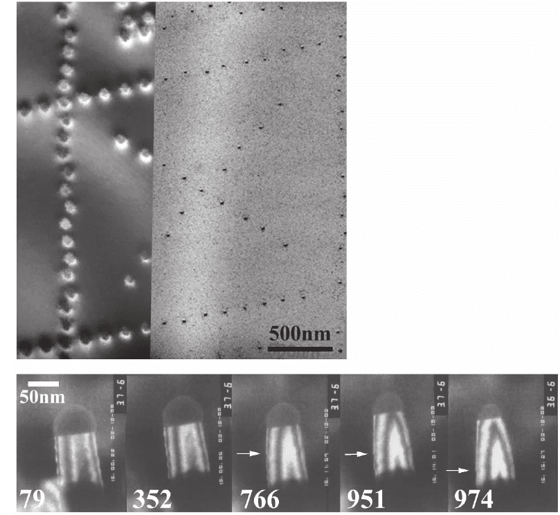
Figure 6–18. Growth on patterned
substrates. (A) Bright fi eld image of
a Si substrate with g = 220, recorded
directly after focused ion beam pat-
terning using an irradiation time of
0.1 ms per feature (left). After anneal-
ing followed by deposition of Ge at
650ºC and 5 × 10
−8
Torr; (g, 3 g) weak
beam image with g = 220 (right),
showing islands (small dots) on the
irradiated areas. (Reprinted with
permission from Kammler et al.,
2003. American Institute of Physics.)
(B) Growth of an epitaxial Si nanow-
ire by the vapour-liquid-solid mech-
anism, at 620ºC and 3 × 10
−6
Torr
Si
2
H
6
with times in seconds indi-
cated. The wire direction is (111) and
the viewing direction is near (01-1)
in this dark fi eld g = 220 series. The
Au-Si droplet is visible on the end of
the wire. As growth continues, the
droplet shrinks due to diffusion of
Au down the wire surface, causing
the wire diameter to decrease with
it.
476 F.M. Ross
to their counterparts in the outside world. Indeed, the relevance of in
situ TEM to the catalyst and microelectronics industries is shown by
the presence of environmental microscopes in several industrial
laboratories.
Opportunities clearly exist for further study. Growth on patterned
surfaces, prepared if necessary with integrated processing tools in the
TEM system, seems a particularly exciting way to examine the funda-
mental processes controlling growth and to fabricate nanoscale objects
with particular properties. Another potentially signifi cant area is the
correlation of structural or kinetic in situ measurements with macro-
scopic properties such as stress. Stress may be measured by depositing
onto a crystalline membrane whose curvature can be observed. This
may provide insight into the important processes associated with
coalescence and grain boundary motion during polycrystalline fi lm
growth, as well as stress relaxation mechanisms in epitaxial fi lms.
4 Magnetic, Ferroelectric, and Superconducting
Materials
It is happily convenient that magnetic and ferroelectric domain walls
and magnetic fl ux vortices can be visualised relatively easily in the
TEM using several different imaging modes. This provides us with the
opportunity to study the dynamics of ferromagnetic and ferroelectric
domain switching, as well as interesting phase transformations associ-
ated with magnetic, ferroelectric, and superconducting materials.
Holography or Lorentz imaging, either in Fresnel or Foucault mode,
can be used to visualise magnetic domain walls and fl ux vortices,
while ferroelectric domains can be observed through their strain fi eld
or defect structure. By applying a varying magnetic or electric fi eld to
the sample, or by changing its temperature, we can modify its magnetic
or ferroelectric structure and correlate the change with the physical
microstructure. Such experiments have provided insight into domain
boundary dynamics, useful in modeling the workings of storage media
or memory elements, as well as providing compelling illustrations of
the motion of fl ux vortices through superconductors.
4.1 Magnetic Domain Motion in Ferromagnetic Materials
The interesting physics and industrially important applications of
magnetic materials have generated a strong in situ experimental effort
in this area. Studies have been motivated by a desire for a basic under-
standing of magnetic phenomena, as well as by the use of magnetic
materials in storage, sensor, and other applications, where the magne-
tization of small regions of a material has to change controllably and
reversibly many times. The most powerful experiments have related in
situ observation of, say, the micromagnetic changes during a hysteresis
cycle to the macroscopic magnetic properties. This allows us to under-
stand, for example, the volume fraction of ferromagnetically or antifer-
romagnetically coupled regions in a material, or the structural reasons
for an imperfect coupling of adjacent layers.
Chapter 6 In Situ Transmission Electron Microscopy 477
To carry out in situ magnetic experiments, two components are
required: the ability to apply a controlled fi eld to the specimen, and
appropriate imaging capabilities for resolving its magnetic structure.
Also useful is the ability to heat or cool the sample so that phenomena
can be studied around the Curie temperature.
For applying a controlled magnetic fi eld, it is preferable if the micro-
scope is designed so that the sample sits in a fi eld-free or low-fi eld
region. However, if a conventional microscope is used, the objective
lens can be switched off, and the magnifi cation this lens usually sup-
plies can be achieved with the other lenses. The controlled magnetic
fi eld can then be applied using coils placed in the column or on the
specimen holder. Several different designs of specimen holders incor-
porating coils have been developed to generate in-plane and out of
plane fi elds, preferably without shifting the beam (for recent examples
see Uhlig et al., 2003 and Yi et al., 2004). An alternative holder geometry
(Park et al., 2005) uses a sharp needle made of a permanent magnet to
produce a strong fi eld near the specimen. A simpler solution that does
not involve modifi cation of the sample holder is to tilt the sample in an
excited objective lens, thereby changing the fi eld it experiences. This
has been used to good effect in the experiments on superconductors
described in Section 4.2.
Several imaging techniques are available for magnetic structures.
These are described in detail elsewhere (Zhu and De Graef, 2001), and
include holography, Lorentz imaging in the Fresnel (easier) or Foucault
(higher resolution) modes, and the differential phase contrast tech-
nique. These techniques have been used extensively to examine static
magnetic structures, but here we discuss only experiments where the
magnetic structure is deliberately changed. Such experiments have
been carried out on single crystals, thin fi lms (often of complex multi-
layers), and patterned magnetic elements.
4.1.1 Thin Film Magnetic Materials
The motion of domain walls in polycrystalline magnetic materials has
been studied for over 30 years to determine how domain walls move
in perfect materials, how they interact, and how grain boundaries and
other defects alter their motion by pinning. For example, macroscopic
hysteresis loops have been correlated with pinning and domain size in
Co/Pt (Donnet et al., 1993), and during magnetization reversals in Nd-
Fe-B, the grain structure has been shown to affect the mechanism and
motion of the domain boundaries (Thompson et al., 1997; Volkov and
Zhu, 2000).
Interesting and complex functionalities can be obtained by fabricat-
ing multilayered materials where each layer has different magnetic
properties. For example, two ferromagnetic materials separated by a
conducting layer can show giant magnetoresistance properties which
have applications in nonvolatile magnetic random access memory and
magnetoresistive read head technologies. When one ferromagnetic
layer is “pinned” by an adjacent antiferromagnetic layer, multistep
hysteresis loops make the structures suitable for spin valves or spin
tunnel junctions. Magnetic tunnel junctions made of two (or more)
478 F.M. Ross
magnetic layers separated by an insulating tunnel barrier can also be
used for storage or sensing. Details of the magnetization reversal
mechanism (for example, whether it occurs by domain nucleation and
growth or by spin rotation), and the relationship of the hysteresis loop
to the microstructure (interface roughness and grain size) are open
questions in these interesting multilayers. For NiFe/Al-oxide/Co and
Co/NiFe/Al-oxide/NiFe multilayers, Lorentz imaging in Fresnel mode
(Yu et al., 2002a, b) showed the process of magnetization reversal by
wall motion and rotation (Figure 6–19), and the relationship of grain
size and texture to domain wall motion. Lim et al. (2002, 2004) exam-
ined reversal in the complex structures such as PtMn/CoFe/Ru/CoFe/
Cu/CoFe/NiFe used in advanced spin valves, while Portier et al. (1999a)
studied the effect of heating on Ta/NiFe/Cu/Co/MnFe/Ta fi lms to
model thermal damage in spin valves. In IrMn/CoFe fi lms, in situ
experiments showed that magnetization reversal must overcome two
energy barriers, explaining the asymmetry in the reversal behavior
and other features of the hysteresis loop, as well as the dependence of
the properties on the layer thicknesses (Y.G. Wang et al., 2002). It is
clear that in situ experiments provide a unique way of studying the
properties of the ever more complex multilayers being developed for
modern read heads and other applications.
4.1.2 Small Magnetic Elements
Since most applications of magnetic materials require the material to be
patterned into small elements, it is naturally useful to examine the
behavior of individual elements in situ. Typically an electron transparent
substrate such as a SiN layer is used, on which magnetic elements of
different shapes and sizes are patterned using electron beam lithogra-
phy. The elements may either be far apart so that they can be studied in
isolation, or near their neighbours so that crosstalk can be observed. It
is possible to perform micromagnetic simulations of the complete struc-
tures, to provide a way to understand the microstructural observations
and relate them to macroscopic measurements of hysteresis loops.
A switching experiment carried out on arrays of Co elements is
shown in Figure 6–20 (Volkov et al., 2004). Such experiments have
allowed the switching fi eld of several materials to be measured (e.g.,
K.J. Kirk et al., 1999). Permalloy (NiFe) elements have been particularly
closely examined (Johnston et al., 1996, McVitie and Chapman, 1997,
Schneider et al., 2001, 2002, 2003, X.X. Liu et al., 2004, Lau et al., 2005).
These studies showed how the different confi gurations of remanent
magnetization and the process of magnetization reversal depend on
the aspect ratio and symmetry of the elements, as well as the tempera-
ture, and how the reversal rate of the fi eld affects the fi nal domain
confi guration. Multilayer elements can also be studied, and by examin-
ing spin valve elements, Portier and Petford-Long (2000) showed that
certain end shapes allowed easier switching by enabling 360º domain
walls to form. Analogous phenomena occur when arrays of holes (anti-
dots) are patterned in an otherwise continuous fi lm. In Permalloy,
antidots alter the domain structure and reversal mechanism (Toporov
et al., 2000).
Chapter 6 In Situ Transmission Electron Microscopy 479
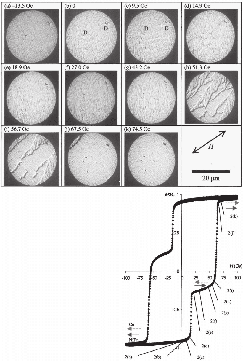
Figure 6–19. Magnetization of a multilayered
fi lm in situ. Lorentz Fresnel images (a–k) of the
magnetization process for an NiFe/Al-oxide/Co
junction fi lm. The direction of the applied fi eld
H is indicated. All images are of the same area.
Also shown is the normalized magnetization vs
applied fi eld for the same fi lm. The correspond-
ing domain structure at different fi eld values
along the hysteresis loop is shown. (Reprinted
with permission from Yu et al., © 2002a. Ameri-
can Institute of Physics.)
480 F.M. Ross
An interesting extension of these in situ studies on magnetic ele-
ments has been the simultaneous measurement of resistivity. Using a
holder in which the resistivity of a single lithographically patterned
element can be measured while its magnetization is changed, Portier
et al. (1997, 1998, 1999b) were able to correlate magnetoresistance with
the magnetic domain structure. Studies on single spin valves made of
multilayers such as NiFe/Cu/Co/NiFe/MnNi allowed magnetoresis-
tance to be correlated with the angle between the magnetization direc-
tions in the ferromagnetic layers. It was also possible to show the
mechanism of magnetization reversal, and to demonstrate the effect of
stray-fi eld coupling, which introduces edge domains, on the reversal
mechanism.
4.1.3 Phase Transitions in Magnetic Materials
The industrial application of materials with giant magnetoresistance
has stimulated study of an unusual class of materials which show
colossal magnetoresistance. These are certain manganites with the
perovskite structure, such as LaSrMnO
3
, which have both ferromag-
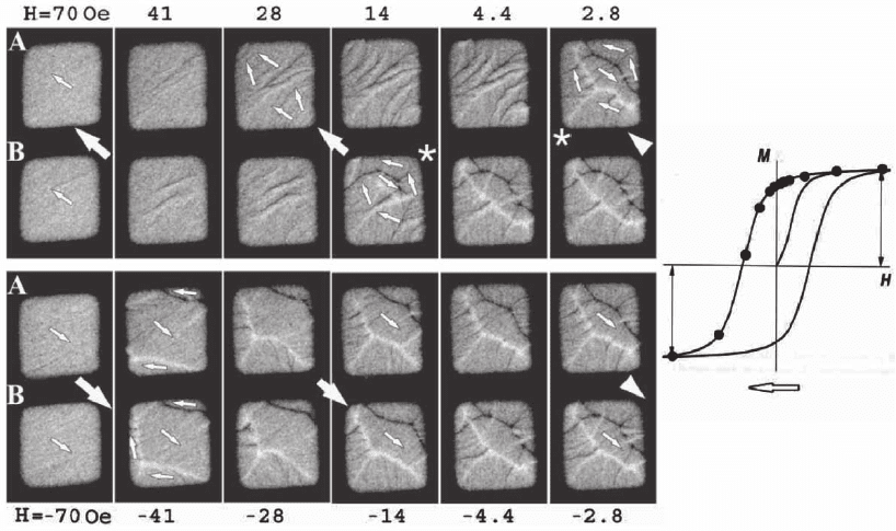
Figure 6–20. Magnetization of small elements. (A, B) In situ magnetization of two Co elements 25 nm
thick and 6?m on a side on an SiN membrane. Images are shown as a function of decreasing applied
fi eld, achieved by tilting the sample in a fi xed normal fi eld excited by the objective lens (160 Oe), and
images are obtained using defocused Fresnel mode. Large and small arrows show the direction of the
applied fi eld and local magnetization, respectively. The nucleation of the reverse domain is indicated
with asterisks. Only the upper semiloop of the hysteresis curve is shown; the reverse process is some-
what alike but differs in the reverse switching fi eld. The processes observed in these images are
coarsening of ripples, due to coherent spin rotation; nucleation and expansion of reverse domains;
wall motion and spin rotation in the remaining domains; expulsion of unfavorable boundary domains;
and edge annihilation. (From Volkov et al., 2004 with kind permission of Taylor and Francis Ltd.)
Chapter 6 In Situ Transmission Electron Microscopy 481
netic and paramagnetic phases. The phase transition and mixed phase
regions in these materials can be studied in situ in a cooling stage using
electron holography. The nucleation of ferromagnetic domains can be
observed on cooling (Yoo et al., 2004a), while in the mixed phase
region, application of a magnetic fi eld creates channels connecting fer-
romagnetic regions, thereby changing the conductivity (Yoo et al.,
2004b). These studies help to explain details of the mechanism of colos-
sal magnetoresistance.
Another interesting class of magnetic materials is ferromagnetic
shape memory alloys such as Ni
2
MnGa and CoNiAl. In these materials,
the shape change may be induced by applying a magnetic fi eld. Again,
in situ TEM using Lorentz imaging and a cooling stage (Murakami et
al., 2002, Park et al., 2003) allows the correlation of the magnetic domain
structure with the grain structure in these materials. Phase transfor-
mations in Ni
2
MnGa are shown in Figure 6–21. In experiments like this
on bulk materials, the effects of varying sample thickness on domain
motion should be considered in order to obtain the most quantitative
results.
4.2 Superconducting Materials
In situ TEM has provided a fascinating glimpse into the dynamics of
superconducting materials. Over the last several years, several groups,
most notably that of Tonomura and coworkers, have observed the pres-
ence and dynamics of vortices in superconductors using in situ tech-
niques. Single vortices, with their magnetic fl ux of h/2e, have an
observable effect on the phase of the imaging electrons. Thus Lorentz
microscopy or holographic techniques can be used to determine their
positions and characteristics. A medium or high voltage TEM with a
cooling stage is used for these studies and the magnetic fi eld can be
conveniently applied to the sample by tilting the sample in the existing
fi eld of the objective lens.
These experiments have provided unique insights into the behavior
of superconducting materials, in particular concerning the formation
of vortex lattices, as well as vortex pinning, which must be controlled
for practical applications of superconductors. Real time imaging has
allowed vortex pinning and dynamics to be related to microstructural
features for several superconducting materials (Figure 6–22). The
motion of vortices was fi rst imaged in Nb foils below 5 K (Figure 6–
22A). The effects of grain boundaries on vortex motion were immediat-
edly visible (Harada et al., 1992, Bonevich et al., 1993; Tonomura, 2002).
The role of dislocations in pinning vortices and nucleating locally
ordered regions of vortices (the Abrikosov lattice) was demonstrated
(Horiuchi et al., 1998). Nb specimens which had been irradiated with
an FIB to produce artifi cial pinning centers showed fascinating vortex
dynamics in which local regions of Abrikosov lattice formed and most
motion took place at the boundaries of such lattices (Matsuda et al.,
1996). Regular arrays of vortices could be formed with period matching
the pinning point period (Harada et al., 1996a). Vortex annihilation was
also observed (Harada et al., 1997) and the interactions between vortices
482 F.M. Ross
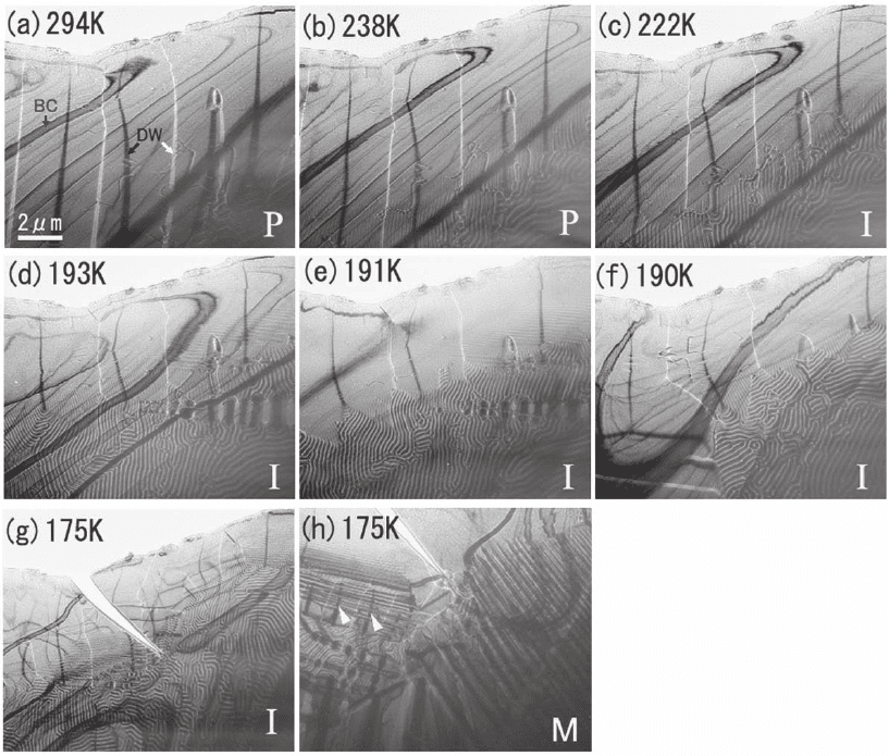
Figure 6–21. Phase transformation in a magnetic material. (a–h) Bright-fi eld images and electron dif-
fraction patterns of the three phases of Ni
2
MnGa: the parent phase at 294K, intermediate phase at
207K, and martensite at 170K (P, I, and M, respectively). Both the crystal structure (via diffraction) and
the magnetic structure (magnetic domains observed by Lorentz microscopy) are visible. BC and DW
represent the bend contours and the domain walls, respectively. (Reprinted with permission from Park
et al., © 2003. American Institute of Physics.)
quantifi ed (Sow et al., 1998) by analyzing their motion through the foil.
Most recently, asymmetric (one-way) motion of vortices has been con-
trolled by FIB-patterning asymmetric channels (Togawa et al., 2005).
For high temperature superconductors, with their potentially wide
range of applications, the pinning of vortices is weak and therefore
particularly important to understand and control. Harada et al. (1996b)
showed that, in these materials, vortex dynamics also depend on the
defects present. The effects of artifi cial pinning centers are highly
temperature dependent, giving useful insight into the different mecha-
nisms active (Tonomura et al., 2001). Furthermore, the vortices adopt
unusual chainlike arrangements in these superconductors (Matsuda et
al., 2001, Tonomura et al., 2002), as shown in Figure 6–22B. It is worth
noting that a high voltage (1 MeV) TEM was required to obtain the
necessary resolution for studying these materials (Tonomura, 2003).
Chapter 6 In Situ Transmission Electron Microscopy 483
4.3 Ferroelectric Phenomena
Ferroelectric domain boundary motion due to an applied electric fi eld
or stress has applications in information storage, and the piezoelectric
properties of these materials make them interesting as sensors, actua-
tors and transducers. Two key issues in the development of ferroelec-
tric devices are fatigue, in other words the change in boundary
dynamics resulting from repeated cycling, and the effects of fi lm thick-
ness and electrode material on boundary dynamics. Polarized optical
A
B
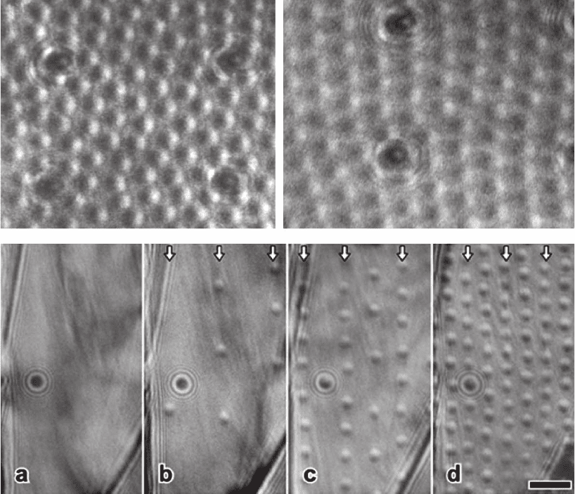
Figure 6–22. Vortex motion in superconductors. (A) Pinning of vortices in Nb. Typical interaction
between the vortex lattice and an array of defects produced in an Nb thin foil by irradiation with a
Ga+ focused ion beam. In the fi rst image, obtained at 4.5K, the pinning site acts as a domain boundary,
while at higher temperatures (8K, lower image) pinning points typically act as cores of edge disloca-
tions in the lattice. (From Harada et al., 1997b with permission from Elsevier.) (B) A series of Lorentz
micrographs of vortices in fi eld-cooled Bi-2212 fi lm at 50 K when a magnetic fi eld Bz perpendicular to
the layer plane is applied. The in-plane magnetic fi eld Bx is fi xed at 5 mT. (a) Bz = 0 (b) Bz = 0.02 mT.
(c) Bz = 0.1 mT. (d) Bz = 0.17 mT. The arrangement of the vertical vortices in chains is marked with
arrows. These chains are caused by interactions with horizontal vortices produced by the in-plane
fi eld. (Reprinted with permission from Tonomura et al., © 2002 by the American Physical Society.)
484 F.M. Ross
microscopy and AFM have been used successfully to investigate the
overall features of boundary motion, but naturally TEM is unparalleled
in its ability to relate the microstructural features within the material
to boundary motion. In situ experiments are usually carried out by
using a specimen holder with electrical connections to apply an electric
fi eld, although domain motion during heating and straining has also
been studied. A heating biasing holder is preferable as it allows domain
dynamics to be studied at different distances from the transition
temperature.
Ferroelectric biasing has been carried out both on mechanically
thinned polycrystalline or single crystal samples, and on thin fi lms
deposited on a substrate. Depending on the material geometry, the
electrical contacts may either both be on top of the sample or one on
each surface. As in other experiments, for quantitative analysis a well
controlled specimen and fi eld geometry is important. Changes in
sample thickness may change the area of domain walls and therefore
infl uence kinetics. Furthermore, as we show below, defects affect wall
motion, so the defects introduced during sample preparation must be
minimized. For all these reasons, experiments on bulk materials may
provide more qualitative information, whereas thin fi lms, especially
on substrates which can be made into electron transparent membranes
with a controlled electrode geometry and with minimal processing,
provide the best opportunity for quantitative results. For example, thin
fi lm studies provide the opportunity to understand the “dead layer,”
in which surface pinning retards domain motion.
Domain motion has been observed under electron beam heating, for
example in K(Ta,Nb)O
3
(Xu et al., 1993), and during straining, for
example in ferroelastic zirconia (Baufeld et al., 1997). Most studies,
however, have used in situ biasing or controlled heating to achieve
domain motion. The most detailed results have come from studies of
BaTiO
3
and related materials. For example, Ren et al. (1994) observed
domain wall motion in PbTiO
3
, while Snoeck et al. (1994) observed
domain growth in BaTiO
3
by tip motion and then by lateral wall motion,
and noted defects along the domain boundaries. Krishnan et al. (1999,
2000, 2002; Figure 6–23) observed different modes of domain wall
motion in BaTiO
3
and KNbO
3
under heating, biasing, and UV irradia-
tion. These studies showed that the motion of 90
o
boundaries depends
on their curvature and on locking interactions with neighboring
domains, and that motion may occur by rippling rather than rigidly.
Interestingly, images showed the presence of trapped charge at curved
or tilted boundaries or at domain tips. The buildup of charge at bound-
aries observed in situ may be important in fatigue.
In relaxor ferroelectrics such as Pb(Mg,Nb)O
3
-PbTiO
3
, cracking is
important in piezoelectric applications. The structures of domain wall
intersections has been characterized in these materials (Tan and Shang,
2004a, b; Tan et al., 2005). Cycling the electric fi eld causes cracking in
the TEM specimens, thereby providing information on crack propaga-
tion pathways along domain walls (Xu et al., 2000; Tan et al., 2000,
2005).