Hoque. Advanced Applications of Rapid Prototyping Technology in Modern Engineering
Подождите немного. Документ загружается.

Application of a Novel Patient - Specific Rapid Prototyping Template in Orthopedics Surgery
131
25 patients (14 male, 11 female, age 17–53 years) with cervical spinal pathology included 10
patients with destabilizing cervical spine injuries, 4 patients with cervical spondylotic
myelopathy, and 11 patients with basilar invagination requiring instrumentation underwent
cervical pedicle screw placement using a novel, patient-specific navigational template
technique. According to this technique, a spiral three-dimensional (3-D) CT scan
(LightSpeed VCT, GE, USA) was performed preoperatively on the cervical spine of each
patient with a 0.625-mm slice thickness and 0.35-mm in-plane resolution. The images were
stored in DICOM format, and transferred to a workstation running MIMICS 10.01 software
(Materialise, Belgium) to generate a 3-D reconstruction model of the desired cervical
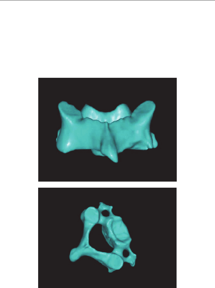
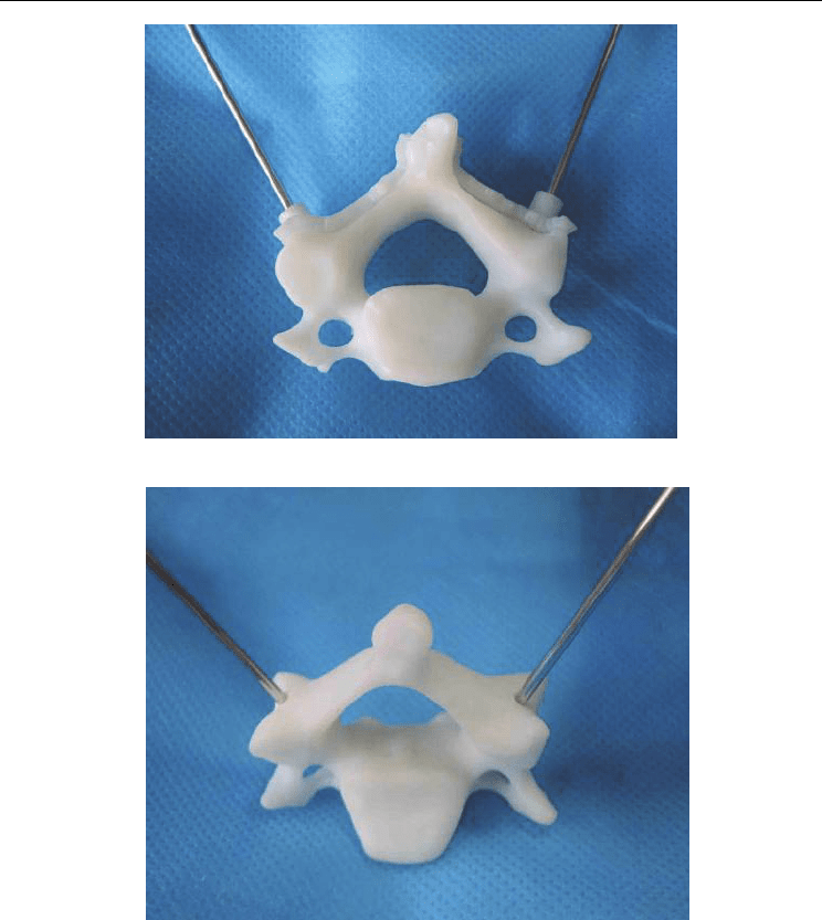
vertebra (Fig. 1).
(a)
(b)
Fig. 1. 3-D model of the cervical vertebra (C3).
a: posterior view ; b: lateral view
Advanced Applications of Rapid Prototyping Technology in Modern Engineering
132
The 3-D cervical vertebral model was then exported in STL format to a workstation running
Reverse Engineer software -UG imageware12.0 (EDS, US), for determining the optimal
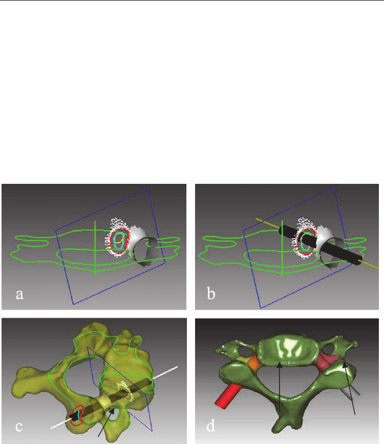
screw size and orientation. Using the UG Imageware software, the pedicles (left and right
pedicle) were projected towards the vertebra and lamina (Fig.2a). As the thickness and cross
section of the pedicle vary along its length, the smaller diameter of the elliptical inner
boundary of the pedicle’s projection was used in determining the maximum allowable
dimension for screw diameter (Fig.2b). This diameter was further used to draw a circle and
projected between the vertebra and the lamina to obtain the optimal pedicle screw trajectory
(Fig.2c,). A 3D vertebral model was reconstructed with a virtual screw placed on both
sides(Fig.2d).
Fig. 2. Analysis of cervical pedicle screw trajectory by the Reverse Engineering software
a: Pedicle and its positive projection; b: the best trajectory of pedicle screw projection;c:
Pedicle screw channel. (arrow) d: Planned screw trajectory (arrows)
Application of a Novel Patient - Specific Rapid Prototyping Template in Orthopedics Surgery
133
Following the determination of the optimal pedicle screw trajectory, a navigational
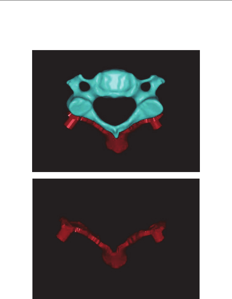
template was constructed with a drill guide on either side. The template surface was
created as the inverse of the vertebral posterior surface, thus potentially enabling a near-
perfect fit. It was also made sure that there was no overlapping of the template onto
adjacent segments (Fig.3).
(a)
(b)
Fig. 3. Design of the navigational template
a: Navigational template fits with the vertebra perfectly; b: The 3-D computer model of
navigational template
Advanced Applications of Rapid Prototyping Technology in Modern Engineering
134
The biomodel of the desired vertbera as well as its corresponding navigational template
were produced in acrylate resin (Somos 14120, DSM Desotech Inc, USA) using
stereolithography – a rapid prototyping (RP) technique (Hen Tong company, China). The
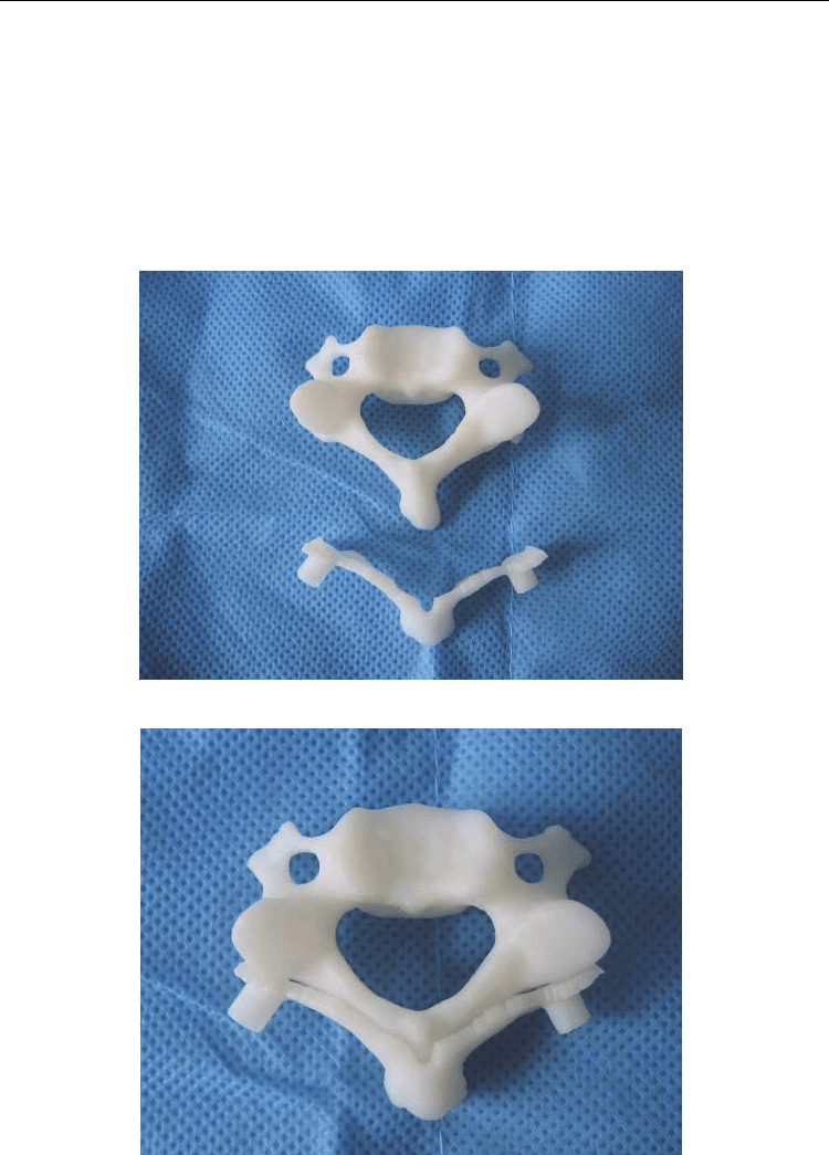
accuracy of the navigational template was examined by visual inspection before surgery.
The biomodel of the vertebra and its corresponding template were placed together, and a
standard electric power-drill was used to drill the screw trajectory into the biomodel of the
vertebra through the template navigation holes. Visual inspection was performed for
identifying any violation (Fig.4)
(a)
(b)
Application of a Novel Patient - Specific Rapid Prototyping Template in Orthopedics Surgery
135
(c)
(d)
Fig. 4. The accuracy of the navigational template was examined by visual inspection
a: RP model of vertebra and navigational template; b: navigational template fits RP model of
vertebra perfectly; c: K wires inserted through navigational template into the pedicles; d:
accuracy of the navigational template examined by visual inspection.
The template was sterilized and used intraoperatively for navigation and for confirming
anatomic relationships. For safety reasons, fluoroscopy was performed intraoperatively
during drilling and insertion of the pedicle screw on the first 3 patients. For the remaining
cases, fluoroscopy was performed only after the insertion of all the pedicle screws, thus
Advanced Applications of Rapid Prototyping Technology in Modern Engineering
136
considerably reducing the exposure time to radiation. After surgery, the positions of the
pedicle screws were evaluated using X-ray and CT scan. An axial image, including the
whole length of each screw, was obtained, and the medial and lateral deviation of the screw
was classified into 4 grades 6. Grade 0, no deviation; the screw was contained in the pedicle.
Grade 1, deviation less than 2 mm or less than half of the screw diameter. Grade 2, deviation
more than 2 mm and less than 4 mm, or half to one screw diameter. Grade 3, deviation more
than 4 mm, or complete deviation.
The accuracy of the navigational template was examined before operation by drilling the
screw trajectories into the vertebral biomodels. Each navigational template was found to be
fitting to its corresponding vertebral biomodel appropriately without any free movement,
and the K wires were found to be inserted through the drill hole through the pedicle and
into the desired vertebra without any violation as found by visual inspection.
During the operation, it was easy to find the best fit for positioning the template manually,
as there was no significant free motion of the template when it was placed in position and
pressed slightly against the vertebral body. As such, the navigational template fulfilled its
purpose for use as in situ drill guide.
A total of 88 screws were inserted into levels C2–C7 with 2-6 screws on each patient. Of
these pedicle screws, 71 were in Grade-0, 14 in Grade-1, 3 in Grade-2, and no screw was in
Grade-3. None of the cases had complications caused by pedicle perforation and especially
there were no injury to the vertebral artery or to the spinal cord, nor was there a need for
revision of pedicle perforation in any of the cases.
In this study, cervical pedicle abnormality existed in five patients. The pedicles (four C2 and
one C7) of these patients were very narrow with a minimum diameter of 3.5mm. Screws of
relatively smaller diameter (3-mm) were chosen for these patients accordingly, and were
placed inside pedicles accurately using navigational templates. (Fig.5)
(a) (b)
Application of a Novel Patient - Specific Rapid Prototyping Template in Orthopedics Surgery
137
(c) (d)
(e) (f)
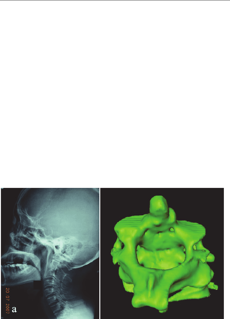
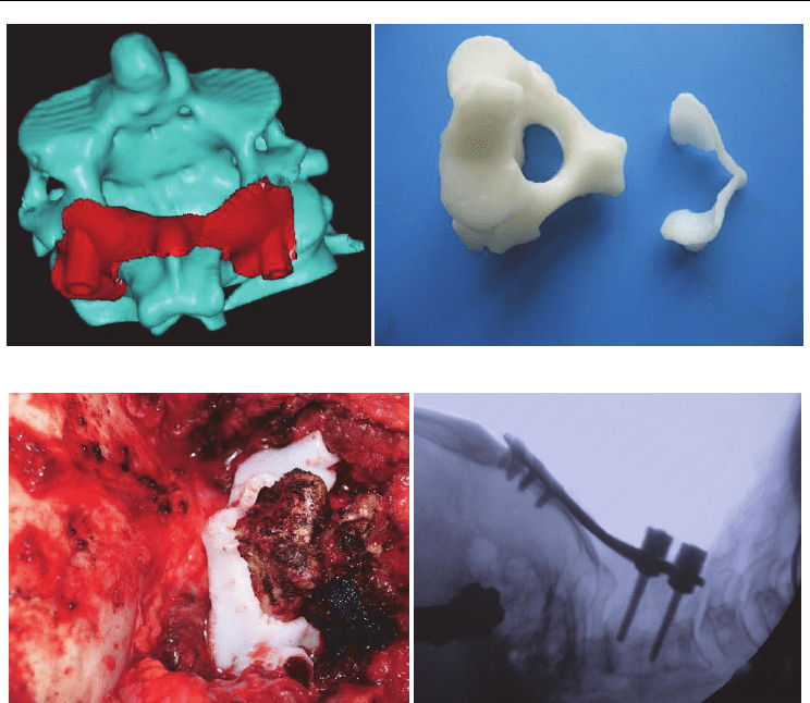
Fig. 5. A female patient was diagnosised of basilar invagination, trans-C2 Pedicle screw
Occipto-Cervical Fusion was done; In this case the C2,3 fusion was observed and the
diameter of left C2 pedicle was only 3.5mm, the C2 pedicle screw was inserted using the
navigational template;
a: the X-ray shows atlantoaxial dislocation; d: 3-D model of C2,3; b,c: Pedicle screw
trajectory and design of the C2 navigational template; d: RP model of C2,3 and navigational
template, e: the navigational template fit the posterior part of C2 perfectly; f: fluoroscopy
show good positioning of pedicle screw;
In another case, the pedicle was extremely narrow in level C2 with a minimum diameter of
only 1.5-mm and therefore the C2 cervical fixation was not performed. The CT data showed
congenital fusion between C2 - C3 and therefore pedicle screw fixation was successfully
performed on level C3 using the drill template.
By using this novel, custom-fit navigational template, the operation time has been
considerably reduced. On an average, each vertebral pedicle screw insertion took about 80
seconds. Fluoroscopy was required only once after the insertion of the entire pedicle screws,
which has considerably reduced the duration of radiation exposure to the members of the
surgical team. Currently the production time for RP model is about 2 days and the cost is
Advanced Applications of Rapid Prototyping Technology in Modern Engineering
138
about $50 per vertebral level. The production time can be brought down to 1 day and the
cost can be reduced to $20 if the RP model of the vertebra is not needed.
2.1.2 A novel patient-specific navigational template for thoracic pedicle placement
Most studies have shown that the rates of misplacement for the free-hand technique are
usually between 28% and 43%, while only a few studies have shown rates of less than 5%.
Hence, the screw breach rate may be dangerously high when the anatomy is altered as in
scoliosis. Lonner et al. [6] suggested that there should be a considerable learning curve for
using the pedicle screws in scoliosis surgery to avoid complications. The need for improved
accuracy and consistency in the placement of thoracic pedicle screws has led to
investigations on the application of computer-navigated spine surgery. Computer-assisted
pedicle screw installation allows for an increased accuracy in using screws, thus decreasing
the incidence of misplaced screws. Considering these difficulties, surgeons must use
whatever techniques they find helpful to create a safe environment when placing thoracic
pedicle screws into the deformed pediatric spine.
16 patients (12 females, 4 males, age 5–18 years) with scoliosis (14 adolescent idiopathic
scoliosis, 2 congenital scoliosis) undergoing spinal deformity correction surgeries using
posterior pedicle screw instrumentation of the thoracic spine formed the study group.
Before the operation, a spiral three-dimensional (3-D) CT scan (LightSpeed VCT, GE, USA)
was performed on the thoracic spine of each patient with 0.625 mm slice thickness and 0.35
mm in-plane resolution. The images were stored in DICOM format and transferred to a
workstation running MIMICS 10.01 software (Materialise company, Belgium) to generate a
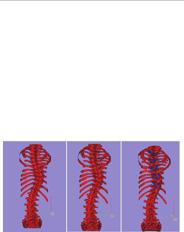
3-D reconstruction model for the desired thoracic vertebra (Fig. 6a). The 3-D vertebral model
(a) (b) (c)
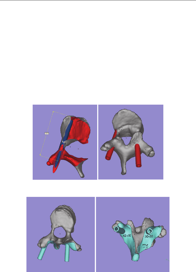
Fig. 6. Trajectory of the pedicle screw and design of the navigational template
a: Three-dimensional model of the scoliosis vertebra; b: Planned screw trajectory (blue). c:
The navigational template was created as the inverse of the posterior vertebral surface
fitting with the vertebra perfectly.
Application of a Novel Patient - Specific Rapid Prototyping Template in Orthopedics Surgery
139
was then exported in STL format and opened in a workstation running Reverse Engineering
(RE) software UG imageware12.0 (EDS, USA)to determine the optimal screw size and
orientation. A screw with a diameter of 5 mm was placed virtually into the 3-D spinal model
on both sides. The virtual screw’s entry point and the trajectory were placed at the center of
the pedicle without violating the cortex.
Pre-operative Planning
According to the type of scoliosis, the fusion level is determined, and the instrumentational
vertebra is chosen. The design and development of the drill template for each vertebra are
also made according to the instrumentational vertebra. The vertebral rotation, axes, length,
and diameter of the pedicle were measured from the pre-operative CT scan. Thus, the length
and diameter of every pedicle screw were decided upon before operation. The concave
periapical (T5-8) pedicles are often deformed and are considered to be the most difficult area
to work on during pedicle screw placement [3,27-29]. If the pedicle is very narrow, an in-
out-in technique can be chosen. (Fig 7).
(a) (b)
(c) (d)
Advanced Applications of Rapid Prototyping Technology in Modern Engineering
140
(e)
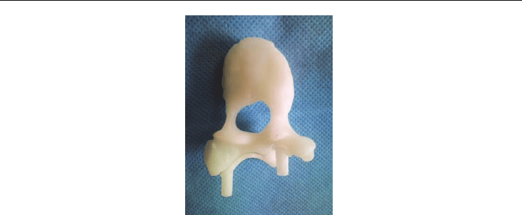
Fig. 7. 3D reconstruction of a single vertebra and the biomodel of the drill template
a: The best trajectory of the pedicle screw and measurement of the length of the pedicle; b:
In-out-in technique in the concave side; c: The navigational template fits the vertebra
perfectly; d: The template can show the location, diameter, and length of the pedicle screw;
e: The navigational template fits the RP model of the vertebra very well.
Operation procedure
The spine was exposed subperiosteally on both sides, up to the tips of the transverse
processes. For the thoracic spine, the soft tissues on the facet joints were thoroughly cleaned
off to ensure better visualization of the bony landmarks. The drill template was then placed
on the spinous, lamina, and transverse processes. The drill template and the corresponding
spinous process were fitted well. A high-speed drill was used along the navigational
channel to drill the trajectory of each pedicle screw. Using a hand drill, the trajectory of the
pedicle screw was carefully drilled to a depth in accordance with the pre-operation plan.
The pedicle screw, the diameter and length of which had been chosen pre-operatively, was
carefully inserted along the same trajectory. After screw placement and correction of
deformity, all exposed laminar surfaces were decorticated, and the autologous iliac crest
bone was grafted.
A total of 168 screws were placed from T2 to T12 in the 16 cases, and post-operative CT
scans were obtained in all 16 patients. About 157 screws were considered intrapedicular,
while 11 screws were considered to have a 0–2 mm breach (1 medial, 10 lateral in which 8
belonged to the planned in-out-in screws). No pedicle screw breached more than 2 mm, and
the overall screw accuracy (<2 mm breach is safe) was 100%. No screws penetrated the
inferior or superior cortex in the sagittal plane.
2.1.3 A Novel Patient-specific Navigational Template for laminar Screw Placement
Instability of the occipitocervical junction requiring surgical stabilization may be treated
with a variety of techniques. The objective is to obtain solid fusion of the involved segments,
which is best achieved by minimizing motion between them. Older methods such as the
Brooks-Jenkins or modified Gallie wiring techniques, are simpler procedures, have been
known for a long time and are associated with failure rates of fusion up to 25%, primarily in
cases with rotational instability. Newer techniques have been described that effectively limit