Kaplan Cynthia G., MD Color Atlas of Gross Placental Pathology
Подождите немного. Документ загружается.


maternal surface and better seen on cross section (Figure 5.19). Over
time the blood breaks down and the infarcts become paler with age
(Figures 5.20). The exact time course for these placental changes to occur
is unknown. When the blood has a means of egress, villous tissue may
not be compressed (Figure 5.21). The blood comprising the clots is
78 Chapter 5 Lesions of the Villous Tissue
B
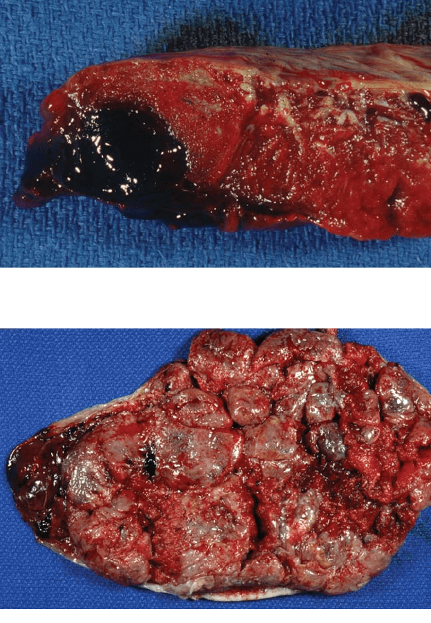
Figure 5.18 (Continued). (B) The three large clots received with the placenta fit
the large and 2 other more subtle depressions. The involved areas of placental
separation will extend well beyond the actual clot.
Figure 5.19. Some retroplacental hemorrhages are not raised above the mater-
nal surface and may not be appreciated until cross-sections are done. Trapping
of maternal blood led to the large retroplacental clot which compressed the
villous tissue. The villi above the blood are solid and pale, having infarcted
from the lack of maternal blood supply. The clot does not show significant
degeneration.

Retroplacental Hemorrhage 79
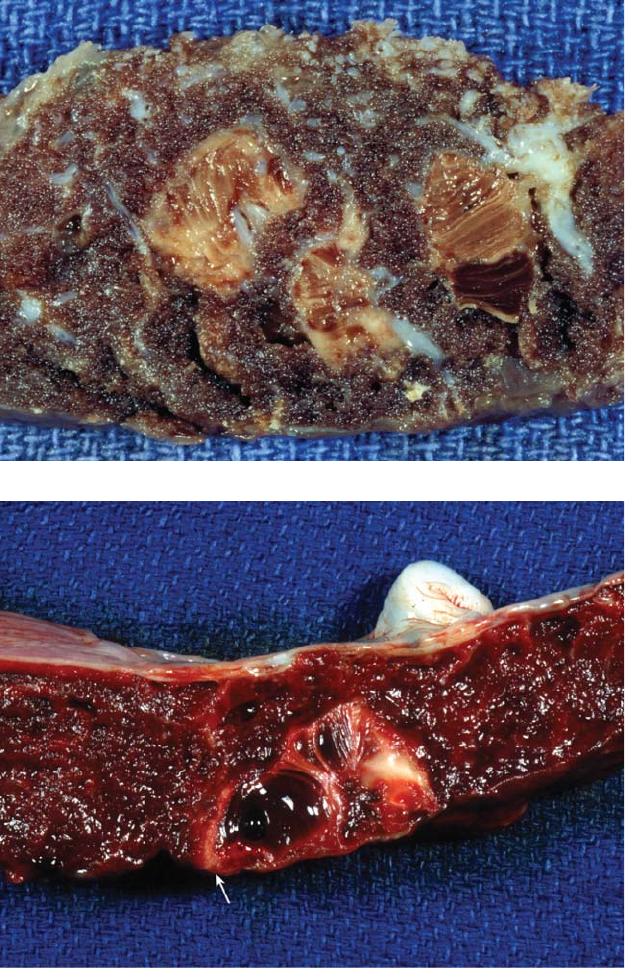
Figure 5.20. If delivery does not occur, the villous tissue will continue through
the usual stages of infarction and the blood clot will degenerate, as can be seen
in this remote retroplacental hemorrhage. Both processes are of roughly similar
ages. If one finds fresh blood overlying old infarction, it suggests a previously
infracted area separated prematurely.
Figure 5.21. In this cross-section of another retroplacental hemorrhage, there is
clot on the surface without villous compression. This occurs if blood has a means
of egress. Again there is pallor and infarction of the villous tissue adjacent to the
clot. This separation involved nearly the entire placenta and led to fetal demise.
A small old yellow infarct near the fetal surface suggests preexisting vascular
disease.

largely maternal, occasionally with some fetal bleeding. Retroplacental
hematomas occur both centrally and at the margin of the placenta and
overlap villous tissue (Figure 5.22).
If placental delivery is delayed or incomplete, one may see fresh retro-
placental hemorrhage with slightly adherent blood clot and villous col-
lapse. This, of course, has no implications for the infant. True marginal
hemorrhage will also have no fetal effects. It is peripheral, with the aggre-
gate of blood extending onto the membranes, not separating the placenta
(Figure 5.23, Figure 5.24). It is a fundamentally different process, related
to marginal sinus hemorrhage.
80 Chapter 5 Lesions of the Villous Tissue
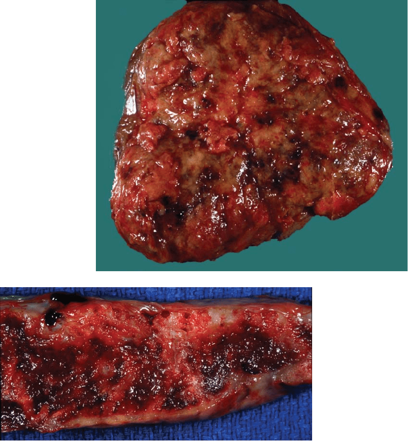
Figure 5.22.
Hemorrhage is most
commonly seen
at the placental
margin and many
separations start in
that region. This
fresh hemorrhage
undermines the
villous tissue and the
overlying area is in
an early stage of
infarction.
Figure 5.23.
Marginal
hemorrhage is
present in this
preterm placenta.
It extends onto the
membranes and not
the villous tissue.
The brown color of
the blood indicates
it is breaking down.
These hemorrhages
come from marginal
sinus bleeding
and not placental
separation. Note
the pale attached
decidua on the
maternal surface.

Intervillous Thrombi
Intervillous thrombi occur in the intervillous space in central areas of the
placenta.The earliest thrombi are fresh red clots, which progress through
laminated thrombi to old white lesions (Figure 5.25, Figure 5.26). No
true organization occurs. Intervillous thrombi contain both fetal and
Intervillous Thrombi 81
Figure 5.24. This large marginal hemorrhage occurred in an immature placenta.
The fetal surface is yellow-green from severe chorioamnionitis. Many severely
infected pregnancies deliver prematurely. Such placentas often show substantial
hemorrhage at the margin, possibly from necrosis of the infected decidua. This is
likely a peripartum event and not the cause of early delivery. The described asso-
ciation of “abruption” with chorioamnionitis is partially due to cases such as this.
Figure 5.25.
Intervillous thrombi
will be palpable as
firm lesions in the
placental tissue. They
are shinier and more
homogenous in
texture than infarcts.
Thrombi are usually
located in the
midportion of the
placenta, as shown
here by this fresh
lesion with minimal
stranding of fibrin.

maternal red blood cells. They are seen more frequently in hydrops and
other conditions with large friable placentas.The etiology of these lesions
is not clear, but may relate to coagulation at sites of villous damage and
fetal bleeding. Infarction may be present as a rim adjacent to intervillous
thrombi, which apparently interfere with local villous blood supply. Such
associated infarction does not imply maternal vascular disease. Thrombi
may also be present at the base of the placenta, where they do not
indicate premature placental separation (Figure 5.27).
82 Chapter 5 Lesions of the Villous Tissue
Figure 5.26. These
are older thrombi in
a fixed placenta.
Lines of Zahn can
readily be seen.
These do not have a
marginal rim of
infarction.
Figure 5.27. This
basal intervillous
thrombus has several
components of
different ages. The
deep red portion
is fresh while the
layered material is
older. There is a
small marginal rim
of infarction (arrow).
Thrombi occur at the
base of the placenta
and should not be
confused with
retroplacental
hemorrhage.

Fibrin Deposition
Localized areas of perivillous fibrin deposition are seen in virtually all
mature placentas and show an irregular lacelike pattern (Figure 5.28,
Figure 5.29). Although the entrapped villi eventually die, small amounts
of fibrin deposition are not generally thought to be related to fetal or
maternal disease, apparently originating from turbulence in the mater-
nal circulation.
Occasionally fibrin deposition is excessive, diffusely involving half or
more of the villous tissue (Figure 5.30, Figure 5.31). This degree is abnor-
mal, and associated with preterm delivery, growth retardation, and death.
Fibrin Deposition 83
Figure 5.28. This
cross section shows a
thrombotic lesion (left)
and perivillous fibrin
deposition (right). The
shiny old thrombus is
actually an extension
of a small, old,
subchorionic
hemorrhage and not a
true intervillous
thrombus. Note the
irregular outlines of
the fibrin deposition
and its admixture with
normal villous tissue.
There is substantial
calcification in this
area.
Figure 5.29. The white material deposited in this term placenta is fibrin. Such
localized fibrin is common in later gestations. It is deposited in the intervillous
space around villi in a lacelike fashion, and usually is quite hard and shiny.
Although the entrapped villi eventually die, such fibrin deposition is not usually
associated with fetal or maternal disease. At times, relatively large regions are
involved, as shown here. The process, however, is still localized and not of
concern.

84 Chapter 5 Lesions of the Villous Tissue
Figure 5.30. Diffuse fibrin deposition involving more than 50% of the placenta
is considered abnormal, and is associated with prematurity, fetal growth retar-
dation, and death. The etiology of such massive perivillous fibrin deposition is
unknown. This thick, immature, very pale placenta showing a diffuse network of
fibrin was associated with intrauterine demise at 25 weeks of a poorly grown
infant. Such placentas are usually quite firm and may actually be relatively heavy.
Figure 5.31. Viewing sections of the entire placenta will help determine the extent
of the process, as show in another example of massive perivillous fibrin deposi-
tion. The material shows a much coarser pattern, the more typical appearance.

“Maternal floor infarction” is a related lesion (Figure 5.32 to Figure
5.34).This is not true infarction, consisting of a layer of bland fibrin depo-
sition around basal villi. It has similar associations to diffuse perivillous
fibrin deposition and can be recurrent. The etiology of maternal floor
infarction is also unknown. Placentas with excess fibrin, both normal and
pathologic, often show surface and septal cysts due to the trophoblastic
proliferation which occurs in areas of fibrin. The cysts form within the
trophoblastic areas (Figure 4.17, 5.35).
Fibrin Deposition 85
Figure 5.32.
Maternal floor of an
immature placenta
with excess fibrin
deposition is shiny
and appears stiff
showing firm yellow
plaques. Such an
appearance is
suggestive of
maternal floor
infarction, which is
better visualized on
cut sections.
Figure 5.33. “Maternal floor infarction” is a recurring lesion is associated with
growth retardation and death. In this process there is a layer of fibrin deposited
at the base for 3 mm to 4 mm. Basal villi are entrapped and die, but it is not true
infarction. This occurs in combination with some degree of diffuse perivillous
fibrin deposition, as shown here. This placenta is from a live-born infant.

86 Chapter 5 Lesions of the Villous Tissue
Figure 5.34. This more dramatic example of maternal floor infarction is from a
25 week stillborn.
Figure 5.35. This thin-walled cyst is located within a septum of a term placenta.
Such cysts also occur on the surface (Figure 4.17). They develop within solid
trophoblastic regions and are often adjacent to fibrin deposition. Cysts do not
appear to be associated intrinsically with any pathology.

Avascular Villi
The presence of avascular vill (villous atrophy) implies interruption of
the fetal blood supply. After fetal demise, the entire placenta undergoes
this change if delivery does not occur. It does not infarct since maternal
perfusion continues. Occlusion of part of the fetal circulation, as with
thrombosis, will lead to zones of atrophic avascular villi, often recogniz-
able grossly (Figure 5.36). Such change may reflect more diffuse fetal
thrombotic processes in utero, with the potential for vascular disruptive
lesions. Early microscopic change in villi includes vascular breakdown
progressing to complete stromal fibrosis (avascular villi) (Figure 5.37).
These histologic changes take days to weeks to develop.
Avascular Villi 87
Figure 5.36. Fixed placenta with irregular pale area of atrophy. The light color
comes from the lack of fetal blood and fibrosis within the affected villi. No gross
thrombosis was noted, which is not unusual; however, fetal vascular thrombi are
often found on histology. The tissue is not collapsed or hard and feels similar to
adjacent villi.
