Kim Y.J. (Ed.) Advanced Environmental Monitoring
Подождите немного. Документ загружается.

324 A. Zitova et al.
O’Mahony F.C. and Papkovsky D.B. (2006), Rapid high-throughput assessment of aerobic bac-
teria in complex samples by fluorescence-based oxygen respirometry, Appl. Env. Microbiol.,
72, 1279–1287.
O’Riordan T.C., Buckley D., Ogurtsov V., O’Connor R., and Papkovsky D.B. (2000), A cell
viability assay based on monitoring respiration by optical oxygen sensing, Anal. Biochem.,
278, 221–227.
Papkovsky D.B. (2004), Methods in optical oxygen sensing: Protocols and critical analyses, Oxyg.
Sens., 381, 715–735.
Papkovsky D.B. (2005), Respirometric Screening Technology (RST), Screen. – Trends Drug
Discov., 6, 46–47.
Sillanpaa M. and Oikari A. (1996), Assessing the impact of complexation by EDTA and DTPA on
heavy metal toxicity using microtox bioassay, Chemosphere, 32, 1485–1497.
Vanhaecke P. and Persoone G. (1984), The arc-test: A stanardized short term routine toxicity test with
Artemia nauplii. Methodology and evaluation, Ecotoxicol. Test. Marine Environ., 2, 143–157.

Chapter 25
Omics Tools for Environmental Monitoring
of Chemicals, Radiation, and Physical Stresses
in Saccharomyces Cerevisiae
Yoshihide Tanaka
1
, Tetsuji Higashi
1
, Randeep Rakwal
2
, Junko Shibato
2
,
Emiko Kitagawa
2
, Satomi Murata
2
, Shin-ichi Wakida
1
, and Hitoshi Iwahashi
2
Abstract The yeast Saccharomyces cerevisiae is one of the most characterized
eucaryotes and its complete genome sequence was published in 1986. Thus, this
organism is a good candidate for biological environmental monitoring. Omics
(genomics, proteomics, metabolomics) technology is being applied to biological
studies from prokaryotes to humans. We are applying omics technologies to
environmental monitoring using yeast cells, medaka, rice, rat, and mouse. In this
report, we focus on yeast omics as tools of environmental monitoring for chemi-
cals, radiation, and physical stresses in yeast. For genomics studies, we use com-
mercially available DNA microarrays. We analyzed the expression profiles for
highly induced or repressed genes, the functional characterization of induced and
repressed genes, and cluster analysis. The list of highly induced and repressed genes
can help to identify candidate biomarkers and strongly induced functions; however,
this may reflect only a small part of the full stress response. The functional char-
acterization studies, on the other hand, can help elucidate the mechanisms of stress
response. Cluster analysis allows comparison of different environmental stress
conditions. For proteomics studies, classical two-dimensional electrophoresis and
peptide sequencing or mass spectrometry were used for monitoring stress induced
and modified proteins. However, protein turnover ratio and especially degradation
rates were slow in yeast cells and the induction levels of proteins do not always
reflect the physiological status of the cell. The role of proteomics in yeast cells
must be focused on modification of proteins. For metabolomics studies, capillary
electrophoresis/mass spectrometry (CE/MS) was used for the separation and iden-
tification of metabolites. This new methods have high potential for the evaluation
of environmental stress.
Keywords: Saccharomyces cerevisiae, environmental stress, OMICS, genomics,
proteomics, metablomics, DNA microarray, CE/MS
1
Human Stress Signal Research Center (HSS), National Institute of Advanced Industrial
Science and Technology (AIST), 1-8-31 Midorigaoka, Ikeda, Osaka 563–8577, Japan
2
HSS, AIST, Tsukuba West, 16-1 Onogawa, Tsukuba, Ibaraki 305–8569, Japan
325
Y.J. Kim and U. Platt (eds.), Advanced Environmental Monitoring,
325–337.
© Springer 2008
326 Y. Tanaka et al.
25.1 Introduction
At present, more than 25 million materials are registered in the Chemical Abstracts
database, and it is estimated that more than 10,000 synthetic chemicals are accu-
mulating in the environment every year. Despite the fact that these industrial
chemicals have given us numerous benefits, there is no doubt that they have
damaged the environment. The chemicals being dispersed on the earth should be
carefully controlled to prevent their adverse effects. In fact, many chemicals can
be detected from environmental samples; however, only 10% of those chemicals
can be identified by current technology (Suzuki and Utsumis 1998). Ten percent
is an inadequate number to protect the environment. Furthermore, not only chemi-
cal but also physical and biological stresses including radiation, temperature, and
pathogens impact humans and ecological systems. Thus, we have to understand
the effects of these environmental stresses on biological systems, which necessi-
tates monitoring these stresses.
Bioassays are used for the assessment of environmental pollution and risk
assessment of environmental stresses (Celemedson et al. 1996). In bioassay sys-
tems, safety is estimated by monitoring biological responses to environmental
stress. One of these studies is the Multicenter Evaluation of in vitro. Cytotoxicity
program, organized by the Scandinavian Society for Cell Toxicology (Celemedson
et al. 1996). The investigators compared LD50 data obtained in vivo (whole organism)
and IC50 obtained in vitro (bioassay). They found a correlation between these
parameters and defined the concept of “basal cytotoxicity”. Basal cytotoxicity can
be understood as the generalized toxic effect to cellular components, functions and
biosynthesis that are universal to all cell lines. On the other hand, the Ames test is
well known as one of the most powerful methods for monitoring the mutagenicity
of environmental samples (Reifferscheid and Heil 1996). In this system, mutants of
Salmonella typhimurium are grown in a minimum medium and mutagenicity is
estimated according to the frequency of back mutation. As the frequency of back
mutation is dependent on DNA damage, we can estimate the mutagenicity of
chemicals or environmental stress.
An extensive literature exists on bioassay systems that include tests by Ames,
Microtox, Umu, and others (Celemedson et al. 1996). Each system can be used for
estimating effects by environmental stress; however, the information that can be
estimated is limited to the degree of toxicity or mutagenicity. Information concerning
the nature of the environmental stress remains unavailable. In addition, bioassay
systems sometimes mistakenly identify natural products as the toxic substance
(data not shown). Although it is important to quantify the degree of effects in the
environment, information concerning the nature of stress is essential for risk assess-
ment and prevention. Bioassay systems are required that can be used for predicting
the mechanism of environmental stress.
We proposed “multiple-end-point bioassays” several years ago (Iwahashi 2000).
The report introduces “multiple-end-point bioassay” systems that are based on
stress sensitivities of microorganisms, responses of one kind of organism, and
25 Omics Tools for Environmental Monitoring 327
microarray technology. Microorganisms are screened to identify strains that are
sensitive to specific stresses and the sensitivity of the isolated strain is then used for
characterizing unknown chemicals or environmental samples. The “multiple-end-
point bioassay” based on one kind of organisms are system using one organism and
many kinds of endpoints such as growth inhibition, viability, induction of stress
proteins, prion curing mutagenicity, cytoplasmic mutagenicity, and chromosomal
mutagenicity. Using these endpoints we tried to characterize chemicals and envi-
ronmental stresses (Iwahashi 2000). DNA microarray technology was also intro-
duced as the candidate for the “multiple-end-point bioassay” (Iwahashi 2000).
In recent years, DNA microarray technology has developed rapidly and been
widely adopted as a tool for understanding biological systems at the genomic level
(Momose and Iwahashi 2001). Furthermore, this technology can be combined with
proteomics and metabolomics technology. Proteomics is essentially based on the
analysis of proteins using two-dimensional electrophoresis. This technology provides
information on the expression levels of hundreds of proteins as well as protein
modifications. Metabolomics is based on the extensive database of analysis of
metabolites using NMR or CE/MS, and this is relatively new technology for biolo-
gists. A combined approach, omics technology, provides data on DNA, mRNA,
proteins, and metabolites and thus allows the development of a robust “multiple-
end-point bioassay”. In this report, we describe the development of a combined
omics approach (genomics, proteomics and metabolomics) for environmental
monitoring for of chemicals, radiation, and physical stresses in yeast.
25.2 Materials and Methods
25.2.1 Strains and Growth Conditions
Saccharomyces cerevisiae S288 C (MATaSUC2 mal mel gal2 CUP1) was grown in
YPD medium (1% Bacto Yeast Extract, 2% polypeptone, 2% glucose) at 25°C
according to the procedure outlined by Kitagawa et al. (2002).
25.2.2 Stress Conditions
For chemical and radiation treatment, yeast cells growing exponentially were
exposed for 2 h as follows: 35 ppm paraquat (Iwahashi 2006), saturated vitamin E
(Iwahashi 2006), 0.16% supiculisporic acid (Kurita et al. 2004), 10% dimethylsul-
foxide (Murata et al. 2003), 16 Gy gamma ray (Kimura et al. 2006), 25 ppm chlo-
roacetaldehyde (Iwahashi 2006), 250 ppm capsaicin (Kitagawa et al. 2002), 5 µM
thiuram (Kurita et al. 2002), 5 mM manganese chloride (Iwahashi 2006), 10 µM
cadmium and 2.5 µM thiuram (Iwahashi 2006), 0.7 mM mercury(II) chloride
328 Y. Tanaka et al.
(Kimura et al. 2006), 0.01%, sodium dodecyl sulfate (Sirisattha et al. 2004a),
×1,500 dilution of roundup high-load (Sirisattha et al. 2004b), 15 µM cyclohex-
imide (Iwahashi 2006), 10 mM hydrogenperoxide(H2O2 in Fig. 25.1) (Iwahashi
2006), 2 mM lead chloride (Iwahashi 2006), 1.5% pentane (Fujita et al. 2004),
5 mM thorium nitrate (Murata et al. 2006a), 400 ppm gingerol (Iwahashi 2006),
1.5 µM fluazinam (Iwahashi 2006), 5 mM 2-aminobenzimidazole (Iwahashi 2006),
0.5 mM benzpyren (Iwahashi 2006), 50 µM pentachlorophenol (Iwahashi 2006),
2 ppm zineb (Kitagawa et al. 2003), 2 ppm maneb (Kitagawa et al. 2003), 75 µM
thiuram (Iwahashi 2006), 10 µM TPN (Kitagawa et al. 2003), 20 µM cadmium and
5 µM thiuram (Iwahashi 2006), 0.3 mM cadmium chloride (Momose and Iwahashi
2001), and 0.3 µM methylmercury(II) chloride (Iwahashi 2006).
For gas treatment, yeast cells growing exponentially were transferred to high
pressure vessels (Iwahashi 2006) and pressured using compressed gas cylinders for
2 h as follows: 10 MPa air (Iwahashi 2006), 40 MPa nitrogen (Matsuoka et al.
2005), 0.5 MPa oxygen (Iwahashi 2006).
For physical stress treatment, yeast cells growing exponentially were frozen at
−80°C for 7 days (Freeze in Fig. 25.1) (Odani et al. 2003), then treated as follows:
40 MPa at 4°C for 12 h (40 MPa 4°C in Fig. 25.1) (Iwahashi et al. 2003), 180 MPa at
4°C for 0 min (180 MPa 4°C in Fig. 25.1) (Iwahashi et al 2003), and 30 MPa 25°C
for 2 h. These cells were allowed to recover for 60 min at 25–30°C. Cold shock treat-
ment (Cold in Fig. 25.1) entailed a shift of exponentially growing yeast cells from
25 °C to 4 °C for 6 h (Iwahashi et al 2005), while pressure shock treatment (40 MPa
Pressure Shock in Fig. 25.1) shifted exponentially growing yeast cells under atmos-
phere pressure to 40 MPa for 2 h (Iwahashi 2006), and followed by incubation under
30 MPa or 10 MPa for 16 h (30 MPa 25 °C Growth or 10 MPa 25 °C growth in Fig.
25.1) (Iwahashi 2006). Heat shock treatment was carried out by shifting exponen-
tially growing yeast cells from 30 to 43°C for 2 h (Iwahashi et al. 1995).
For environmental samples A–E and the incinerator sample in Fig. 25.1, YPD
medium were made with an environmental sample replacing the DW. These YPD
media were filter sterilized (Kim et al. 2004; Murata et al. 2006b). Exponentially
growing yeast cells were transferred to the YPD medium made of environmental
sample.
25.2.3 DNA Microarray Analysis
Each microarray, spotted on a glass slide for hybridization with labeled mRNA
probes, represented almost all ORFs of yeast (5,809∼5,819 genes; depending on the
lot, DNA Chip Research Inc. Yokohama, Japan). Extraction of total RNA, mRNA
purification, labeling with Cy3 or Cy5, and hybridization were described previ-
ously [Kim et al. 2004; Momose and Iwahashi 2001). A Scan Array 4000 laser
scanner (GSI Lunomics, Billeria, MA, USA) was used to acquire hybridization
signals. Array images were analyzed with Gene Pix 4000 (Inter Medical, Nagoya,
Japan). Cluster analysis of the mRNA expression profiles after the combination
25 Omics Tools for Environmental Monitoring 329
treatment was according to Murata et al. using the GeneSpring ver. 4.2.1 software
(Silicon Genetics, CA, USA) (Momose and Iwahashi 2001; Kitagawa et al. 2002,
2003; Kurita et al. 2002, 2004; Iwahashi et al. 2003, 2005; Murata et al. 2003,
2006a, b; Odani et al. 2003; Kim et al. 2004; Sirisattha et al. 2004a, b; Matsuoka
et al. 2005; Iwahashi 2006; Kimura et al. 2006).
25.2.4 Two-Dimensional Electrophoresis
Two-dimensional electrophoresis was carried out essentially according to O’Farrell’s
method (1975) and extraction of proteins, labeling with [H
3
] leucine, and staining
with CBB were described previously (Iwahashi et al. 1995).
25.2.5 Capillary Electrophoresis/Mass Spectrum
(CE/MS) Analysis
A Beckman P/ACE MDQ capillary electrophoresis system (Beckman Coulter,
Tokyo, Japan) was connected to an Esquire 3000 plus ion trap mass spectrometer
(Bruker Daltonics, Yokohama, Japan) through an electrospray ionization (ESI)
source (Agilent Technologies Japan, Tokyo, Japan). Yeast extracts were prepared
after the stress conditions by filtration of yeast cells (0.45 µm membrane filter),
extraction of metabolites with cold methanol, and a second filtration (Microcon,
5 kDa cut-off, Millipore, Bedford, MA, USA) (Sato et al. 2004). After prefreezing
the filtered solution at −80°C for 2 h, lyophilization was carried out at 25°C under
vaccum (10 Pa) overnight in a lyophilizer (model FRD-MINI, Asahi Techno Glass,
Chiba, Japan). The residue was dissolved in 25 µl of water/methanol (1:1, v/v).
Analytical conditions were established based on a previously reported method (Soga
et al. 2003; Sato et al. 2004).
25.3 Results and Discussion
25.3.1 Genomics Technology for the Assessment of Stress
Response Through mRNA Expression Levels
Figure 25.1 shows cluster analysis of expression profiles obtained after the stress treat-
ments (Momose and Iwahashi 2001; Kitagawa et al. 2002, 2003; Kurita et al. 2002,
2004; Iwahashi et al. 2003, 2005; Murata et al. 2003, 2006a, b; Odani et al. 2003; Kim
et al. 2004; Sirisattha et al. 2004a, b; Matsuoka et al. 2005; Iwahashi 2006; Kimura et
al. 2006). This calculation is based on the correlation factors between the treatments.
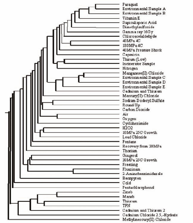
330 Y. Tanaka et al.
We may select calculation methods and the calculations were mainly based on the
Euclidean distance (distance between the treatments) or Pearson CC (direction or
angle between the treatments). In any calculation what we can obtain is the similari-
ties among the expression profiles after stress treatments. Expression profiles
reflect the effect of stress on cells and the effect must be specific to the stress treat-
ments. We may speculate that clustering represents similar responses among
stresses. Correlation factors for the expression profiles must be high among chemi-
cals that cause similar damages or responses. For example, zineb, maneb, and
thiuram belong to dithiocarbamate fungicides and they have similar chemical
Fig. 25.1 Cluster analysis of the various environmental stress treatments to yeast cells. Each
stress conditions was described previously (see text)

25 Omics Tools for Environmental Monitoring 331
structures (Kitagawa et al. 2003). Thus, these chemicals were expected to cause
similar damages or similar cellular response (expression profile). As shown in Fig.
25.1, Zineb, Mneb, and Thiuram cluster and we may conclude that these chemicals
cause similar damages or responses. Thus, cluster analysis allows us to understand
the effect of stress on cells.
Table 25.1 shows the list of induced genes after the stress of “Recovery from
40 MPa 4C” treatment. This information is useful to understand the damage to cells
by the stress treatment and the scavenging mechanism used to decrease stress.
The stress of “Recovery from 40 MPa 4C” treatment seems to induce genes of
HSPs, proteasome, and ubiquitin. We may then speculate that pressure effects pro-
tein metabolism. The list of highly induced and repressed genes can help to identify
candidate biomarkers and strongly induced functions, however this may reflect
only a small part of the full stress response.
Table 25.1 List of induced genes in the cells of recovery condition after exposure yeast cells to
40 MP of hydrostatic pressure for 16 h
Systematic Fold of T-test Common
name induction P-value name Description from MIPS
YER103w 13.1 0.001 SSA4 Heat shock protein of HSP70
family, cytosolic
YFL014w 7.0 0.000 HSP12 Heat shock protein
YLR216c 5.6 0.002 CPR6 Member of the cyclophilin family
YGR142w 5.1 0.009 BTN2 Gene/protein is elevated in a btn1
mutant
YBR067c 4.8 0.018 TIP1 Esterase
YBR072w 4.5 0.038 HSP26 Heat shock protein
YGR286c 4.0 0.015 BIO2 Biotin synthetase
YHR138c 3.9 0.003 Protein involved in vacular fusion
YER142c 3.9 0.008 MAG1 3-Methyladenine DNA glycosylase
YNL274c 3.8 0.009 Putative hydroxyacid dehydrogenase
YMR002w 3.6 0.009 Unknown localised to cytoplasm
and nucleus
YDR059c 3.4 0.001 UBC5 E2 Ubiquitin-conjugating enzyme
YDL100c 3.4 0.012 ARR4 Involved in resistance to heat and
metal stress
YER143w 3.3 0.008 DDI1 Induced in response to DNA
alkylation damage
YOR007c 3.3 0.054 SGT2 Glutamine-rich cytoplasmic protein
YJL026w 3.2 0.029 RNR2 Ribonucleoside-diphosphate
reductase
YER012w 3.2 0.019 PRE1 20S Proteasome subunit C11(beta4)
YER004w 3.2 0.009 Found in Mitochondrial Proteome
YJL001w 3.2 0.004 PRE3 20S Proteasome subunit (beta1)
YDL007w 3.2 0.002 RPT2 26S Proteasome regulatory subunit
YGR037c 3.1 0.017 ACB1 Acyl-coenzyme-A-binding protein
YLR303w 3.0 0.025 MET17 O-Acetylhomoserine sulfhydrylase

332 Y. Tanaka et al.
Table 25.2 shows the functional categories of induced genes after the stress of
“Recovery from 40 MPa 4C” treatment. These categories were configured accord-
ing to the functions of each gene by MIPS (Munich Information Center for Protein
Sequences). There were 120 genes that were induced more than twofold by the
“Recovery from 40 MPa 4C” treatment (Iwahashi et al. 2003) and it is not easy to
factor out the meaning of the induction of 120 genes. Making a list of functional
categories help us to understand the induced function for the stress treatment. Table
25.2 shows that the categories of “protein fate” and “cell rescue, defense and
virulence” were significantly activated and priority of the categories of “protein
fate”, “cell rescue, defense and virulence”, “protein with binding function”, and
Table 25.2 Functional categories of Induced genes by high pressure stress
Category and subcategory Entry
*
Induced
*
Frequency
*
Share
*
Metabolism**
Amino acid metabolism
**
243 9 3.7 7.4
Nitrogen and sulfur metabolism 96 3 3.1 2.5
Nucleotide metabolism 227 4 1.8 3.3
Phosphate metabolism 414 14 3.4 11.5
C-compound and carbohydrate metabolism 504 14 2.8 11.5
Lipid, fatty acid and isoprenoid metabolism 272 6 2.2 4.9
Metabolism of vitamins 163 4 2.5 3.3
Secondary metabolism 77 1 1.3 0.8
Energy 365 13 3.6 10.7
Cell cycle and DNA processing 1001 14 1.4 11.5
Transcription 1063 6 0.6 4.9
Protein synthesis 476 4 0.8 3.3
Protein fate 1137 58 5.1 47.5
Protein with binding function 1034 45 4.4 36.9
Protein activity regulation 238 7 2.9 5.7
Cellular transport 1031 30 2.9 24.6
Cellular communication 234 1 0.4 0.8
Cell rescue, defense and virulence 548 32 5.8 26.2
Interaction with the cellular environment 458 8 1.7 6.6
Interaction with the environment 5 0 0.0 0.0
Transposable elements 124 0 0.0 0.0
Development (systemic) 70 1 1.4 0.8
Biogenesis of cellular components 854 11 1.3 9.0
Cell type differentiation 449 7 1.6 5.7
Unclassified proteins 2038 11 0.5 9.0
*
Entry: number of genes grouped in the category or subcategory
Induced: number of genes induced by stress
Frequency: percentage of induced genes in the category
Share: percentage of induced genes of category in total induced gene number
**
Capitals are functional category and lower case is subcategory

25 Omics Tools for Environmental Monitoring 333
“cellular transport” were also significant among the induced genes. This informa-
tion helps us to understand the induced functions of 120 kinds of genes and also
suggests that pressure treatment affects protein metabolism and transport. We may
also focus on subcategories within the main categories. The subcategories were also
configured according to the functions of each gene by MIPS. Table 25.3 was
focused on the subcategory in the category of “protein fate”. In the “protein fate”
category, it is clear that “protein degradation” is significantly activated. This sug-
gests that high pressure stress caused protein denaturation, necessitating upregula-
tion of protein degradation pathways.
25.3.2 Proteomics Technology for Protein Modifications
Figure 25.2 shows the two-dimensional (2-D) gel electrophoresis results after heat
shock treatment (43°C for 2 h). The 2-D gels clearly show that the two conditions
produce different patterns of protein expression. However this result came from the
Table 25.3 Induced genes by stress in subcategories of proteinfate
Subcategory Entry
*
Induced
*
Frequency
*
Share
*
Protein folding and stabilization 91 12 13.2 9.8
Protein targeting, sorting and
translocation
277 8 2.9 6.6
Protein modification 606 28 4.6 23.0
Modification with fatty acids
30 0 0.0 0.0
Modification with sugar residues
68 0 0.0 0.0
Modification by phosphorylation
186 1 0.5 0.8
Modification by acetylation,
deacetylation
69 0 0.0 0.0
Modification by ubiquitination,
deubiquitination
77 6 7.8 4.9
Modification by ubiquitin-related
proteins
20 0 0.0 0.0
Posttranslational modification of amino
acids
24 1 4.2 0.8
Protein processing (proteolytic) 88 19 21.6 15.6
Assembly of protein complexes 196 7 3.6 5.7
Protein degradation 250 40 16.0 32.8
Cytoplasmic and nuclear protein
degradation
186 31 16.7 25.4
Lysosomal and vacuolar protein
degradation
23 2 8.7 1.6
*
Entry: number of genes grouped in the category or subcategory
Induced: number of genes induced by stress
Frequency: percentage of induced genes in the category
Share: percentage of induced genes of category in total induced gene number