Lax Alistair J. Bacterial protein toxins: Role in the interference with cell growth regulation (Бактериальные токсины белков: роль в регуляции роста клеток)
Подождите немного. Документ загружается.

P1: IwX
052182091Xc06.xml CB786/Lax 0 521 82091 X November 4, 2005 2:30
126
lu
´
ıs j mota and guy r cornelis
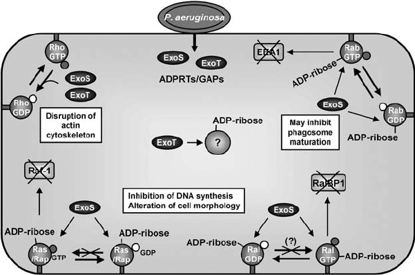
Figure 6.4. The action of P. aeruginosa ExoS and ExoT on small GTP-binding proteins.
ExoS and ExoT are highly similar proteins exhibiting both ADPRT and GAP activity. ExoS
ADPRT activity targets the small GTP-binding proteins H-Ras (Ras in the figure), Rap1B
(Rap), RalA (Ral), and Rab5 (Rab). In the case of Ras and Rap (Ras/Rap in the figure),
ADP-ribosylation by ExoS inhibits the interaction of the Ras proteins with their GEFs, and
this prevents the interaction with downstream effectors such as Raf-1. In the case of Ral
and Rab, ADP-ribosylation by ExoS has been shown to inhibit their interaction with
downstream effectors RalBP1 and EEA1. ADP-ribosylation of Ras may also prevent the
activity of RalGDS, a specific GEF for Ral, and, thus, interfere with GEF-catalyzed RalGDP
to RalGTP exchange (Fraylick et al., 2002). The cellular target of the ADPRT activity of
ExoT (0.2–1% of that of ExoS) is unknown. The GAP activity of both ExoS and ExoT is
displayed towards RhoGTPases, such as Cdc42, Rac1, and RhoA.
them, ExoS and ExoT, exert their effects on signalling mediated by small
GTP-binding proteins (Figure 6.4).
ExoS is a bifunctional protein containing an amino-terminal domain that
possesses GAP activity (Goehring et al., 1999), and a carboxyl-terminal do-
main displaying ADP-ribosyltransferase (ADPRT) activity (Frank, 1997). The
action of ExoS on epithelial cells causes a decrease in DNA synthesis, long-
term alterations in cell morphology, microvillus effacement, and loss of the
ability to re-adhere to plastic surfaces (Olson et al., 1999), thus affecting cell
viability. These effects seem to be largely due to the ADPRT activity of ExoS
(Fraylick et al., 2001). In vitro, this ADPRT activity is dependent on a eukary-
otic protein termed FAS (factor activating ExoS) and is exerted preferentially
P1: IwX
052182091Xc06.xml CB786/Lax 0 521 82091 X November 4, 2005 2:30
127
type iii–delivered toxins
on small GTP-binding proteins (Frank, 1997). In vivo,ExoS has been shown to
ADP-ribosylate Ras (H-Ras, Rap1B, and RalA) and Rab proteins (Figure 6.4)
(Barbieri et al., 2001; Fraylick et al., 2002; McGuffie et al., 1998; Riese et al.,
2001). ExoS ADP-ribosylation of H-Ras inhibits the interaction with its spe-
cific GEF, Cdc25, and consequently inhibits the nucleotide exchange reac-
tion catalysed by Cdc25 (Ganesan et al., 1999). Because only GTP-bound
Ras is capable of interacting with its downstream effectors, this disrupts
Ras-mediated signal transduction (Ganesan et al., 1998). Accordingly, in vivo
ExoS-mediated ADP-ribosylation of H-Ras inhibits the ability of the GTPase
to interact with its downstream effector Raf-1 (Vincent et al., 1999). The mod-
ification of H-Ras by ExoS correlates directly with its ability to inhibit DNA
synthesis (McGuffie et al., 1998).
ExoS ADP-ribosylation of Rap1B also inhibits the interaction between
Rap1B and its GEF, C3G, which could modulate the oxidative-burst in neu-
trophils or signal transduction through integrin-mediated pathways (Riese
et al., 2001). ExoS ADP-ribosylation of RalA alters its ability to bind its down-
stream effector RalBP1 (Fraylick et al., 2002). Similarly, the action of ExoS
on Rab5 inhibits the interaction of the GTPase with its effector EEA1 (early
endosome antigen 1) (Barbieri et al., 2001), which is predicted to affect phago-
some maturation. Therefore, ExoS may also inhibit phagocytosis through its
action on Rab.
ExoS displays GAP activity towards RhoA, Rac1, and Cdc42, both in vitro
and in vivo (Figure 6.4) (Goehring et al., 1999; Krall et al., 2002). Transfec-
tion of the amino terminal part of ExoS, or delivery of ExoS by the TTSS of
P. aeruginosa in eukaryotic cells, disrupts actin filaments (Krall et al., 2002;
Pederson et al., 1999). In addition, type III secretion delivered ExoS confers
phagocytosis resistance against macrophages (Frithz-Lindsten et al., 1997).
The primary structure of ExoT is 75% identical to that of ExoS. However,
the ADPRT activity of ExoT is only 0.2–1% of that of ExoS (Frank, 1997),
and ExoT does not interfere with cell viability or with Ras signalling (Sundin
et al., 2001). ExoT disrupts the cytoskeletal architecture of epithelial cells
(Vallis et al., 1999), and inhibits the uptake of P. aeruginosa by epithelial cells
and macrophages (Garrity-Ryan et al., 2000). These activities are partially
dependent on the GAP activity that ExoT exhibits towards RhoA, Rac1, and
Cdc42 both in vitro and in vivo (Figure 6.4) (Kazmierczak and Engel, 2002;
Krall et al., 2000). Furthermore, the GAP activity of ExoT is required for the
ability of the protein to inhibit lung epithelial wound repair in vitro (Geiser
et al., 2001). Since point mutations at the arginine finger of ExoT result
only in intermediate defects, it is predicted that its ADPRT activity may also
contribute to cell rounding and anti-internalisation activities (Sundin et al.,
P1: IwX
052182091Xc06.xml CB786/Lax 0 521 82091 X November 4, 2005 2:30
128
lu
´
ıs j mota and guy r cornelis
2001). As yet, the host protein(s) that ExoT ADP-ribosylates has not been
identified.
TheEPEC Effectors Tir and Map Also Modulate
Small RhoGTPases
Central to enteropathogenic E. coli (EPEC) – mediated disease is its colonisa-
tion of the intestinal epithelium. EPEC reside extracellularly on the surface of
the infected epithelial cell, where they deliver toxins by using a TTSS. At least
four effector proteins are delivered by EPEC into target cells, Tir (translocated
intimin receptor), Map (mitochondrial-associated protein), EspG, and EspF.
A recent study indicates that Tir and Map modulate RhoGTPase signalling
pathways.
Tir is by far the best studied of the EPEC effectors (reviewed by Goosney
et al., 2000). After its delivery into the target cell, Tir is inserted into the plasma
membrane, where it serves as a receptor for the bacterial outer membrane
intimin. The interaction between Tir and intimin triggers signalling events
that subvert the normal regulation of the host cell cytoskeleton and lead to
the formation of the pedestal-like structures that characterise EPEC infection
of epithelial cells. This process is not dependent on RhoGTPases, although
Tir has a putative GAP motif (Kenny et al., 2002). However, EPEC binding to
HeLa cells also induces a Tir-independent cytoskeletal rearrangement charac-
terised by early and transient formation of filopodia-like structures at sites of
infection (Kenny et al., 2002). Filopodia formation requires the Map effector,
a protein that targets host mitochondria where it appears to disrupt mem-
brane potential (Kenny and Jepson, 2000). Map-mediated filopodia formation
is dependent on Cdc42, which is activated by an unknown mechanism, and is
independent of Map targeting to the mitochondria (Kenny et al., 2002). Inter-
estingly, in addition to the formation of the pedestal structures, Tir binding
to intimin also downregulates filopodia formation (Kenny et al., 2002). This
suggests that Tir, like SptP, YopE, ExoS, and ExoT, could indeed have a GAP
activity. However, the consequences of the Tir and Map manipulation of
Cdc42 activity for EPEC pathogenesis are unclear.
Type III – Delivered Kinases and Phosphatases Interfering
with Signalling Pathways
Phosphorylation cascades play an important role in many animal signal trans-
duction pathways. For example, the MAPK signalling pathways (Garrington
and Johnson, 1999), the phosphatidylinositol 3-kinase (PI3K) pathway
P1: IwX
052182091Xc06.xml CB786/Lax 0 521 82091 X November 4, 2005 2:30
129
type iii–delivered toxins
(Vanhaesebroeck and Alessi, 2000), and the phosphorylation cascade that
activates NF-κB (Karin and Lin, 2002) are each involved in the control of
a multitude of cellular processes. These pathways are obvious targets for
bacterial virulence factors. We have already described SopE from Salmonella,
which acts on MAPK signalling pathways through its GEF activity on RhoGT-
Pases (see above). However, in addition, some of the type III effectors are
themselves kinases and phosphatases, thus interfering directly on host phos-
phorylation signalling cascades. Kinases and phosphatases delivered by TTSS
into eukaryotic cells have been identified in Salmonella, Shigella, and Yersinia.
Yersinia YopH, a Tyrosine Phosphatase That Inhibits Innate
and Adaptive Immune Responses
YopH is among the most powerful protein tyrosine phosphatases (PTPases)
known and it has long been known to contribute to the ability of Yersinia
to resist phagocytosis by macrophages (Rosqvist et al., 1988). When injected
into J774 macrophages, YopH dephosphorylates p130
Cas
and disrupts focal
adhesions (Hamid et al., 1999). Other YopH targets in J774 macrophages are
the Fyn-binding protein Fyb (Hamid et al., 1999) and the scaffolding protein
SKAP-HOM (Figure 6.3) (Black et al., 2000). These two proteins interact with
each other and become tyrosine phosphorylated in response to macrophage
adhesion. It is likely that the action of YopH against focal adhesions, p130Cas,
and Fyb, is relevant to the antiphagocytic action (Deleuil et al., 2003; Persson
et al., 1999). YopH also suppresses the oxidative burst in macrophages (Bliska
and Black, 1995), and protects against phagocytosis by PMNs (Grosdent et al.,
2002; Visser et al., 1995).
Recent observations have shown that YopH also contributes to the
downregulation of the inflammatory response (Figure 6.3) (Sauvonnet et al.,
2002). Upon infection, macrophages release the monocyte chemoattractant
protein 1 (MCP-1), a chemokine involved in the recruitment of other
macrophages to the sites of infection. In fact, the MCP-1 mRNA levels are
downregulated in macrophages infected with Y. enterocolitica, and this in-
hibition is dependent upon YopH. MCP-1 synthesis is known to be under
the control of the PI3K pathway, which is involved in the control of multi-
ple cellular processes (Vanhaesebroeck and Alessi, 2000). Consistently, with
its negative action on MCP-1 mRNA levels, YopH was shown to abrogate
the PI3K-dependent activation of protein kinase B (PKB, also called Akt)
(Sauvonnet et al., 2002). The site of action of YopH on this cascade is still
unknown but it is likely to be a tyrosine-phosphorylated receptor upstream
from PI3K (Figure 6.3). Furthermore, YopH (together with YopE and YopJ/P)
also counteracts YopB-stimulated pro-inflammatory signalling in infected
P1: IwX
052182091Xc06.xml CB786/Lax 0 521 82091 X November 4, 2005 2:30
130
lu
´
ıs j mota and guy r cornelis
epithelial cells, and is likely that this is dependent on its action on the PI3K
pathway (Viboud et al., 2003). In addition, YopH also seems to incapaci-
tate the host adaptive immune response. T and B cells transiently exposed
to Y. pseudotuberculosis are impaired in their ability to be activated through
their antigen receptors. YopH appears to be the main effector involved in
this block of activation (Yao et al., 1999), which most likely also involves the
PI3K/PKB pathway (Sauvonnet et al., 2002). In agreement with these observa-
tions, recombinant YopH introduced into human T lymphocytes was shown
to dephosphorylate the primary signal transducer for the T cell receptor, the
Lck tyrosine kinase (Alonso et al., 2004).
SptP, a PTPase Delivered by Salmonella SPI-1 TTSS
As already pointed out, the GAP domain of the SptP protein reverses the
cytoskeleton changes induced by Salmonella.Inaddition, the bifunctional
SptP protein appears to downregulate the Salmonella-induced activation of
JNK and ERK (Fu and Gal
´
an, 1999; Murli et al., 2001). At least in the case of
inhibiting ERK activation, this capacity seems to rely on the PTPase activity
of SptP (Murli et al., 2001). Therefore, SptP may be a general downregulator
of the cellular responses stimulated by Salmonella, and its PTPase activity
may play an important role in this process. Clearly, the tyrosine phosphatase
activity of SptP is not exerted on proteins phosphorylated by MAPKs, as these
are serine/threonine protein kinases. To date, only the intermediate filament
protein vimentin was identified as a potential target of the SptP PTPase
activity (Murli et al., 2001). However, other target(s) of the PTPase remain
to be identified, as it is unclear how the action of SptP on vimentin could
contribute to reversing Salmonella-induced cellular responses.
SopB/SigD and IpgD, the Inositol Phosphatases from
Salmonella and Shigella
As mentioned before, SopB displays inositol phosphatase activity both in vitro
and in vivo.Inaddition to stimulating actin cytoskeleton reorganisation, SopB,
through its inositol phosphatase activity, has been associated with the mod-
ulation of chloride secretion induced by Salmonella infection (Norris et al.,
1998), and is known to be essential for the activation of PKB that is observed
upon infection of epithelial cells by Salmonella (Steele-Mortimer et al., 2000).
The modulation of chloride secretion, which directly contributes to diarrhoea,
appears to be a consequence of products of inositol metabolism, in particular
Ins(1,4,5,6)P
4
(Feng et al., 2001). With respect to PKB, the mechanism of ac-
tivation by SopB and its importance in Salmonella pathogenesis are unclear.
More recently, SopB was shown to be responsible for the disappearance of

P1: IwX
052182091Xc06.xml CB786/Lax 0 521 82091 X November 4, 2005 2:30
131
type iii–delivered toxins
phosphatidylinositol (PtdIns)(4,5)P
2
at the invaginating regions of the ruffles
induced by Salmonella. This reduces the rigidity of the membrane skeleton
and is thought to promote the formation of the Salmonella-containing vac-
uoles (Terebiznik et al., 2002).
The Shigella effector IpgD is 59% similar and 41% identical to SopB at the
amino acid level. IpgD has been shown to be involved in the modulation of the
host cell response after contact of Shigella with epithelial cells (Niebuhr et al.,
2000). Furthermore, purified IpgD displays PtdIns phosphatase activity in
vitro and in infected cells (Niebuhr et al., 2000) (Marcus et al., 2001), and also
activates PKB in an inositol phosphatase-dependent fashion (Marcus et al.,
2001). In vitro, IpgD displays the greatest activity towards PtdIns(4,5)P
2
, and
the transformation of PtdIns(4,5)P
2
into PtdIns(5)P by IpgD is responsible
for dramatic morphological changes of the host cell, which are thought to
promote Shigella entry (Niebuhr et al., 2002).
YpkA/YopO, the Yersinia type III Delivered Protein Kinase
YpkA (for Yersinia protein kinase A) (called YopO in Y. enterocolitica)isalso
an effector that modulates the dynamics of the cytoskeleton (Hakansson et al.,
1996), and that contributes to Yersinia resistance to phagocytosis (Grosdent
et al., 2002). It is an autophosphorylating serine-threonine kinase (Galyov
et al., 1993), which shows some sequence and structural similarity to RhoA-
binding kinases (Dukuzumuremyi et al., 2000), but which becomes active
only after interacting with actin (Juris et al., 2000). In addition to being an
activator of YpkA, actin can also function as an in vitro substrate of the ki-
nase. YpkA/YopO interacts with, but does not phosphorylate, RhoA and Rac1
irrespective of the nucleotide bound, and apparently without affecting the
guanine-nucleotide exchange capacity (Dukuzumuremyi et al., 2000). How-
ever, the kinase target and the exact mode of action of YpkA/YopO remain
unknown (Figure 6.3).
The Modulation of Death Pathways and Inflammation
by Type III Effectors
Several bacteria harbouring TTSS induce cell death in eukaryotic cells, partic-
ularly in macrophages. The process has been well studied in Yersinia, Shigella,
and Salmonella, where the type III effectors YopJ/P, IpaB, and SipB, respec-
tively, trigger cell death. Type III effectors from P. aeruginosa and EPEC
were also reported to promote cell death, but in these cases the molecules
and death pathways involved have not been thoroughly characterised. In the
case of Yersinia and Shigella,itisclearly established that macrophage cell
P1: IwX
052182091Xc06.xml CB786/Lax 0 521 82091 X November 4, 2005 2:30
132
lu
´
ıs j mota and guy r cornelis
death occurs by apoptosis. However, while apoptosis is normally considered
an immunologically silent death process unaccompanied by inflammation,
Shigella- and Salmonella-induced macrophage cell death triggers characteris-
tically acute inflammatory responses. In contrast, Yersinia-induced apoptosis
is accompanied by a downregulation of the inflammatory response, at least
in cell culture systems. Therefore, cell death induction by type III effectors,
in addition to allowing the elimination of key immune cells and thus pro-
moting bacterial proliferation, may be a way by which bacteria modulate the
host inflammatory response.
The NF-κB transcription factor is central to the modulation of cell sur-
vival and immune and inflammatory responses (Karin and Lin, 2002). NF-κB
exists in the cytoplasm of resting cells in an inactive form associated with
inhibitory proteins termed inhibitor-kappa B (IκB). After cell stimulation,
NF-κB activation is achieved through the phosphorylation of IκBbythe IκB
kinase (IKK) complex. The components of the IKK complex that regulate the
NF-κB pathway include IKKα and IKKβ, which are activated by morphogenic
and pro-inflammatory signals, respectively. Phosphorylated IκBisthen se-
lectively ubiquitinated, which targets its degradation by the proteasome. This
allows NF-κBtotranslocate to the nucleus and activate transcription of genes
involved in immune responses, including several cytokines.
Yersinia YopJ/P
YopJ (YopP in Y. enterocolitica) counteracts the normal pro-inflammatory re-
sponse in various cell culture systems (Boland and Cornelis, 1998; Denecker
et al., 2002; Schesser et al., 1998). This capacity results from the ability of
YopJ/P to inhibit the MAPK pathways JNK, p38, and ERK1 and 2, and also
the NF-κB signalling pathway (Figure 6.5). Furthermore, inhibition of the
MAPK pathways abrogates phosphorylation of CREB, a transcription factor
involved in the immune response (reviewed by Orth, 2002).
YopJ/P binds directly to the members of the superfamily of MAPK ki-
nases (MKKs), blocking their phosphorylation and consequent activation
(Orth et al., 1999). Similarly, YopJ/P interacts with IKKβ, presumably also
preventing its phosphorylation and activation (Orth et al., 1999). Thus, YopJ/P
acts by preventing activation of MKKs and IKK in the MAPK and NF-κB sig-
nalling pathways, respectively (Figure 6.5). The exact molecular mechanism
by which YopJ/P acts is still elusive, but its secondary structure resembles
that of an adenovirus cysteine protease. The presumed protease catalytic triad
of YopJ/P is required for the ability of YopJ/P to inhibit the MAPK and NF-κB
signalling pathways (Orth et al., 2000). Based on the observations that over-
expression of YopJ/P results in a decrease of SUMOylated and ubiquitinated
P1: IwX
052182091Xc06.xml CB786/Lax 0 521 82091 X November 4, 2005 2:30
133
type iii–delivered toxins
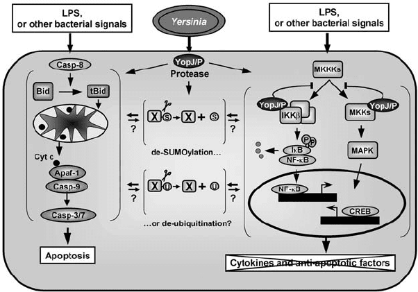
Figure 6.5. Action of Yersinia YopJ/P on NF-κB, MAPK, and apoptosis signalling
pathways. YopJ/P binds to IKKβ and MKKs and block their activation through
phosphorylation by upstream MKK kinases (MKKKs). This inhibits the activity of the
NF-κB and CREB transcription factors, thus, preventing transcription of anti-apoptotic
genes and of pro-inflammatory cytokines. YopJ/P also induces apoptosis in macrophages,
either by directly activating a death pathway or indirectly by blocking the synthesis of
anti-apoptotic factors together with bacterial LPS/ lipoproteins apoptotic signalling. The
apoptotic cascade is most probably triggered by caspase-8 activation, leading to Bid
cleavage. The subsequent translocation of tBid to the mitochondria induces the release of
cytochrome c, which binds to the apoptotic protease activating factor-1 (Apaf-1) and leads
to recruitment and activation of caspase-9 that, in turn, activates executioner caspases-3
and -7. The YopJ/P protease may act on SUMOylated and/or ubiquitinated proteins, but
the relevance of these activities for YopJ/P function is unclear.
proteins, it was proposed that YopJ/P could be an ubiquitin or a SUMO pro-
tease (SUMO are ubiquitin-like modifiers that are involved in stabilisation
or destabilisation of proteins) (Orth, 2002; Orth et al., 2000). It is unclear
how a de-SUMOylating activity could contribute to the ability of YopJ/P to
disrupt MAPK and NF-κB signalling, but the NF-κB pathway is regulated at
two distinct points by ubiquitination (Orth, 2002).
YopJ/P induces apoptosis in macrophages, but not in other cell types
(Mills et al., 1997; Monack et al., 1997). This apoptosis is accompanied by
cleavage of the cytosolic protein Bid, the release of cytochrome c from the mi-
tochondria, and the activation of caspase-3, -7, and -9 (Figure 6.5) (Denecker
P1: IwX
052182091Xc06.xml CB786/Lax 0 521 82091 X November 4, 2005 2:30
134
lu
´
ıs j mota and guy r cornelis
et al., 2001). The apoptosis-inducing activity of YopJ/P is lost when the cys-
teine residue forming the catalytic triad responsible for its putative protease
activity is replaced by a threonine (Denecker et al., 2001). It is very tempting
to speculate that YopJ/P could induce apoptosis by cleaving a pro- or anti-
apoptotic factor. However, it has not been possible to demonstrate this and it is
unclear how a de-SUMOylating or de-ubiquitinating activity could be related
to the induction of a death pathway. It is even unclear whether apoptosis re-
sults from a YopP-induced early cell death signal, or from the YopP-blockage
of the NF-κB signalling, coupled to cellular activation by bacterial lipopolysac-
charide (LPS) (Denecker et al., 2001; Ruckdeschel et al., 2001). On one hand,
it seems clear that YopJ/P transfected macrophages undergo apoptosis in a
significant percentage. On the other hand, this percentage of cell death is
considerably increased by LPS treatment (Ruckdeschel et al., 2001).
Shigella IpaB
Macrophages can engulf Shigella, but instead of successfully destroying the
bacteria in the phagosome, they succumb to apoptotic death. The Shigella-
induced macrophage apoptosis has been described both in vitro and in vivo
(Zychlinsky et al., 1992; Zychlinsky et al., 1996). The IpaB type III-delivered
toxin is necessary and sufficient to induce apoptosis (Chen et al., 1996b;
Zychlinsky et al., 1994). Following the escape of Shigella from the macrophage
phagosome, IpaB is secreted into the cytoplasm where it binds to caspase-1.
Furthermore, in vivo studies using different knockout mice showed that IpaB-
induced apoptosis is absolutely dependent on caspase-1 activation (Hilbi et al.,
1998). IpaB-induced caspase-1–dependent apoptosis of macrophages triggers
an acute inflammatory response, characterised by a massive influx of PMNs
that infiltrate the infected site and destabilise the epithelium (Sansonetti,
2001). Caspase-1 is also known as interleukin (IL)-1-converting enzyme, due
to its ability to cleave pro-IL-1β and the related cytokine pro-IL-18 into their
mature active forms. Accordingly, IpaB-activated caspase-1 can cleave the
two cytokines, which are then released from the dying macrophage (Hilbi
et al., 1998). Therefore, Shigella-induced macrophage cell death is central
to the early triggering of inflammation. This inflammatory response causes
significant tissue destruction and facilitates further invasion by the bacte-
ria, but inflammation is ultimately responsible for controlling the infection
(Sansonetti, 2001).
Salmonella SipB
In vitro studies indicate that Salmonella spp. induces cell death in macro-
phages (Chen et al., 1996a; Lindgren et al., 1996; Monack et al., 1996). The
P1: IwX
052182091Xc06.xml CB786/Lax 0 521 82091 X November 4, 2005 2:30
135
type iii–delivered toxins
process by which Salmonella induces cell death is not totally clear, as fea-
tures of both apoptotic and necrotic death have been described (Boise and
Collins, 2001). The TTSS SPI-1 SipB protein is highly similar to the Shigella
IpaB protein, and SipB seems to promote rapid cell death by a mechanism
resembling that of IpaB (Hersh et al., 1999). However, macrophages from
casp-1 knockout mice are still susceptible to Salmonella-induced cell death
in a SipB-dependent way, although with delayed kinetics (Jesenberger et al.,
2000). Furthermore, Salmonella also kill macrophages independently of SipB
and of SPI-1 TTSS, most likely by mechanisms involving the SPI-2 TTSS
(van der Velden et al., 2000), but the bacterial toxin(s) involved remain to be
identified. Quite recently, it was suggested that SipB promotes autophagy
and cell death by disrupting mitochondria (Hernandez et al., 2003).
P. aeruginosa ExoU, ExoS, PcrV, PopB, and PopD
The delivery of ExoU by P. aeruginosa TTSS into host cells induces a cytotoxic
phenotype in tissue culture models and a fatal outcome in an acute lung
infection model (Finck-Barbancon et al., 1997). The cell death mechanism
mediated by ExoU in macrophages and epithelial cells has the characteris-
tics of necrosis rather than apoptosis (Hauser and Engel, 1999). Recently, it
was shown that ExoU is a phospholipase and that its cytotoxic action can be
blocked by different phospholipase A2 inhibitors (Phillips et al., 2003; Sato
et al., 2003). In addition, P. aeruginosa induces apoptosis in macrophages
and epithelial cells in an ExoU-independent, but TTSS-dependent fashion,
and P. aeruginosa clinical isolates that do not express ExoS or ExoY also in-
duce apoptosis (Hauser and Engel, 1999). In contrast to this observation,
P. aeruginosa-mediated apoptosis of epithelial and fibroblast cell lines was
shown to require the ADPRT activity of ExoS (Kaufman et al., 2000). In ad-
dition, the pore forming activity of the translocators PcrV, PopB, and PopD
has been reported to promote the death of macrophages and neutrophils
(Dacheux et al., 2001).
Enteropathogenic E. coli EspF
EPEC induces cell death in epithelial host cells by a process that resembles
apoptosis in some ways, but not in others (Crane et al., 1999). The EPEC type
III effector EspF disrupts host intestinal barrier function, thus contributing
to diarrhoea (McNamara et al., 2001), and in addition it appears to mediate
cell death (Crane et al., 2001). Because cell death promoted by EspF has
features of pure apoptosis (Crane et al., 2001), this indicates that EPEC may
also trigger cell death independently of EspF. To date, the apoptotic pathway
induced by EspF is unknown.