Lowenthal G., Airey P. Practical Applications of Radioactivity and Nuclear Radiations
Подождите немного. Документ загружается.

lead (Z = 82) until E
g
> 2 MeV (Figure 3.7(a)). Following pair production in
suf®ciently large detectors, the electron pair is normally absorbed, resulting
in a full energy pulse. If one or both electrons escape, this leads to the
formation of so-called single or double escape peaks to be discussed in
Section 6.4.2.
Although the kinetic energy of charged particles is absorbed continuously
3.4 Properties of gamma rays and X rays 71
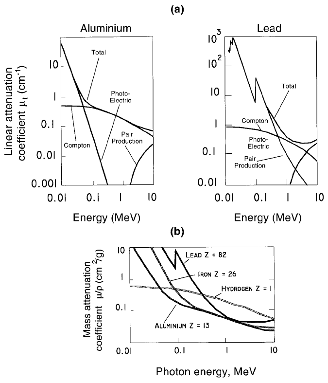
Figure 3.7. Total linear and mass attenuation coef®cients (m
t
cm
71
and m
m
cm
2
/g)
for g rays. (a) m
t
values for aluminium (Z = 13, r = 2.70 g/cm
3
) and lead (Z = 82,
r = 11.3 g/cm
3
), showing the three mutually independent componen ts. (b) The mass
attenuation coef®cient m
m
for the elements H, Al, Fe and Pb. Note the different
function for hydrogen.

along their path in large numbers of individually very small steps (Section
1.3.5), g rays can pass millions of atoms between interactions, so helping to
explain the relatively high penetrating power of these rays.
3.4.4 Photon attenuation, an overview
As just noted, photon attenuation occurs as the sum of three mutually
independent components: photoelectric (pe), Compton (C) and pair produc-
tion (pp), though m
pp
is zero at g ray energies below 1022 keV. Writing m
t
(cm
71
) for the total linear attenuation coef®cient one has,
m
t
= m
pe
+ m
C
+ m
pp
. (3.2)
Properties of radiations72
Figure 3.8. (a) A Compton scatter event. The g ray loses part of its energy to the
atomic electron which scattered it out of its path. (b) The energies of the Compton
edge E
C
and the backscatter peak E
R
as functions of the g ray energy, E
g
. It is seen
that E
C
+E
R
= E
g
(CoN, 1988).
Figure 3.7(a) shows m
t
and its three components for aluminium and lead, with
m
t
depending not only on E
g
and on the Z number of the attenuator but also
on its physical density.
Figure 3.7(a) shows that the E
g
and Z dependence is particularly strong for
m
pe
. It has long been known (Bush, 1962, Section 9.7), that:
m
pe
! Z
4
/(E
g
)
3
. (3.3)
m
pp
is zero below 1022 keV and remains a minor component of m
t
, even
when 3 MeV g rays are attenuated in lead (Figure 3.7(a)). By contrast, the
Compton component m
C
is relatively independent of both E
g
and Z.At
1 MeV, when Compton scatter is seen to dominate for all elements m
C
is
about 4.5 times larger for lead than for aluminium, which is closely equal to
the ratio of their physical densities.
Gamma ray applications are often in the range of energies where Compton
scatter is dominant. With m
t
(cm
71
) being approximately proportional to r
(g/cm
3
), introduction of the mass attenuation coef®cient m
m
followed de®ned
as m
m
=(m
t
/r)cm
2
/g (Figure 3.7(b)). The coef®cient m
m
is then almost
independent of r, which is particularly useful in the Compton range of
energies as will be seen in Section 3.4.6.
3.4.5 Attenuation equations for narrow beam geometry
Narrow beam geometry has to be mentioned here even though details will be
introduced in the next chapter.
Whenever practical, g rays emitted from a source should be collimated, as
shown in Figure 4.6(a), a procedure known as narrow beam geometry. This is
employed to prevent g rays which lose energy during Compton scatter
(Figure 3.8(a)) from reaching the detector, so reducing the peak-to-total
ratio. If there is no collimation, one refers to broad beam geometry (Figure
4.6(b)), the additional number of detected g rays being known as build-up
(Figure 4.6(c)). Since the extent of build-up is dif®cult to predict g ray
attenuation should, whenever possible, be carried out in narrow beam
geometry and this will be assumed in what follows unless otherwise stated.
There are situations, notably during g ray applications in open country, when
build-up is dif®cult to prevent or could even be an asset (Section 8.2.4).
Given narrow beam geometry and a total linear attenuation coef®cient m
t
,
gamma rays lose their intensity I (measured in gamma rays per square
centimetre per second, g/cm
2
/s) in accordance with:
7dI/dt = m
t
6I, (3.4)
3.4 Properties of gamma rays and X rays 73
where dt is a small distance in the material and m
t
depends on the three
components shown in Eq. (3.2). Hence:
I
t
= I
0
exp (7m
t
6t), (3.5)
where I
0
is the intensity of the unattenuated beam and I
t
its intensity after
travelling t cm in the material of interest. Equation (3.5) is employed for all
attenuation calculations that rely on m
t
or any of its components.
Photon beams employed for applications frequently have to pass through a
number of different materials before reaching the detector, each with its own
m
t
and Dt. Writing Eq. (3.5) as:
ln (I
0
/I
t
)=m
t
Dt (3.6)
the term m
t
Dt is expanded as required, say to i terms, so obtaining:
ln (I
0
/I
t
)=m
t
1
6Dt
1
+ m
t
2
6Dt
2
+ + m
t
i
6Dt
i
. (3.7)
These equations apply for narrow beam geometry.
The magnitude of attenuations is conveniently estimated with the help of
the half thickness t
1/2
. On substituting I
t
= 0.5I
0
in Eq. (3.5), one obtains:
t
1/2
= ln2/m
t
= 0.693/m
t
, (3.8)
with t
1/2
values depending on the same variables as m
t
. Half value thicknesses
for three commonly used materials are shown in Table 7.3.
3.4.6 Photon attenuation measurements using m
m
When replacing m
t
(cm
71
)bym
m
(cm
2
/g) (Section 3.4.4), it is necessary to
replace the linear distance t cm by the surface density SD (g/cm
2
)
(Section 3.3.5) to ensure a dimension-free exponent. Equation (3.5) then
becomes:
I
SD
= I
0
exp (7m
m
6SD). (3.9)
Figure 3.7(b) shows that m
m
values are very similar for all light elements
(Z < 35) at g ray energies between about 0.5 and 2.0 MeV and for nearly all
stable elements for g ray energies between 1 and 2 MeV.
Working with, say 1.33 MeV g rays from
60
Co and with metal strips which
are identical in thickness, any fault in their internal structure which affects
their physical density, e.g. a void due to a faulty weld, can be identi®ed from
differences in the attenuation of the g rays relative to that in a suitably chosen
standard. This is so because the g rays that traversed an air space (void)
within the metal travel a different distance in SD terms than the rays that did
Properties of radiations74
not encounter voids. This effect is also the basis of g ray radiography to be
discussed in Section 7.2.1.
Although m
m
is almost independent of Z between about 0.5 and 2 MeV,
this is not so below 0.1 MeV (Figure 3.7(b)) which makes the attenuation of
low-energy g or X rays a sensitive monitor, e.g. for the detection of chemical
impurities in foils of metals or plastics. Accurate attenuation measurements
at low photon energies can only be made on suf®ciently thin samples.
Only hydrogen (Z = 1) does not conform to the general pattern (Figure
3.7(b)). The marked difference between m
m
for hydrogen and for other
molecules with signi®cant hydrogen content relative to hydrogen-free mate-
rials is extensively employed in many applications.
3.5 Pulse height spectra due to alpha particles and gamma rays
3.5.1 The response of detectors
Alpha and beta particle emissions signal nuclear decays between sharply
de®ned energy levels belonging respectively to the parent and daughter
nuclide. Why the interaction of b
7
or b
+
particles in energy-sensitive
detectors fails to produce peaks was stated in Section 3.3.4. However, a
particles carry away the entire energy liberated by an a particle emitting
decay, e.g. the decay of radium-226 to radon-222 (Figure 1.5), except for
recoil energies left with the emitting atoms. If all a particles from a source
reach the detector without losing energy between source and detector
(Section 3.2.2), they give rise to pulses of identical height (except for random
¯uctuations), so yielding a narrow full energy peak (Section 3.5.2).
The situation is more complex for g rays. Most nuclear decays occur via
several excited states commonly de-excited by g rays (but also in part by
conversion electrons, see Section 3.6.2) leading to multi-energy g ray spectra
(Section 3.4.1). Gamma ray detectors should be able to resolve peaks due to
closely spaced g ray energies which is effectively done by high resolution
semiconducting detectors (Figure 3.9(b),(c)).
Although the energy resolution of NaI(Tl) detectors at room temperature
is 20 to 40 times lower than that of germanium detectors at liquid air
temperature (Figure 3.10), the former have the advantage of being consider-
ably less expensive for a comparable size, plus they are easier and cheaper to
operate.
3.5 Pulse height spectra 75
Properties of radiations76
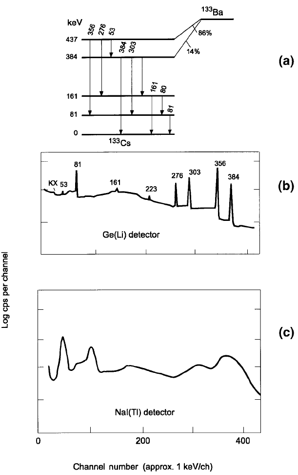
Figure 3.9. (a) The decay of barium-133 (T
1/2
= 10.6 y) by electron capture,
followed by nine g rays. (b) The
133
Ba spectrum obtained with a Ge(Li) detector, and
(c) with a NaI(Tl) detect or (Debertin and Helmer, 1988, Figure 4.25). The latter
detector clearly displays low-energy photons which are barely shown by the Ge(Li)
detector. Graphs (b) and (c) have the same energy scales.
3.5.2 Alpha particle spectra
The preferred method for obtaining a particle spectra makes use of ion
implanted silicon detectors (Section 5.5.4). These are small crystals mounted
in chambers which can be evacuated and which are ®tted to permit accurately
reproducible source±detector geometry. The crystals range in thickness from
about 0.1 mm upwards to several millimetres.
The range of 10 MeV a particles in silicon is only about 0.1 mm so that a
0.1 mm or at most a 0.3 mm thick silicon detector, with source and detector
mounted in vacuum, satis®es requirements for spectrometry when employing
a particles from naturally occurring radionuclides (Figure 1.5). To minimise
3.5 Pulse height spectra 77
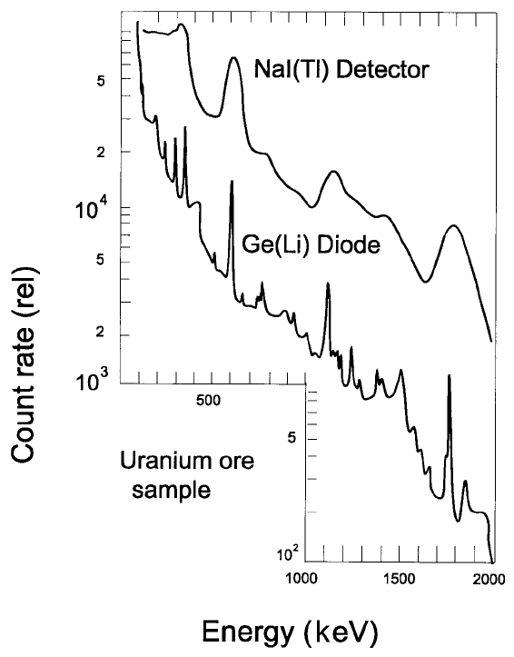
Figure 3.10. A comparison of g ray spectra due to a uranium ore sample obtained
with a Ge(Li) detector and a NaI(Tl) detector.
energy losses in the source, it should be ultrathin and be prepared with as low
an activity as acceptable for the required accuracy. Figure 3.6(e) shows a
particle spectra (FWHM~15 keV) due to polonium radioisotopes which each
emit only a single a particle energy. Only the peaks are shown, there being no
identi®able features at lower energies.
When employing a particle spectrometry routinely, e.g. to identify a
particle emitting pollutants from coal ®red power stations which normally
emit measurable concentrations of uranium, thorium and their daughters,
this can often be done satisfactorily when the FWHM values of the a particle
spectra are in the range 50 to 100 keV. The equipment is then cheaper and
easier to operate.
3.5.3 Gamma ray spectra
As was outlined in Section 3.4.3, g rays are detected in three ways ± via
photoelectric and Compton interations and pair production. Pair production
will be further dealt with in Section 6.4.2. The other two processes are
responsible for g ray spectra such as those shown in Figures 3.4(d) and 3.6(f ).
Relatively simple spectra such as these are the preferred type for industrial
applications. Figure 3.6(f ) shows a typical spectrum for gamma rays that did
not interact by pair production (Section 3.4.3). It also shows the effects of
random ¯uctuations in pulse heights due to the statistics of g ray interactions
in the detector and adjacent materials. These random interactions account
for the difference between the theoretical and observed spectrum shown in
the ®gure. If spectra are obtained in a broad beam geometry, interpretations
become more dif®cult (Section 4.4.4).
The multiple interactions of g rays even when emitted at identical energies
results in a pulse height distribution from near zero energy to a maximum
exceeding the emitted g ray energy due to the quasi-Gaussian shape of the full
energy peak shown in the ®gure. The fraction of pulses reaching the full
energy peak depends principally on the energy with which the g rays were
emitted and the conditions prevailing during the measurement. The ratios of
pulses in the peak to pulses in the entire spectrum (peak-to-total ratio)
obtained in optimum conditions as functions of the g ray energy and the
dimensions of NaI(Tl) detectors are shown in Figure 4.5 in the next chapter.
Coming to the near zero and low-energy pulses shown e.g. in Figure 3.6(f),
they are due predominantly to photoelectric absorption of g rays which had
been scattered to low energies (Figure 3.7(a)). On the other hand, the energies
of the pulses due to Compton scatter for a given initial energy extend from
low values upwards to the so-called Compton edge. Here again, the statistics
Properties of radiations78
of interactions cause the observed spectrum to show broad rather than sharp
edges. The Compton edge is due to electrons dislodged when g rays are fully
backscattered within the detector and adjacent material (Figure 3.8(b)).
Further details about the formation of g ray spectra go beyond the scope of
this book, except as regards backscattered g rays (1508 to 1808 ) lead to pulses
in the so-called backscatter peak shown in Figure 3.4(d). This peak is small as
is the backscatter peak in Figure 3.6(f ) since these spectra were obtained in a
relatively scatter-free environment. However, these peaks can become domi-
nant in spectra due to strongly scattered radiations (Section 3.9.1).
The energies of the Compton edge (E
C
) or the backscatter peak (E
B
) are not
always easily identi®ed, especially so in multi gamma ray spectra. However, if
the position of one of them is known, that of the other can be calculated since
E
C
+ E
B
= E
g
, the energy of the interacting g ray (Figure 3.8(b)).
3.6 Electron capture (EC), gamma rays and conversion electrons
3.6.1 EC decays and their use as quasi-pure gamma ray emitters
Electron capture (EC) decays occur (Figure 3.11) when an atomic electron is
pulled into the nucleus of its atom where it can be pictured to turn a proton
into a neutron. EC decays are the inverse of b
7
decays. An EC decay is a
primary transformation, decreasing the Z number of the parent by one unit
(see Figure 3.11) without the emission of primary radiations (except for
neutrinos), though there are follow-on radiations emitted by the daughter
nuclide.
Between 80 and 90% of EC decays originate in the K shell of their atoms,
the shell nearest to the nucleus. The resultant vacancies are at once ®lled by
electrons from beyond the K shell. The energy liberated during these transfers
is emitted mainly either as KX rays or as K-Auger electrons (Figure 3.12).
Each electron transfer from an outer to an inner shell leads to the release of
binding energy which appears either as a ¯uorescent X ray (Section 3.9.1) or
as the kinetic energy of an Auger electron. The ratio of the intensities of
¯uorescent X rays to those of Auger electrons is known as the ¯uorescent
yield, denoted v, which is a function of the atomic number Z of the element
of interest (Section 3.9.3). For each element ¯uorescent X ray energies are
signi®cantly high for interactions in the K shell (v
K
), but for outer shells they
are commonly too low to be measured except by specialists. The dependence
of v
K
on Z is illustrated in Figure 3.12.
The decay energy liberated during the EC process is carried away by
ordinarily unobservable mono-energetic neutrinos, each neutrino carrying
3.6 Electron capture, g rays and conversion electrons 79
away all the decay energy and not only part of it as happens during decays by
b particle emissions (Section 3.3.4).
For a few radionuclides decaying by EC, decays go direct to the ground
state of the daughter. However, as a rule decays proceed to one or more
excited states. If there is only a single excited state, de-excited by no more
than one or two g rays with other follow-on radiations being of much lower
energy, the radionuclide could be labelled a quasi-pure g ray emitter.
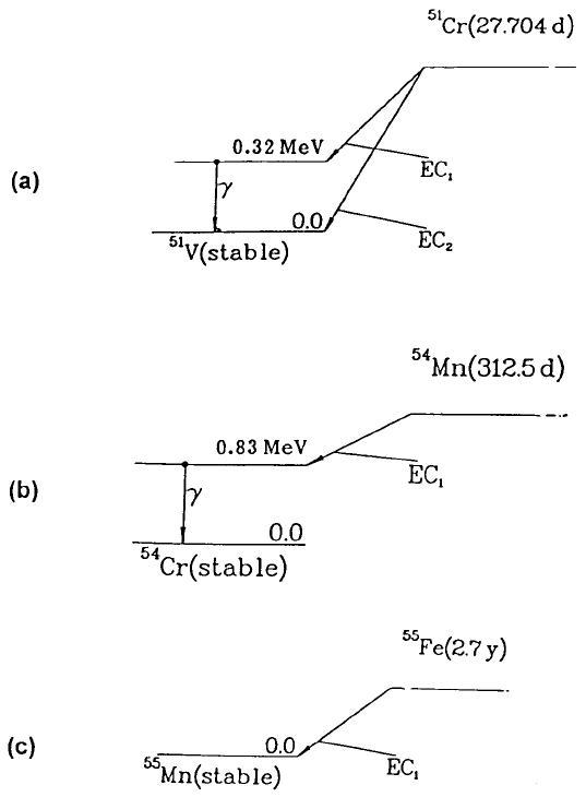
Figure 3.11 shows the decay schemes for the EC decays of chromium-51
and manganese-54, two examples of quasi-pure g ray emitters which are often
Properties of radiations80
Figure 3.11. Decay schemes of three electron capture decays followed (for two of
them) by a single g ray. (a) Chromium-51 to vanadium-51 (E
g
= 320 keV, f
g
= 9.86%).
(b) Manganese-54 (E
g
= 835 keV, f
g
= 99.98%). (c) Iron-55 (no gammas). Based on
Lagoutine et al. (1978).