Murray J. Clifford. Angiogenesis Protocols - Methods in Molecular Medicine, Vol. 46
Подождите немного. Документ загружается.

Microvessel ECs from Adipose Tissue 215
2. Materials
2.1. Equipment for EC Isolation, Characterization, and Culture
2.1.1. Hardware
1. A class II laminar flow cabinet is essential for all procedures involving the pro-
cessing of tissue and cultured cells in order to maintain sterility and protect the
operator.
2. Phase contrast microscope for observing cell cultures.
3. Light microscope equipped with epifluorescence for immunocytofluorescent
characterization of the cells.
4. Scalpels, scissors, and forceps are required for the isolation procedure and should
be sterilized prior to use by autoclaving at 121°C for 30 min.
2.1.2. Sterile Plasticware
1. Tissue culture flasks (25 and 75 cm
2
).
2. Large plastic dishes (e.g., Bioassay dishes, Nunc, Naperville, IL, USA).
3. 30 mL universal tubes.
4. 50 mL centrifuge tubes.
5. Lab-Tek multiwell glass chamber slides (Nunc, Naperville, IL, USA).
2.1.3. Antibodies
There are many commercially available antibodies against endothelial markers.
1. Monoclonal antibodies against human PECAM-1, E-selectin (e.g., clones 9G11
and 13D5 respectively, R&D Systems, Abingdon, Oxon, UK), and vWF (e.g.,
clone F8/86 Dako, High Wycombe, Bucks, UK).
2. Fluorescein isothiocyanate (FITC)-conjugated goat antimouse secondary anti-
bodies (Sigma, Poole, Dorset, UK).
2.2. Preparation of Anti-PECAM-1 Coated Magnetic Beads (see
Note 1
)
Mix 0.1–0.2 mg of mouse anti-PECAM-1 monoclonal antibody in sterile
calcium-magnesium free Dulbecco’s phosphate buffered saline (PBS/A) con-
taining 0.1% bovine serum albumin (BSA) (PBS/A+0.1% BSA) per 10 mg of
Dynabeads-M450 (Dynal UK, Wirral, UK) precoated with antimouse IgG
2
(see
Note 2). Incubate on a rotary stirrer for 16 h at 4°C. Remove free antibody by
washing four times for 10 min, and then overnight in PBS/A+0.1% BSA.
PECA-beads maintain their activity for more than 6 mo if sterile and stored at
4°C. However, it is necessary to wash the beads with PBS/A plus 0.1% BSA to
remove free antibody prior to use.
Magnet: A suitable magnet is required for the cell selection system
employed. We use the Dynal Magnetic Particle Concentrator-1 (MCP-1;
Dynal), which will accept 30 mL universal tubes.
216 Hewett
100-µm nylon filters: Cover the top of a polypropylene funnel ~10 cm with
100 µm nylon mesh filter (Lockertex, Warrington, Cheshire, UK) and sterilize
by autoclaving.
2.3. Solutions for Cell Microvessel EC Isolation and Culture
1. 10% BSA solution: Dissolve 10 g of bovine serum albumin (BSA) in 100 mL of
PBS/A, filter sterilize, and store at 4°C.
2. 2% Antibiotic/antimycotic solution: Dilute antibiotic/antimycotic solution
(Sigma, Poole, Dorset, UK) containing 10,000 U/mL penicillin, 10 mg/mL strep-
tomycin and 25 µg/mL amphotericin B in PBS/A.
3. Collagenase (type-II) solution: Dissolve type-II collagenase (Sigma) at 2000 U/mL
in Hanks’ basal salt solution (HBSS) containing 0.5% BSA, sterile filter, aliquot,
and store at –20°C.
4. Trypsin/EDTA solution: Dilute 2.5 % stock solution of trypsin derived from por-
cine pancreas (Sigma) in sterile PBS/A to give a 0.25% solution and add 1 mM
(0.372 g/L); ethylenediaminetetraacetic acid (EDTA), ensure that it has fully dis-
solved, sterile filter, aliquot and store at –20°C. Trypsin/EDTA solution should
be stored at 4°C while in use.
5. Gelatin solution: Dilute stock (2%) porcine gelatin solution (Sigma) in PBS/A to
give a 0.2% solution and store at 4°C. To coat tissue culture dishes add 0.2%
gelatin solution (~10-mL/75-cm
2
flask) and incubate for 1 h at 37°C or overnight
at 4°C. Remove the gelatin solution immediately prior to plating the cells.
6. Growth medium (see Note 3): Supplement M199 (with Earle’s salts) with
14 mL/L of 1 M N-[2-hydroxyethyl] piperazine-Nv-[2-hydroxy-propane]
sulphonic acid (HEPES) solution, 20 ml/l of 7.5 % sodium hydrogen carbonate
solution, 20 mL/L 200 mM L-glutamine solution and mix 1/1 with Ham F12
nutrient mix (Sigma). To 680 mL of M199/Ham F12 solution add 20 mL of peni-
cillin (100 U/mL), streptomycin (100 mg/mL) solution (Sigma), 1500 U/L of
heparin (Leo Laboratories, Princess Risborough, Bucks, UK), 300 mL of iron-
supplemented calf serum (see Note 4) (CS) (Hyclone, Logan, Utah, USA), 5 ng/mL
basic fibroblast growth factor (bFGF) and 20 ng/mL epithelial growth factor
(EGF; see Note 5; PeproTech EC Ltd, London, UK). Store at 4°C for no more
than 1 month.
3. Methods
3.1. Isolation of Human Adipose Microvessel Endothelial Cells
3.1.1. Collection of Tissue
A suitable large sterile container is required for the collection of adipose
tissue obtained during breast or abdominal (see Note 6) reductive surgery. The
fat can be processed immediately or stored for up to 48 h at 4°C.
Microvessel ECs from Adipose Tissue 217
3.1.2. Isolation of Adipose Microvessel Endothelial Cells
1. Working under sterile conditions in a class II cabinet, place the tissue on a large
sterile dish (e.g., Bioassay dish, Nunc) and wash with 2% antibiotic/antimycotic
solution. Avoiding areas of dense (white) connective tissue (which are often
prevalent in breast tissue) and large (visible) blood vessels, scrape the fat free
from the connective tissue fibers with two scalpel blades.
2. Chop the fat up finely and aliquot 10–20 g into sterile 50 mL centrifuge tubes.
Add 10 mL of PBS/A and 5–10 mL of the type-II collagenase solution. Shake the
tubes vigorously to further break up the fat and incubate with end-over-end mix-
ing on a rotary stirrer at 37°C for approximately 1 h. Following digestion the fat
should have broken down and no spicules should be evident.
3. Centrifuge the digests at 500 g for 5 min, discard the fatty (top) layer, and retain
the cell pellet with some of the lower (aqueous) layer. Add PBS/A and recentri-
fuge 500 g for 5 min.
4. Re-suspend the cell pellet in 10% BSA solution and centrifuge (200 g, 10 min).
Discard the supernatant and repeat the centrifugation in BSA solution. Wash the
pellet with 50 mL of PBS/A. Viewed under the light microscope the tissue digest
should contain obvious microvessel fragments in addition to single cells and
debris.
5. Resuspend the pellet obtained in 5 mL of trypsin/EDTA solution and incubate for
10–15 min with occasional agitation at 37°C. Add 20 mL Hanks’ balanced salt
solution (HBSS) containing 5% CS (HBSS + 5%CS) and mix thoroughly to neu-
tralize the trypsin. We have found it advantageous to break up the microvessel
fragments and cell clumps further with trypsin/EDTA as this reduces the number
of contaminating cells coisolated with ECs during PECA-bead purification.
6. Filter the suspension through 100 µm sterile nylon mesh to remove large frag-
ments of sticky connective tissue. Centrifuge the filtrate 700 g, 5 min and resus-
pend the resulting pellet in ~1–2 mL of ice-cold HBSS + 5%CS.
7. Add approximately 50 µL of PECA-beads and incubate for ~20 min at 4°C
with occasional agitation (see Note 7). Add HBSS + 5%CS to a final volume of
10–12 mL, mix thoroughly, and select the microvessel fragments using the MCP-1
(Dynal) for 3 min. Repeat the cell selection process a further 3–5 ×, i.e., resus-
pend in 12 mL of HBSS + 5%CS, wash, and reselect the microvessel fragments
(see Note 8).
8. Suspend the selected cells in growth medium and seed at high density onto 0.2%
gelatin-coated 25 cm
2
tissue culture flasks and incubate at 37°C in a humidified
atmosphere of 5% CO
2
with the lids loose.
3.1.3. Adipose Microvessel Cells in Culture
Following the PECA-bead selection procedure there should be obvious small
microvessel fragments and single cells coated with Dynabeads under light

218 Hewett
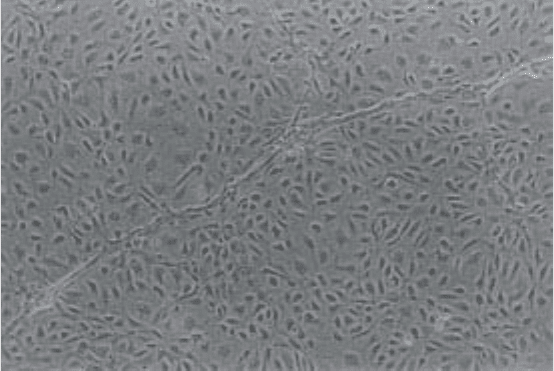
microscopy (see Note 9; Fig. 1). After 24 h the cells adhere to the flasks and
also start to grow out from any microvessel fragments present to form distinct
colonies. Human mammary microvessel endothelial cells (HuMMEC) isolated
using this technique grow to confluence forming cobblestone contact-inhibited
monolayers within 10–14 days depending on the initial seeding density. The
life span of cultures appears to be dependent on the individual tissue. We have
successfully cultured these cells to passage 8 without observable change in
morphology but routinely use them for experiments between passage 3 to 6.
3.1.4. Endothelial Cell Morphology in Culture
Cobblestone morphology is very typical of ECs derived from many tissues
and they are usually readily distinguished from the fibroblastoid contaminat-
ing cell populations (see Note 10). However, a more elongated morphology
with cells forming ‘swirling’ monolayers is often observed following stimula-
tion of ECs with growth factors. When cultured on gel matrices such as
Matrigel ECs will form “capillary-like” networks within a few hours of plat-
ing. This phenomenon also occurs in many types of microvessel ECs after sev-
eral days at confluence. However, it should be noted that the formation of
“capillary-like” structures is not an exclusive property of ECs in culture.
Fig. 1. Photomicrographs of postconfluent monolayer of HuMMEC (passage 2)
demonstrating typical cobblestone morphology and tube formation on the surface of
the monolayer.
Microvessel ECs from Adipose Tissue 219
3.1.5. Cell Culture
Subculture: Maintain the microvessel ECs at 37°C, 5% CO
2
changing the
medium every 3–4 d. When confluent HuMMEC are routinely subcultured with
trypsin/EDTA solution, onto 0.2% gelatin-coated dishes at a split ratio of 1/4.
1. Discard the old medium and wash the EC monolayer with 5–10 mL of PBS/A to
remove any remaining serum. Add a few mL of trypsin/EDTA solution, wash it
over the cell monolayer, remove the surplus leaving the cells just covered and
incubate at 37°C. Monitor the cells under the microscope until they have fully
detached (see Note 11). This can be readily achieved by striking the flask sharply
to dislodge and break up cell aggregates.
2. Add sufficient growth medium to achieve a split ratio of 1/4 and plate the cells
onto gelatinized flasks.
Cryopreservation of cells: Suspend ECs at approx 2 × 10
6
/mL in growth
medium containing 10% (v/v) dimethyl sulphoxide (Sigma) and dispense into
suitable cryovials. Cool the vials to –80°C at 1°C/min and store under liquid
nitrogen (see Note 12).
3.1.6. Maintaining the Purity of Endothelial Cultures
To maintain the purity of cultures it may be necessary to reselect the ECs
with PECA-beads and/or minor manual ‘weeding’ performed under sterile con-
ditions in a flow hood using a light microscope. Reselection with PECA-beads
can be performed as described above (Subheading 3.1.2., item 7) following
removal of the cells from flasks using trypsin/EDTA solution (Subheading
3.1.5.). Provided that there are clear morphological differences between con-
taminating cells and the endothelial cells (see Note 10), it is relatively straight-
forward although time consuming to remove them. Manual weeding should be
performed with the stage of a phase contrast microscope within a class II cabi-
net to ensure sterile conditions. A needle or Pasteur pipet is used to carefully
remove contaminating cells from around EC colonies. The medium is discarded
and the adherent cells washed with several changes of sterile PBS/A to remove
all remaining dislodged contaminating cells.
3.2. Characterization of Endothelial Cells
There are many different criteria on which EC identification may be based
and these have been extensively reviewed (2,3,5,6,19). Many of these markers/
properties are not unique to ECs and several may be required to confirm endothel-
ial identity. ECs may demonstrate heterogeneity in expression of cell markers
220 Hewett
and therefore a lack of a given endothelial marker may not preclude the endot-
helial origin of isolates (2). It is often useful to demonstrate the absence of
markers, such as stress fibers staining with smooth muscle _-actin and the
intermediate filament protein, desmin, that are characteristic of smooth muscle
cells and pericytes (20), both potential endothelial culture contaminants.
3.2.1. Key Endothelial Cell Markers
Over recent years, more specific endothelial markers have emerged, such as
PECAM-1 (13), E-selectin (14), ICAM-2, and VE cadherin (5), and they have
increased the ease of endothelial characterization. Here we focus on von
Willebrand Factor (vWF), PECAM-1, and E-selectin, which we believe to be
useful for the rapid identification of ECs. For more detailed literature on these
endothelial markers there are several reviews that cover in depth the character-
ization of ECs (2,3,5,6,19).
vWF is only expressed at significant levels in ECs, platelets, megakaryo-
cytes, and syncytiotrophoblast. In ECs it is stored in the rod-shaped Weibel-
Palade bodies that produce the characteristic punctate perinuclear staining.
These organelles are present in human ECs isolated from large vessels (1,6)
but have been reported to be scarce or absent in capillary endothelium from
various species (2,4). However, typical granular perinuclear staining for vWF
has been reported in human kidney, dermis (9), synovium, lung, stomach,
decidua, heart, adipose tissue, and brain (8) microvessel ECs.
PECAM-1 is constitutively expressed on the surface of ECs (>10
6
molecules/
cell), and to a lesser extent in platelets, granulocytes, and a sub-population of
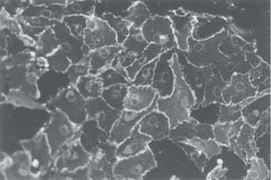
CD8+ lymphocytes (13). PECAM-1 staining of ECs in vitro is characterized
by typical intense membrane fluorescence at points of cell-cell contact (Fig. 2).
E-selectin expression appears to be unique to ECs (14). Although it is not
constitutively expressed by the majority of ECs, E-selectin expression is
induced following stimulation with inflammatory cytokines such as tumor
necrosis factor-_ (TNF_) or interleukin-1` (IL-1`) (14). Absent on unstim-
ulated controls, intense E-selectin expression is induced by pretreatment for
4 h with 10 ng/mL of TNF_ reaching a maximum at 4–8 h before returning to
background levels.
3.2.2. Immunocytofluoresent Characterization of Endothelial Cells
Immunocytoflourescence represents a simple rapid technique to character-
ize ECs. Outlined below is a staining protocol that we have routinely used for
this purpose.
1. Preparation of ECs on glass slides. Multiwell glass chamber slides are extremely
useful for this purpose as multiple tests can be performed on the same slide con-

Microvessel ECs from Adipose Tissue 221
serving both reagents and cells. The cells are cultured on chamber slides that
have been pretreated for 1 h with 5 µg/cm
2
bovine fibronectin (Sigma) in PBS/A
or 0.2 % gelatin (Subheading 2.3. item 5). When sufficient cells are present
discard the medium and wash the cells twice with PBS/A prior to fixation. A
range of fixatives can be employed depending on the activity of the antibody
required. We routinely use acetone fixation that is suitable for most antibodies or
formaldehyde. For acetone fixation, place the slides (see Note 13) in cold acetone
(–20°C, 10 min), air dry and store frozen. Alternatively, fix cells in 3% formalde-
hyde solution for 30 min at room temperature. As formaldehyde does not
permeabilize the plasma membrane 0.1% Nonidet P-40 or Triton X-100 can be
added to detect an internal antigen.
2. Immunocytoflourescent staining
a. Warm up slides to room temperature and wash with PBS/A (2X 5 min). To
prevent nonspecific binding of the secondary antibody block slides for
20 min with 10% normal serum from the species in which the secondary anti-
body was raised.
b. Incubate slides with predetermined or the manufacturers’ recommended dilu-
tion of primary antibody in PBS/A for 60 min at room temperature.
c. Wash slides with PBS/A (3X 5 min) and incubate with the appropriate FITC-
labelled secondary antibody at 1/50 dilution in PBS/A for 30 min, 1 h at room
temperature.
d. Wash slides in PBS/A (3X 5 min), mount in 50% (v/v) glycerol in PBS/A.
Stained slides can be stored for several months in the dark at 4°C.
Fig. 2. Immunofluorescent staining for platelet EC adhesion molecule-1 (PECAM-1/
CD31) in HuMMEC.
222 Hewett
Controls: To avoid false positives generated by nonspecific binding of sec-
ondary antibodies it is essential to include a negative control of cells treated as
described above but with PBS/A substituted for the primary antibody. It is also
useful to include control cell types such as fibroblasts or smooth muscle cells
and previously characterized ECs to act as negative and positive controls
respectively.
E-selectin: Most ECs do not constitutively express E-selectin and so must be
incubated with a suitable inflammatory cytokine such as TNF_ or IL-1` in growth
medium for 4–6 h to induce its expression. We routinely use 10 ng/mL recom-
binant human TNF-_. Unstimulated cells should also be included as controls.
3.2.2. Other Properties of Adipose Microvessel Endothelial Cells
We have extensively examined the expression of many endothelial markers
in our isolated microvessel ECs. These cells possess typical endothelial cell
characteristics including scavenger receptors for acetylated low density lipo-
protein, and functional angiotensin-converting enzyme at high levels. There
has been considerable interest in the endothelial-restricted receptor tyrosine
kinases (RTK) involved in EC differentiation and proliferation. Vascular EC
growth factor/vascular permeability factor (VEGF) (16) is a key angiogenic
factor and is unique in that it demonstrates pleiotropic activities specifically on
ECs, including stimulation of proliferation and induction of procoagulant
activity. All the microvessel EC types that we have cultured express the VEGF
receptor family Flt-1, Flt-4, and KDR/Flk-1 (16,17) and proliferate and express
increased tissue factor in response to VEGF. Similarly the Tie family of recep-
tors (Tie-1 and Tie-2/Tek) are also expressed on these cells (17). As more reli-
able antibodies become available against these endothelial receptors they
should be useful for EC characterization and selection.
3.3. Isolation of Endothelial Cells from Other Vascular Beds
We have adapted this basic method for the selection of adipose microvessel
ECs for the isolation of ECs from other tissues. Here we briefly outline modi-
fications that have been made for the isolation of human lung (12) and stomach
ECs (18).
3.3.1. Microvessel Endothelial Cells from Human Lung
Although rich in ECs lung is composed of many cell types and is often more
difficult to obtain. We have successfully used normal lung from transplant
donors and diseased tissue from transplant recipients. To ensure that only
microvascular ECs are harvested, a thin strip of tissue at the periphery of the
lung is used. As the amount of tissue available is usually limited, and the
subsequent yield of cells very low after Dynabead selection, it is better to
Microvessel ECs from Adipose Tissue 223
select ECs after a few days in culture before they became overgrown with con-
taminating cells.
1. Cut small peripheral sections of lung (3–5 cm long, ~1 cm from the periphery)
and wash in antibiotic/antimycotic solution.
2. Dissect the underlying tissue from the pleura and chop it up very finely using a
tissue chopper.
3. Wash the ‘mince’ above a sterile 20-µm nylon mesh to filter out red blood cells
and debris.
4. Incubate the retained material overnight in growth medium containing 2 U/mL
dispase (Boerhinger Mannheim, Mannheim, Germany) on a rotary stirrer at 37°C.
5. Pellet the digest, resuspended in ~5 ml of trypsin/EDTA solution and incubate at
37°C for 15 min.
6. Add growth medium and remove fragments of undigested tissue by filtration
through 100 µm nylon mesh.
7. Pellet and resuspended the cells in growth medium and plate onto gelatin-coated
dishes.
8. Monitor the cultures daily, trypsinize, and select the ECs using PECA-beads
before they became overgrown by contaminating cells.
3.3.2. Human Stomach Microvessel Endothelial Cells
For isolating microvessel ECs cultured from stomach biopsies and whole
stomach from organ donors (18):
1. Expose the stomach mucosa and wash with antibiotic/antimycotic solution.
2. Dissect the mucosa from the underlying muscle, chop into 2–3 mm pieces and
incubate in 1 mM EDTA in HBSS at 37°C in a shaking water bath for 30 min.
3. Transfer the pieces of mucosa to collagenase (type-II) solution containing 0.1%
BSA for 60 min, and then trypsin/EDTA solution for 15 min, in a shaking water
bath at 37°C.
4. Using a blunt dissecting tool scrape the mucosa and submucosa from white
fibrous tissue.
5. Suspend the mucosal tissue in HBSS + 20% CS and wash through 100 µm nylon
mesh.
6. Centrifuge the filtrate (700g, 5 min) and resuspend the pellet in ~12 mL of HBSS
+ 5% CS. Proceed with PECA-bead selection (Subheading 3.1.2., step 5).
4. Notes
1. We have found PECA-beads to be more reliable for purification of ECs (12) than
using tosyl-activated Dynabeads directly coated with UEA-1 (9).
2. Precoated Dynabeads (and CELLelection™ beads; see Note 8) from Dynal are
available carrying various secondary antibodies and are very convenient. How-
ever, antiimmunoglobulin-coated beads can be prepared using Tosyl-activated
Dynabeads-M50 as follows: Incubate the secondary antibody (150 µg/mL) in
224 Hewett
0.17 M sodium tetraborate buffer (pH 9.5; sterile filtered) with tosyl-activated
Dynabeads-M450 for 24 h on a rotary stirrer at room temperature. Wash the beads
4X for 10 min and then overnight in PBS/A+0.1% BSA on a rotary stirrer at
4°C before proceeding to coat them with the primary antibody as described
(Subheading 2.2.).
3. We have found that this M199/Ham F12 based recipe works well for a range of
ECs but researchers may wish to optimize the medium further. There are a num-
ber of specialized EC growth media described in the literature such as MCDB131.
Among several commercial sources of optimized EC medium, Clonetics
(Clonetics Corp., San Diego, CA) supply a low-serum medium based on
MCDB131.
4. Iron-supplemented calf serum (CS) supports the growth of ECs very well and
represents an economical alternative to fetal calf serum.
5. We routinely use recombinant basic fibroblast growth factor (bFGF) and epider-
mal growth factor (EGF) instead of endothelial growth supplement because we
have found them to be more consistent in supporting EC proliferation.
6. Omental adipose tissue obtained through general abdominal surgery can also be
used with this method. Care should be taken to remove the fat from the omental
membranes that are covered with a layer of mesothelium. Using PECA-beads we
have not found mesothelial cell contamination to be a problem.
7. The cell PECA-bead suspension is incubated at 4°C during the purification steps
to minimize nonspecific phagocytosis of Dynabeads.
8. We do not routinely remove Dynabeads following cell selection. However, it
may be necessary to remove the Dynabeads if for example the cells are required
for flow cytometry. This problem can be overcome by using CELLelection™
Dynabeads (Dynal) coated with the anti-PECAM-1 antibody to select the ECs. In
this system antibodies are conjugated to the Dynabead via a DNA linker that can
be cleaved with DNase-1 to release the beads from the cells following selection.
9. Dynabeads are internalized within –24 h of selection and are diluted to negligible
numbers/cell by the first passage through cell mitosis (9). Consistent with the
original observations of Jackson and colleagues (9,16) with UEA-I-coated
Dynabeads we have not observed any adverse effects on the adherence, prolifera-
tion, or morphology of ECs following PECA-bead selection (12).
10. The major contaminating cell population that we have observed in unselected
adipose EC cultures demonstrate a distinct fibroblastic morphology.
11. We have found that it is important for EC viability to rapidly remove the ECs
from the surface of flasks because they seem to be very sensitive to trypsin expo-
sure. It has been suggested that the use of a trypsin inhibitor to neutralize the
tryptic activity immediately following detachment from the flask may prolong
life span of ECs. We have used mung bean trypsin inhibitor (Sigma) for this
purpose although we have not examined its effect on endothelial viability.
12. We have not observed an obvious decrease in EC viability following storage in
liquid nitrogen for over 6 yr.
