N?lting B. Methods in Modern Biophysics
Подождите немного. Документ загружается.

4.1 Fourier transform and X-ray crystallography 81
(a) Im(F1(h,k))
(b) Re(F1(h,k))
Fig. 4.32
(
a
) Imaginary part of the Fourier transform of the array of Fig. 4.31a. (
b
) Real
part of the Fourier transform of the array of Fig. 4.31a
82 4 X-ray structural analysis
(a) Phase(F1(h,k))
(b) |Phase(F1(h,k))|
Fig. 4.33
Phase (
a
) and absolute of the phase (
b
) of the Fourier transform of the array of
Fig. 4.31a, calculated from imaginary and real parts of the Fourier transform
4.1 Fourier transform and X-ray crystallography 83
(a) f2(x,y)
(b) |F1(h,k)| – |F2(h,k)|
Fig. 4.34
(
a
) Representation of the array from Fig. 4.31 with an additional heavy atom.
(
b
) Difference of the absolutes of the Fourier transforms of native array (Fig. 4.31a) and
heavy atom replaced array (Fig. 4.34a). When we compare Figs. 4.33b and 4.34b, we can
see a connection between phases and amplitude differences. Note that |F| = ((Im(F))
2
+
(Re(F))
2
)
0.5
; phase(F) = arctan(Im(F)/Re(F)), where "Im" and "Re" stand for imaginary and
real parts, respectively
4.1.2.5 Calculation of the electron density and refinement
Software for the calculation of the initial electron density from the diffraction data
and the refinement of structures is being rapidly developed by several academic
institutions and often supplied for free. It may be found on the internet, e.g., by
searching with the keywords “protein crystallography software”.
84 4 X-ray structural analysis
4.1.2.6
Cryocrystallography and time-resolved crystallography
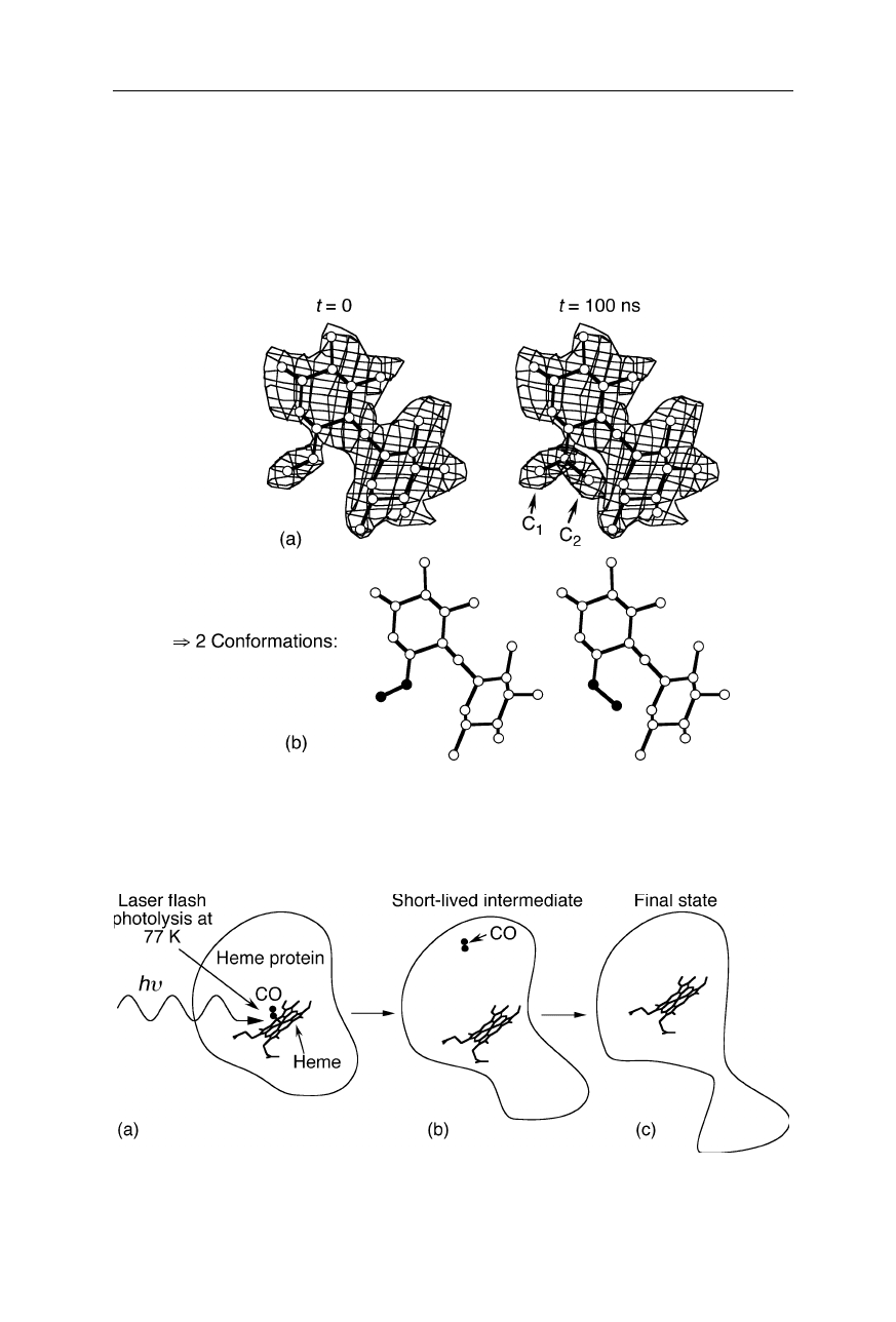
Short-living conformational intermediates in the microsecond and nanosecond
time scale have been resolved by time-resolved crystallography (Srajer et al.,
1996; Genick et al., 1997; Fig. 4.35) and cryocrystallography (Schlichting et al.,
2000; Petsko and Ringe, 2000; Wilmot and Pearson, 2002; Fig. 4.36). Time-
Fig. 4.35
Example for time-resolved crystallography. 100 ns after initiation of a confor-
mational change, the electron density indicates the occurrence of two conformations, C
1
and C
2
Fig. 4.36
Example for cryocrystallography. The CO is flashed off the heme group of the
heme protein. This initiates a conformational transition which is detected, e.g., at –196
o
C
4.2 X-ray scattering 85
resolved crystallography interprets time-dependent electron density maps and can
offer detailed structural information on short-lived intermediates under near-
physiological conditions. In cryocrystallography, reactions are induced and meas-
ured at a low temperature. At the very low temperatures of flash photolysis and
acquisition of the diffraction pattern in the experiment shown in Fig. 4.36, the
reaction kinetics of the conformational changes is slowed down by many orders of
magnitude. This enables to determine the coordinates of structural intermediates
that would normally be too short-lived to be resolved by X-ray crystallography.
4.2 X-ray scattering
4.2.1 Small angle X-ray scattering (SAXS)
Small angle X-ray scattering serves for the elucidation of microstructural
information in amorphous materials on length scales ranging from a few Å to a
few
µ
m (Figs. 4.37 and 4.38). Fig. 4.39 is an illustration of the setup for SAXS.
Significant effort is undertaken to enable the measurement at very small angles.
Since there is an about reciprocal relationship between distance separation of
scattering points (
∆
x
) and the scattering angle (
θ
), this measurement is essential to
obtain sufficient information in the relatively large length scale compared with the
wavelength (
λ
) of the X-rays (
∆
x
≈
0.5
λ
sin
–1
(
θ
/2)). For details on X-ray optics
see also the previous section.
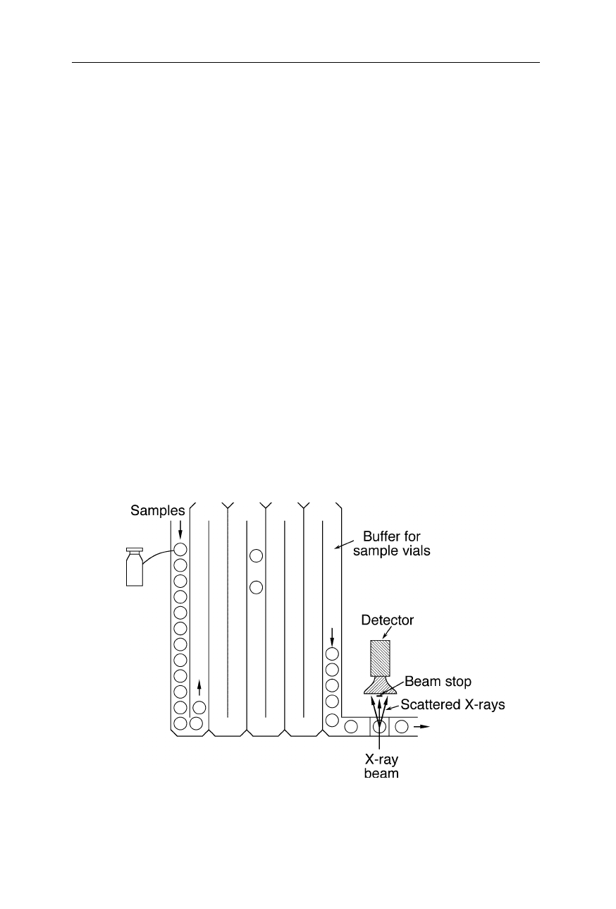
Fig. 4.37
Automatic SAXS and wide angle X-ray scattering analysis of biological sam-
ples. A large number of vials is automatically sampled and production faults immediately
are detected and responded to

86 4 X-ray structural analysis
SAXS measurements revealed that
(a) an unliganded aspartate transcarbamoylase adopts a T-quaternary structure
(Fetler et al., 2002),
(b) the axial period of collagen fibrils is 65.0 ± 0.1 nm in healthy human breast
regions, and 0.3 nm larger in cancer-invaded regions (Fernandez et al., 2002),
(c) flax cellulose microfibrils probably have a cross section of 10
×
50 Å
2
(Astley
and Donald, 2001),
(d) microfibrils with an axial repeating period of approximately 8 nm are present
in the major ampullate silk from the spider
Nephila
(Miller et al., 1999; Riekel and
Vollrath, 2001), and
(e) the ATPase domain of SecA has dimensions of approximately 13.5 nm
×
9.0 nm
×
6.5 nm (Dempsey et al., 2002).
SAXS revealed information regarding the conformational diversity and size
distribution of unfolded protein molecules (Kamatari et al., 1999; Panick et al.,
1999a; Garcia et al., 2001; Choy et al., 2002), and was used in a large number of
protein-folding and peptide-folding studies to obtain information about size
changes (e.g., Chen et al., 1998; Panick et al., 1998, 1999b; Arai and Hirai, 1999;
Segel et al., 1999; Kojima et al., 2000; Russell et al., 2000; Aitio et al., 2001;
Canady et al., 2001; Katou et al., 2001; Muroga, 2001; Tcherkasskaya and
Uversky, 2001). SAXS is one of the very few methods which can directly monitor
structural changes of small virus particles (Sano et al., 1999; Perez et al., 2000).
Fig. 4.38
Diffraction pattern of a cell suspension. SAXS can serve to obtain a “finger-
print” of a biological specimen which helps to identify unknown biological samples
4.2 X-ray scattering 87
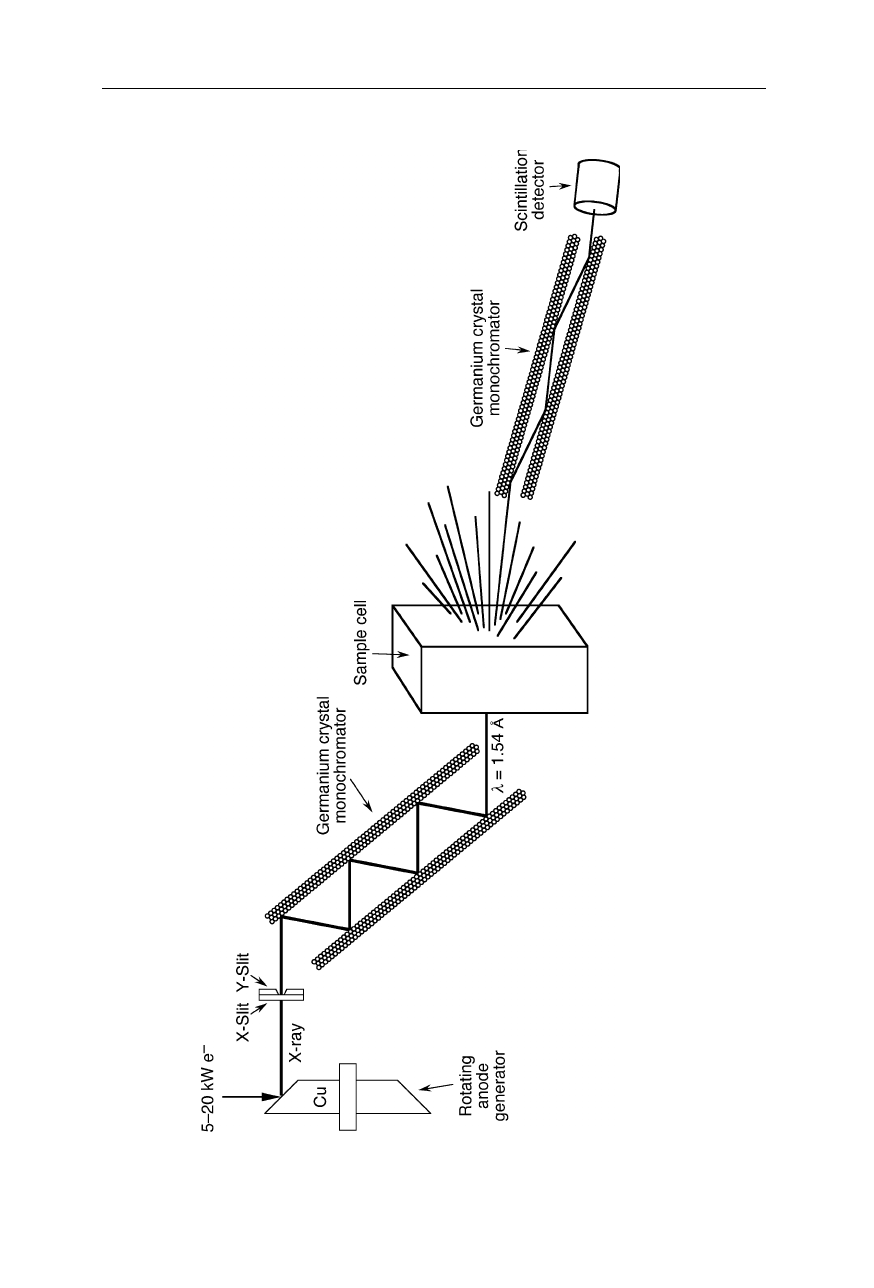
Fig. 4.39 Setup for small angle X-ray scattering (SAXS). The X-ra
y beam from a rotating anode generator is passed through a crystal
monochromator that selects a wavelength. The monochromatic X-ray beam is passed though the sample cell, and scattered X-rays a
t very
small angels are passed through a second crystal monochroma
tor and then detected with a scintillation detector. For many appli
cations,
this set-up may be simplified, e.g., by choosing fewer reflections in the monochromators which yields a higher intensity of X-r
ays
88 4 X-ray structural analysis
Distance constraints derived from SAXS measurements can be used to filter
candidate protein structures for the purpose of protein structure prediction (Zheng
and Doniach, 2002). In some cases even low resolution solution structures of
proteins were obtained solely from SAXS data (Chacon et al., 1998; Shilton et al.,
1998; Bada et al., 2000; Maruyama et al., 2001; Scott et al., 2002) or SAXS data
combined with neutron scattering data (Egea et al., 2001). SAXS can reveal the
structure of bones (Rinnerthaler et al., 1999) and structural changes in bones due
to diseases (Grabner et al., 2001). The method was used to obtain information
about conformational changes of bacterial cell wall enzymes upon binding to a
substrate (Schönbrunn et al., 1998), and structural changes in artificial biological
membranes (Riske et al., 2001). SAXS results on human dentin, which is a
complex composite of collagen fibers and carbonate-rich apatite mineral phase,
are consistent with nucleation and growth of an apatite phase within periodic gaps
in the collagen fibers (Kinney et al., 2001).
4.2.2 X-ray backscattering
The property of X-rays to penetrate materials is used in many biophysical applica-
tions, ranging from for the purpose of determination of the molecular weight of
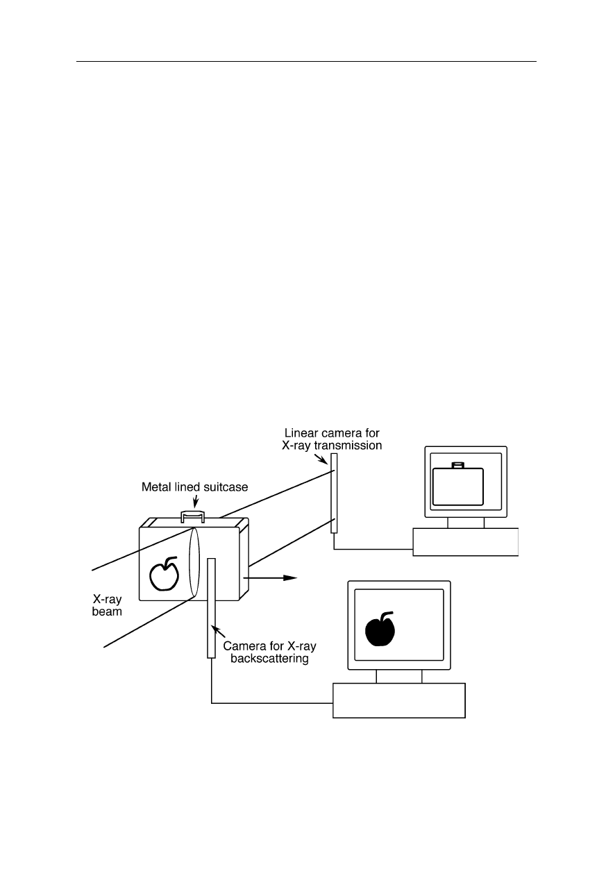
Fig. 4.40
Detection of biological and other organic material behind a metal layer with
X-ray backscattering: since the absorption of biological material is much smaller than that
of metal, the biological material is difficult to detected in single-wavelength X-ray
absorption measurements. X-ray backscattering provides much better contrast in this ap-
plication. However, the quantum efficiency of X-ray scattering is low and thus relatively
large expositions and sensitive cameras must be used

4.2 X-ray scattering 89
proteins to X-ray backscattering for the purpose of detection of organic material
hidden in metal containers (see, e.g., Fig. 4.40).
A problem of detection of organic material, such as illicit drugs and explosives,
by X-ray absorption is their low absorption coefficient compared with metals and
the possibility to camouflage the material, e.g., by embedding it in other organic
material, such as flour or sugar. X-ray backscattering offers a good contrast for
the detection of such powdery material (Fig. 4.40). The main disadvantage is the
low backscattering coefficient compared with transmission coefficient of most
organic samples. Thus, a significantly higher exposure compared with X-ray
transmission is usually required.
5 Protein infrared spectroscopy
Infrared spectroscopy is based on the infrared absorption of molecules and is,
compared with crystallography, a relatively simple and inexpensive tool for the
global characterization of molecular conformations and conformational changes of
proteins and other biomolecules. Depending on the measurement technique, scan-
ning infrared (IR) spectrometers, Fourier transform infrared (FTIR) spectrometers,
and single wavelength infrared apparatuses are distinguished (see Sect. 5.1).
Typically the most interesting spectral region for biomolecules is
ν
= 400 –
4000 cm
–1
, where the wavenumber,
ν
, is defined as
ν
≡
1/wavelength. Infrared ac-
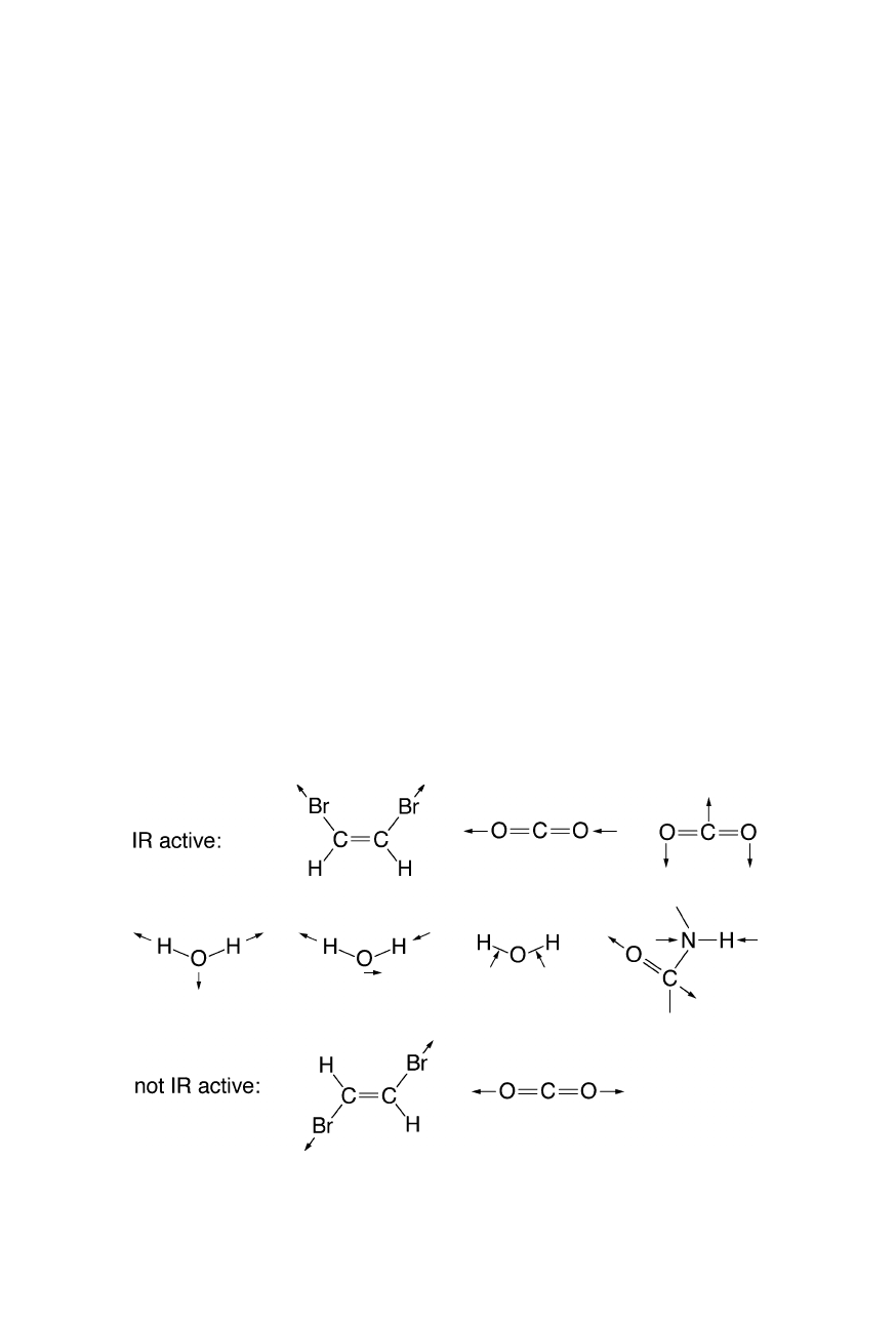
tivity requires a change of dipole moment upon excitation (Fig. 5.1). For proteins
the amide chromophore absorption in the region of 1500 cm
–1
–1700 cm
–1
(
≈
6
µ
m
wavelength) is particularly important for the assessment of secondary structure
content and structural changes. Regarding the resolution of protein secondary
structure, the information content of IR and FTIR spectroscopy is comparable with
that of circular dichroism (see, e.g., Nölting et al., 1997b; Nölting, 2005), and
regarding the resolution of features of the tertiary structure of proteins, IR and
FTIR are often inferior, and yet IR is much easier to apply on a fast time scale and
for remote sensing (see, e.g., LIDAR in Sect. 5.1.3).
Fig. 5.1
Example of infra-red active and non-active vibrations. Note that infra-red activity
requires a change of dipole moment