Reed S.J.B. Electron microprobe analysis and scanning electron microscopy in geology
Подождите немного. Документ загружается.

9.2 Mounting
9.2.1 The SEM ‘stub’
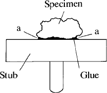
Specimens for SEM examination are commonly mounted on a ‘stub’, which
takes the form of a disc, usually made of aluminium and typically about 1 cm
in diameter, with a spigot for attachment to the stage mechanism (Fig. 3.9).
(Sometimes graphite stubs are used, to minimise X-ray background, especially
for particulates.) The sample is glued to the stub and coated to provide
conduction, as described in Section 9.5. A quick alternative method of attach-
ment is to use double-sided sticky tape, though this is to be avoided if possible.
Quick-setting glue can also be used when speed is important. Mounting
materials and adhesives should have a low vapour pressure so that the instru-
ment vacuum is not adversely affected. Whatever arrangement is used, there
must be an electrical path to the holder: if necessary this should be provided by
applying carbon or silver paint (Fig. 9.2). For small specimens such as micro-
fossils the use of a vacuum-compatible wax has some advantages (Finch,
1974). The wax is warmed so that it flows over the surface of the stub and
the specimens are pressed into the wax while it is soft. For some purposes it is
desirable to mount an electron-microscope grid with numbered bars on the
stub, so that individual specimens can be relocated by means of their ‘grid
reference’. The special problems arising in the mounting of soil samples have
been treated in detail by Lohnes and Demirel (1978).
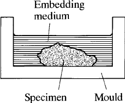
9.2.2 Embedding
For some specimens (e.g. ore minerals) it is not necessary to use thin sections;
instead, embedding in a solid block is appropriate. The sample is placed in a
mould made of non-stick material, and the embedding medium in liquid form
poured in (Fig. 9.3). Alternatively, a metal or plastic ring can be temporarily
Fig. 9.2. An SEM specimen mounted on a ‘stub’; note that conductive coating
may not connect across overhanging regions (a), which should therefore be
painted with silver or carbon ‘paint’.
9.2 Mounting 155
stuck down to serve the same purpose, remaining part of the mount after the
medium has set. Bakelite (supplied as a powder and polymerised by applica-
tion of pressure and heat) can be used for embedding, but epoxy resins that are
either cold setting or require only relatively mild heating are less likely to cause
damage to the specimen. Bubbles can be removed by applying moderate
vacuum. There is some advantage in using a conducting medium, such as
epoxy resin filled with fine carbon or metal particles.
9.2.3 Thin sections
In many applications, thin sections are required so that viewing by transmitted
light is possible. Preparation is as for ordinary thin sections in the first stages,
the rock slice being attached to the glass slide using one of the special epoxy
resins with suitable optical properties that are available. The slice is ground to
a thickness somewhat greater than the 30 mm final thickness required, before
commencing polishing. Long microscope slides can be shortened by cutting
the ends off, in order to fit the specimen holder more conveniently. Also
sometimes used, especially in the USA, are 1-inch (25.4-mm)-diameter round
sections.
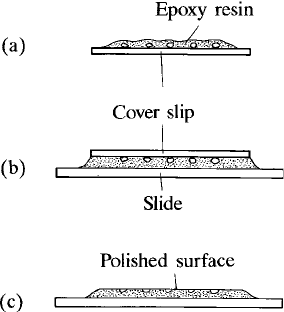
9.2.4 Grain mounts
Special techniques are required for mounting small grains. One possibility is to
mix the grains with embedding medium and set in a mould. Alternatively, they
may be pressed into a thin layer of epoxy resin on a glass cover slip. After
setting, this is inverted and mounted on a glass slide, and the cover slip ground
away, leaving the grains lying in one plane ready for polishing (Fig. 9.4). To
avoid clumps, mixing with crushed graphite particles of similar size prior to
embedding is an effective way of keeping the grains separate.
Fig. 9.3. Embedding a specimen for polishing: liquid medium (e.g. epoxy resin)
is poured into a ‘non-stick’ mould and allowed to set (with heating if necessary).
156 Sample preparation
For SEM work, polishing might not be required. In this case, grains can be
scattered onto a sticky surface or partly dried carbon paint, etc., or alterna-
tively suspended in a liquid, drops of which are transferred to the substrate and
then evaporated (in the latter case, it may be necessary to de-coagulate the
particles with the aid of an ultrasonic bath). Suitable substrate materials
include beryllium (which gives minimal X-ray emission), carbon (which is
almost as good in this respect, but is difficult to obtain with a very smooth
surface) and silicon (which can be highly polished, but emits more X-rays,
which is a drawback in some applications.)
9.2.5 Standards
Methods used for mounting standards are essentially similar to those already
described. Usually fairly small (e.g. a few millimetres) pieces of standard
materials are used, so a number can be mounted in the specimen holder at
one time. Standards can be prepared individually, allowing selection of those
required for each application, or alternatively a single block containing many
standards can be used. This saves space, but obtaining a good polish on a wide
variety of materials simultaneously is difficult. Prepared standard blocks are
obtainable from commercial suppliers. It is convenient for commonly used
standards to be mounted in the specimen holder semi-permanently. Those
needed only occasionally for specific applications can be loaded temporarily
for calibration and then removed to make space for specimens.
Fig. 9.4. Mounting small grains: (a) grains embedded in a thin layer of epoxy
resin on a cover slip; (b) embedded grains mounted on a microscope slide; and
(c) cover slip ground away and exposed grains polished.
9.2 Mounting 157
9.3 Polishing
For X-ray analysis, mapping and BSE imaging it is extremely desirable to avoid
topographic effects: specimens therefore should be flat and well polished.
Polishing procedures for ore microscopy can be adapted to rocks consisting
predominantly of silicates. Starting with a flat ground surface, polishing is
carried out with progressively finer grades of abrasive (typically carborundum
or emery for the coarser grades and diamond or alumina in the later stages).
Paper or woven nylon laps are preferable to cloth with a ‘nap’, since they have
less tendency to produce surface relief between minerals of different hardnesses.
Either rotating or vibrating motion of the lap is used, the former being prefer-
able. The specimens should be thoroughly cleaned after each stage, to avoid
transfer of abrasive material in pores and cracks. For soft phases a final hand
polish using very fine alumina may be necessary. A single-stage polishing
technique using only alumina has been described by Allen (1984). Special
procedures are required for electron backscatter diffraction studies (Section
4.8.3), the damaged surface layer left by conventional polishing being removed
by a final polish with alkaline colloidal silica slurry (Lloyd et al., 1981).
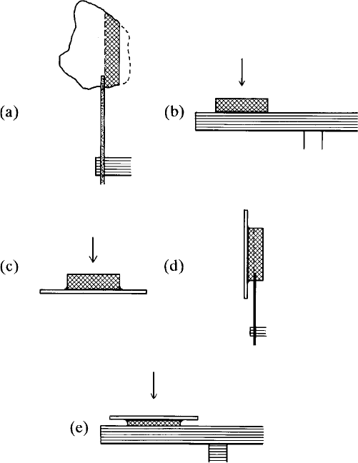
Polished thin sections are usually made by polishing a section that has
previously been ground to a thickness somewhat greater than 30 mm(Fig.9.5).
However, the final thickness is then poorly constrained, which is undesirable for
polarised-light microscopy. This can be avoided by polishing one face of the
rock slice first, temporarily mounting it face down on a glass slide, grinding off
surplus material to give a thickness of 30 mm, mounting permanently with epoxy
resin on another glass slide (ground face down), and finally removing the first
slide(seeFig.9.5).Afterpolishing,specimensshouldbecleanedbywashingina
solvent that does not attack the mounting medium (e.g. ethanol or petroleum
ether), preferably using an ultrasonic bath to dislodge remnants of polishing
materials. When surface contamination is especially important (e.g. for light-
element analysis) plasma cleaning is desirable (Isabell et al., 1999).
9.4 Etching
Chemical etching enables chemical and crystallographic differences to be con-
verted into topography that can be observed in secondary-electron images. (It is
inappropriate for quantitative EMPA, for which flat, smooth, surfaces are
required, and etching may alter surface composition.) Carbonates can be etched
with dilute hydrochloric acid (1–5%), acetic acid (20%), or EDTA, the most
delicate effect being obtained with the last two. In some cases heavy etching to
remove carbonate cement, leaving exposed grains of quartz etc. for SEM study,
158 Sample preparation
is appropriate. Quartz grains may be etched with concentrated hydrochloric
acid for studies of surface texture. Etching of polished sections with hydrofluoric
acid can be used to reveal textures of quartzitic sandstones, for example, and fine
exsolution textures in silicates (Section 4.6.3). The sections can be suspended
above an acid bath, to be etched by the fumes, or immersed for a stronger effect.
(The glass slide may be protected by covering it with paraffin wax.)
9.5 Coating
Most geological samples, being non-conductors of electricity, require a con-
ductive coating to prevent charging under electron bombardment unless this is
Fig. 9.5. Preparation of a polished thin section: (a) a slice of rock is cut with a
diamond saw and trimmed to size; (b) one face of the slice is ground and
lapped; (c) the slice is attached to a glass slide and lapped face down;
(d) surplus material is cut off with a fine diamond saw; and (e) the surface is
ground and polished leaving 30 mm thickness.
9.5 Coating 159
avoidedbyselectingalowacceleratingvoltage(Section5.6.7)orbyusingan
‘environmental’ or ‘low-vacuum’ SEM (Section 3.10.2). The preferred coating
element for X-ray analysis is carbon, because it has a minimal effect on the
X-ray spectrum. It is also the best choice for cathodoluminescence studies.
However, it is not ideal for SEM imaging, owing to its low secondary-electron
yield. For this purpose a metal such as gold is preferable, or alternatives with
finer grain structure, such as gold–palladium alloy, chromium or iridium, but
these are less suitable for X-ray analysis and BSE imaging. Gold coating is also
advantageous for fibrous clay minerals, which tend to collapse under the
electron beam when coated with carbon (Purvis, 1991).
9.5.1 Carbon coating
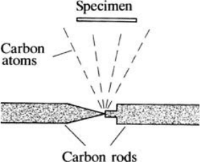
The usual method of coating with carbon is to place the specimen in a vacuum
chamber with a carbon evaporation source consisting of pointed carbon rods
(3–6 mm in diameter) in contact (Fig. 9.6). A current of about 100 A is passed
through the rods for a few seconds, causing carbon to be evaporated from the
region where the rods are in contact. The pressure should be less than approxi-
mately 10
4
torr, as obtained with either a diffusion pump or a turbo pump.
(Carbon films produced under poor vacuum conditions are ‘sooty’ and lack
adhesion.) Since the evaporated carbon atoms travel in straight lines, this
coating method is suitable only for flat specimens, not for irregularly shaped
objects, better coverage of which can be obtained by rotating the sample
during coating.
The optimum thickness of carbon is about 20 nm. The thickness can be
controlled approximately by using a fixed current and evaporation time. It can
also be estimated from the colour of the coated surface of a polished metal such
as brass (Kerrick, Eminhizer and Villaume, 1973): orange corresponds to
Fig. 9.6. Evaporation of carbon to give conductive coating on sample, using
pointed carbon rods and large cu rrent.
160 Sample preparation
15 nm, indigo red to 20 nm, blue to 25 nm and bluish green to 30 nm. More
accurate monitoring can be achieved by means of a quartz crystal forming part
of an electronic oscillator circuit, with one surface exposed to the evaporant, the
oscillator frequency being used to indicate coating thickness. Other methods of
thickness monitoring are based on the electrical resistance or optical density of
the coating. Specimens should be equi-distant from the carbon source in order
to ensure that uniformity of coating thickness is attained.
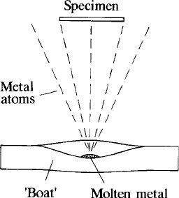
9.5.2 Metal evaporation
Although carbon is usually the preferred coating material for samples that are
to be analysed, the enhanced thermal conductivity obtained with a metal coat
is advantageous in reducing the effects of electron bombardment for certain
types of sample (Section 8.7). Gold, as used for SEM samples, is best avoided
because it interacts strongly with electrons and X-rays on account of its high
atomic number. Alternatives such as aluminium, copper and silver are there-
fore sometimes used. Coating with these metals can be carried out by vacuum
evaporation, using a wire basket made of tungsten, or a ‘boat’ made from
molybdenum sheet, heated by passing a large electric current (Fig. 9.7). In
some cases sputter coating may be used instead (see the next section).
9.5.3 Sputter coating
This is a convenient method for producing coatings of metals, including gold
and gold–palladium alloys. Since a relatively poor vacuum is used, sputtered
atoms are strongly scattered by gas molecules and travel in all directions,
Fig. 9.7. Evaporation of metal (e.g. Ag, Al) by heating in a molybdenum
‘boat’ carrying a large current.
9.5 Coating 161
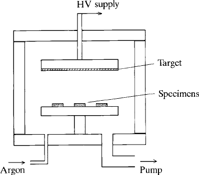
which is advantageous for coating specimens of irregular shape. The ‘diode’ type
of sputter coater is shown in Fig. 9.8. The chamber is first evacuated with a
rotary pump; argon is then admitted to give a pressure of about 10
1
torr. When
a high voltage is applied across the electrodes a discharge occurs and the metal
foil target is bombarded with ions, which remove atoms by sputtering. These are
deposited onto the specimen, the coating thickness being determined by the
discharge current and time. The current is controlled by varying the argon
pressure. The specimen is heated significantly by electron bombardment,
which can cause damage to fragile materials. ‘Cold’ sputter coaters, in which
the electrons are deflected by a magnet, are available. For high-resolution SEM
work, metal films of better quality can be obtained using a turbo-pumped
sputter coater.
9.5.4 Removing coatings
It may be desirable to remove a coating applied for SEM or EMP work, for
example to allow unhindered examination by optical microscopy, or to permit a
gold coating applied for SEM examination to be replaced by carbon for X-ray
analysis (or vice versa). Carbon deposited under high-vacuum conditions
adheres strongly to the substrate, but can be removed from polished sections
with a fine polishing medium (e.g. 0.25-mm diamond on a cloth lap). Gold and
other metals can easily be wiped off polished specimens, but traces remaining in
Fig. 9.8. Sputter coating: air is removed from the chamber and replaced by
argon at low pressure; high voltage (HV) applied to the top electrode causes a
discharge in the gas; specimens are coated with metal atoms (e.g. Au) removed
from the target by ‘sputtering’ due to bombardment with argon ions.
162 Sample preparation
cracks, etc., can be troublesome in SEM images. Specimens with strong three-
dimensional topography require a different approach. Gold can be removed
from such samples by treatment with a 10% aqueous solution of sodium cyanide
(Sela and Boyde, 1977), with appropriate precautions in view of its toxicity.
Silver has the advantage that it can be removed easily with photographic
‘Farmer’s reducer’ (Mills, 1988).
9.6 Marking specimens
Specimens must, of course, be marked for identification purposes. Aluminium
SEM stubs can be inscribed with a fine metal stylus, or alternatively written on
with a pen. In the case of blocks of epoxy resin in which opaque specimens are
mounted for polishing, a label can be embedded with the specimen, or else the
identification number can be written or scratched onto the back. For thin
sections a number may be inscribed on the back using a diamond point.
It is also sometimes desirable to mark areas of interest within specimens to
make them easier to find in the electron microprobe or SEM. Ink rings can be
drawn on the back of thin sections for this purpose, if the instrument con-
cerned has transmitted-light viewing facilities. Rings drawn on the front are
naturally appropriate for opaque specimens and when transmitted-light view-
ing is unavailable: for this purpose either conducting ink should be used or else
the carbon coating must be applied after drawing the rings. Ink that is
unaffected by the solvents used for cleaning should be selected. The need for
marking can be obviated by using other approaches described in the following
sections.
9.6.1 Specimen ‘maps’
Finding areas of interest using the optical microscope in the electron micro-
probe can be difficult because of the high magnification and small field of view.
Scanning electron images, which allow much lower magnification, can be used
instead, but relevant features are sometimes not easily identifiable in such
images. Time can therefore be saved by employing strategems described below.
A sketch map or low-magnification photograph of the whole specimen is an
invaluable aid. A ‘macro’ photograph can be used, but scanned digital images
are now preferred. Micrographs of small areas at higher magnifications are
also sometimes useful and can be taken with an ordinary photomicrography
set-up. Opaque grains provide useful ‘landmarks’ in plain-light micrographs
that can easily be recognised either in the built-in microscope (in the case of the
electron microprobe) or in backscattered electron images.
9.6 Marking specimens 163
The need for a ‘map’ may be obviated by using a bench microscope with x
and y coordinate read-out. Full microscope facilities – low- and high-power
objectives, polarised light, etc. – are available for finding areas of interest. The
positions of these can be recorded relative to a reference mark, so that the same
points may be found easily after transfer to the electron microprobe or SEM
(provided that this has calibrated stage movements).
9.7 Specimen handling and storage
Specimens and standards should be kept in a dust-free environment, prefer-
ably in a desiccator. For pure-element standards that oxidise readily and
minerals such as sulphides that tend to tarnish it is desirable to use a vacuum
desiccator. The SEM ‘stubs’ can be stored in plastic boxes available for the
purpose.
Cleanliness should be observed when handling specimens, to avoid degra-
dation of the specimen-chamber vacuum and enhancement of the rate of
deposition of carbon contamination; gloves should preferably be used.
Specimens can be cleaned by washing with a residue-free solvent and wiping
with a tissue. The solvent should be one that does not attack the specimen,
mounting medium, or ink used for labelling. Suitable choices include pet-
roleum ether and ethanol. Dust can be removed with a compressed-air jet.
164 Sample preparation