Reed S.J.B. Electron microprobe analysis and scanning electron microscopy in geology
Подождите немного. Документ загружается.


transitions that obey the rules of quantum theory are allowed: the most sig-
nificant of these are shown in Fig. 2.9.LinesaredesignatedK,L,etc.according
to the shell containing the initial vacancy. Within a given shell, lines in the group
which are most intense are labelled a, those in the next most intense group, b,
etc., and within each group the lines are numbered in order of intensity (approxi-
mately). Typical spectra are shown in Fig. 2.10. (Note that a, b and g are often
used instead of a, b and g on computer screens and print-outs.)
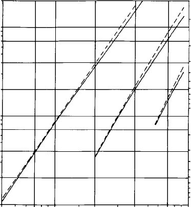
The energy of a given line varies approximately as the square of the atomic
number of the emitting element (Moseley’s law). The energies and wave-
lengths
*
of Ka
1
,La
1
and Ma
1
lines are plotted against Z in Fig. 2.11. For
analysis one is mainly concerned with X-ray energies up to about 10 keV, and
the Ka
1
line is used for the analysis of elements of atomic number up to about
30, above which the La
1
line is used (or Ma
1
for the heaviest elements). Other,
less intense, lines are rarely used for analysis, but sometimes interfere with lines
that are so used, and therefore cannot be ignored. The relative intensities of
these lines depend on the numbers of electrons occupying the energy levels
involved and are fairly constant.
Fig. 2.10. Typical examples of characteristic X-ray spectra (principal lines
only are shown): (a) the K spectrum of potassium (Z ¼ 19); (b) the L spectrum
of silver (Z ¼ 47); and (c) the M spectrum of uranium (Z ¼ 92).
*
Wavelength (l) and energy (E) are related thus: E l ¼ 12 398, in which E is in electron volts and l is in
a
˚
ngstro
¨
m units (1 A
˚
¼ 10
10
m).
2.5 X-ray production 15
For the most part the relative intensities and positions of X-ray lines are
similar for different elements, apart from a shift as a function of atomic
number. However, substantial changes occur in L and M spectra for atomic
numbers below approximately 26 and 65, respectively, owing to incomplete
filling of the relevant shells. As a consequence the number of lines is reduced,
also the relative intensity of the line decreases (disappearing completely in
the case of Ma).
2.6 X-ray absorption
The observed intensity of characteristic X-ray lines may be affected signifi-
cantly by absorption occurring in the sample itself. The effect of absorption in
a thin layer is given by the equation
I ¼ I
0
expðxÞ; (2:4)
where I
0
is the initial intensity, I the intensity after absorption, the ‘mass
absorption coefficient’ (cm
2
g
1
), the density (g cm
3
) and x the path length
(cm). Values of are widely variable, ranging from less than 100, for X-rays
of high energy and absorbers of low atomic number, to more than 10 000, for
1
2
4
7
0.1
0.2
0.4
0.7
10
20107
0.2
0.4
0.7
0.1
4 40 70 100
Z
1
2
4
7
10
20
E (ke
V)
λ (A)
˚
K
L
M
Fig. 2.11. Energy (E) and wavelength (l)ofKa,La and Ma lines (solid lines)
and their excitation energies (dashed lines), versus atomic number (Z).
16 Electron–specimen interactions

X-rays of low energy and absorbers of high atomic number. In the latter case
severe absorption occurs even for x less than 1 mm.
The predominant mechanism of X-ray absorption is the ejection of inner-
shell electrons. On a plot of versus X-ray energy, sharp discontinuities
(‘absorption edges’) occur at energies corresponding to the critical excitation
energies of the different shells of the absorbing element (Fig. 2.12). Below such
an edge the X-rays have insufficient energy to ionise the shell concerned.
2.7 The Auger effect and fluorescence yield
The energy released when an atom ionised in an inner shell returns to its
normal state by electron transitions from outer levels may be used to eject
another bound electron instead of an X-ray photon (the ‘Auger effect’).
The main significance of this is its influence on X-ray intensities, but also it
is the basis of ‘Auger analysis’ (Section 1.4.4). The ‘fluorescence yield’, denoted
by !
K
(for the K shell), is the probability of ionisation being followed by the
emission of an X-ray photon rather than an Auger electron. This increases
rapidly with increasing Z (Fig. 2.13), which tends to compensate for the
decrease in Q
K
with Z already noted. The fluorescence yields of other shells
behave similarly.
2.8 Cathodoluminescence
In some types of sample, electron bombardment stimulates the emission of
light by the process of cathodoluminescence (CL). In a non-metallic material
Fig. 2.12. A schematic representation of the variation of the mass absorption
coefficient as a function of X-ray energy.
2.8 Cathodol uminescence 17

incident electrons cause excitation by raising electrons in the valence band to
the normally empty conduction band, from which they return to their original
state in one or more steps. The surplus energy can be dissipated in various
ways, one of which is the emission of photons (Fig. 2.14). Only relatively low
energies (a few electron volts) are involved, and the wavelengths fall within the
visible region (sometimes extending into the ultra-violet or infra-red).
hν hν h
ν
Conduction
band
Valence
band
(a) (b) (c) (d)
Fig. 2.14. Cathodoluminescence: (a) electron bombardment raises an electron
from the valence to the conduction band; de-excitation may occur directly (b)
or via localised levels in the band gap, associated with defects or impurities (c)
and (d). The photon energy h is the difference in energy between the initial
and final levels.
0.8
0.7
0.6
0.5
0.4
0.3
0.2
0.1
0
010203040
Atomic number (Z
)
Fluorescence yield (
ω
K
)
Fig. 2.13. The atomic-number dependence of the fluorescence yield of the K
shell (!
K
), which is the probability of K-shell ionisation being followed by
characteristic X-ray emission (rather than Auger electron emission).
18 Electron–specimen interactions
Minerals that exhibit CL include diamond, quartz, corundum, rutile, cassiter-
ite, benitoite, willemite, halite, fluorite, spinel, calcite, dolomite, kaolinite,
apatite, barite, strontianite, sphalerite, zircon, feldspar, jadeite, diopside, wol-
lastonite, forsterite and fayalite.
Localised energy levels in the gap between valence and conduction bands,
arising from lattice defects, interstitial ions, or substitutional impurity atoms,
often play an important role in CL emission (see Fig. 2.14). Certain elements
behave as ‘activators’, small concentrations of which are sufficient to produce
CL. Others (notably divalent Fe) have the effect of ‘quenching’ CL emission.
The intensity of some forms of CL emission is strongly influenced by the
density of defects, which is dependent on factors such as temperature of
formation, cooling rate, deformation and irradiation. High defect densities,
however, may suppress CL by promoting alternative modes of de-excitation.
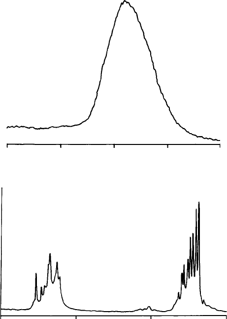
The colour of CL emission depends on the difference in energy between the
states concerned. Most commonly the energy is not narrowly defined and the
emission takes the form of a band (Fig. 2.15(a)). In a few cases line spectra
characteristic of the impurity element are produced (Fig. 2.15(b)).
Excitation of CL is not very sensitive to the beam accelerating voltage, but
sometimes it is advantageous to use a relatively high value (at least 20 kV)
because this enables the electrons to penetrate the non-luminescent damaged
surface layer. Prolonged electron bombardment tends to cause fading, whereby
the intensity declines, sometimes irreversibly. Cooling the specimen below room
temperature significantly increases CL intensity for certain types of sample.
Cathodoluminescence can be detected in SEMs and EMPs with appropriate
light-detection equipment (Section 3.12.2). Owing to the complexity of the
factors governing CL emission in natural materials, unambiguous elemental
analysis is problematic, but nevertheless information not easily obtained by
other means in a range of geological applications can be obtained from CL
images (see Section 4.8.4).
2.9 Specimen heating
The fraction of the energy in the incident electron beam which reappears in the
form of X-rays, light, etc. is small, most being converted into heat in the target.
The temperature rise T can be estimated from the following expression:
T ¼ 4:8E
0
i=ðkdÞ; (2:5)
where E
0
is the incident electron energy (keV), i the current (mA), k the thermal
conductivity (W cm
1
K
1
)andd the beam diameter (mm). For metals k is
2.9 Specim en heating 19
typically in the range 1–4 and under normal conditions T is negligible. On the
other hand, for materials of low thermal conductivity, including many minerals,
thetemperaturemayberaisedsignificantly:forexample,inthecaseofmica
(k ¼ 6 10
3
) the calculated temperature rise is 160 K for E
0
¼ 20 keV, i ¼ 10 nA
and d ¼ 1 mm. The heating effect can be moderated by reducing i or increasing d,
or by using a surface coating of a good conductor (see Section 8.6).
400
500
600
700
800
Wavelength (nm)
(a)
450 550 600500
Wavelength (nm)
(b)
Fig. 2.15. Cathodoluminescence spectra: (a) the emission band caused by Mn
in carbonates etc.; and (b) emission lines caused by rare earths (mainly Dy) in
zircon.
20 Electron–specimen interactions
3
Instrumentation
3.1 Introduction
Scanning electron microscopes and electron microprobes have much in com-
mon, including a source of electrons (an ‘electron gun’), and electron lenses to
focus the beam on the specimen, which together form the ‘column’. Column
design is similar in principle for both types of instrument: descriptions in the
following sections therefore apply equally to both for the most part (differences
are explained where necessary). Beam-deflection coils and electron detectors
enable scanning images to be produced. X-ray spectrometers (described in the
next chapter) are a common SEM accessory and an essential component of the
EMP. Other types of detector are available optionally. Features also described
in the following sections include the vacuum system, specimen stage and, in the
case of EMPs (but not usually SEMs), optical microscope.
3.2 The electron gun
The source of electrons in EMP and SEM instruments is held at a negative
potential (typically 10–30 kV), which accelerates the electrons towards the
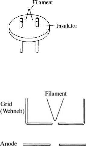
sample. The commonest type of emitter is a tungsten filament (about 0.1 mm
in diameter) bent into a ‘hairpin’ shape and attached to legs mounted on an
insulator (Fig. 3.1). This is heated electrically to about 2700 K, giving electrons
sufficient thermal energy to overcome the potential barrier at the surface. The
grid or ‘wehnelt’ (Fig. 3.2) is held at a negative potential relative to the cathode
and limits the effective emitting area of the filament to the region close to
the tip. The anode consists of an earthed plate with an aperture to let the
beam pass.
The lifetime of the filament is governed by thinning due to evaporation of
tungsten and is strongly temperature-dependent. A filament should last for several
21
weeks if correctly adjusted and operated. When fitting a new filament it should be
accurately centred in the wehnelt aperture and set to the correct height.
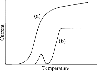
With increasing filament heating current the emission current rises rapidly
at first, but then levels out to a ‘saturated’ value of 50–100 mA. The small
fraction of this current that reaches the specimen behaves similarly, but
reaches a ‘plateau’ at a somewhat higher filament temperature (Fig. 3.3). In
the unsaturated condition, electrons from the sides of the filament as well as from
the tip contribute to the beam, and the focal spot is enlarged. It is therefore
important to operate on the plateau in order to achieve both a small beam
diameter and insensitivity to variations in temperature. The current meter fitted
as standard in EMP instruments may be used for finding the ‘plateau’; SEMs
usually lack such a meter, but saturation can be determined by observing the
brightness of the image. Operating significantly above the ‘knee’ of the curve has
no advan tages and results in reduced filament life. The saturation temperature can
be lowered, and hence filament life increased, by increasing the distance between
the tip of the filament and the wehnelt (the maximum current obtainable is
reduced, but for most purposes this is unimportant).
Fig. 3.1. A tungsten ‘hairpin’ filament used as the electron source in SEMs etc.
Fig. 3.2. An electron gun (triode type): electrons emitted by the filament are
accelerated towards the anode; the grid (or ‘wehnelt’) controls emission from
the filament tip.
22 Instrumentation
3.2.1 High-brightness electron sources
Replacing the tungsten filament by lanthanum or cerium hexaboride (LaB
6
or
CeB
6
) gives an order-of-magnitude increase in brightness (and a correspond-
ing increase in current for a beam of given diameter), but higher vacuum is
required. In a ‘field emission’ (FE) source, electrons are drawn from an
extremely fine tungsten point by a strong electric field. Field emitters are
very sensitive to vacuum conditions and require ultra-high vacuum. They
can be used at room temperature, but operation at an elevated temperature
has the advantage of minimising gas adsorption, hence giving more stable
emission. The ‘Schottky’ type of field emitter has a tip of larger radius, which
is coated to enhance electron emission. Its somewhat lower brightness is
compensated by higher maximum beam current and less extreme vacuum
requirement. Field emission sources offer the best available performance for
high-resolution SEM, but their higher cost is less justifiable for X-ray analysis
and other modes of operation, for which relatively high beam current is
desirable and resolution is limited mainly by spreading in the sample rather
than by beam diameter.
3.3 Electron lenses
The effective source diameter obtained with the conventional (tungsten-filament)
electron gun is about 50 mm. Magnetic electron lenses, each consisting of a coil
of copper wire carrying a direct current, surrounded by an iron shroud, project
a demagnified image of the source onto the surface of the specimen. The
Fig. 3.3. The relationship between the filament temperature and the current
(a) emitted by the electron gun and (b) reaching the specimen; the normal
operating point is just above the ‘knee’ of the latter curve.
3.3 Electron lenses 23
electromagnetic field is contained within the iron except where there is a gap, in
the region of which the field on the axis rises sharply to a peak. Interaction with
this field causes electrons to be deflected towards the axis, giving properties
analogous to those of convex glass lenses used for focussing light. The strength
of the lens can be controlled by varying the current in the coil.
For a single lens the demagnification factor equals the source-to-lens dis-
tance divided by the lens-to-specimen distance. To obtain sufficient overall
demagnification, several (typically three) lenses are used (Fig. 3.4). (Fewer
lenses are required with a field emission source, in view of the small source
diameter.) The first two lenses in such a system are usually known as ‘con-
densers’. In SEMs the final (‘objective’) lens is commonly of the ‘pinhole’ type
(as shown in Fig. 3.4), in which the lower polepiece has a small internal
diameter, which minimises the magnetic field in the region of the specimen.
A smaller focal spot can be achieved with an ‘immersion’ lens, in which the
bore of the polepiece is large enough to allow a small specimen to be raised into
Electron
gun
First
condenser
lens
Second
condenser
lens
Final
lens
Specimen
Final aperture
Spray aperture
Fig. 3.4. A schematic diagram of a three-lens probe-forming column as used
in SEMs etc.: lenses produce a demagnified image of the electron source;
aperture diaphragms intercept the unwant ed part of the beam.
24 Instrumentation