Townsend Courtney M.Jr., Evers B. Mark. Atlas of General Surgical Techniques: Expert Consult
Подождите немного. Документ загружается.

CHAPTER 2 • Modifi ed Radical Neck Dissection Preserving Spinal Accessory Nerve 25
Facial vessels
Marginal mandibular nerve
FIGURE 2 –3
Incision is over the hyoid bone
Submental fibro-fatty tissue
Anterior belly of digastric muscle
FIGURE 2 –4

26 Section I • Head and Neck and Endocrine Procedures
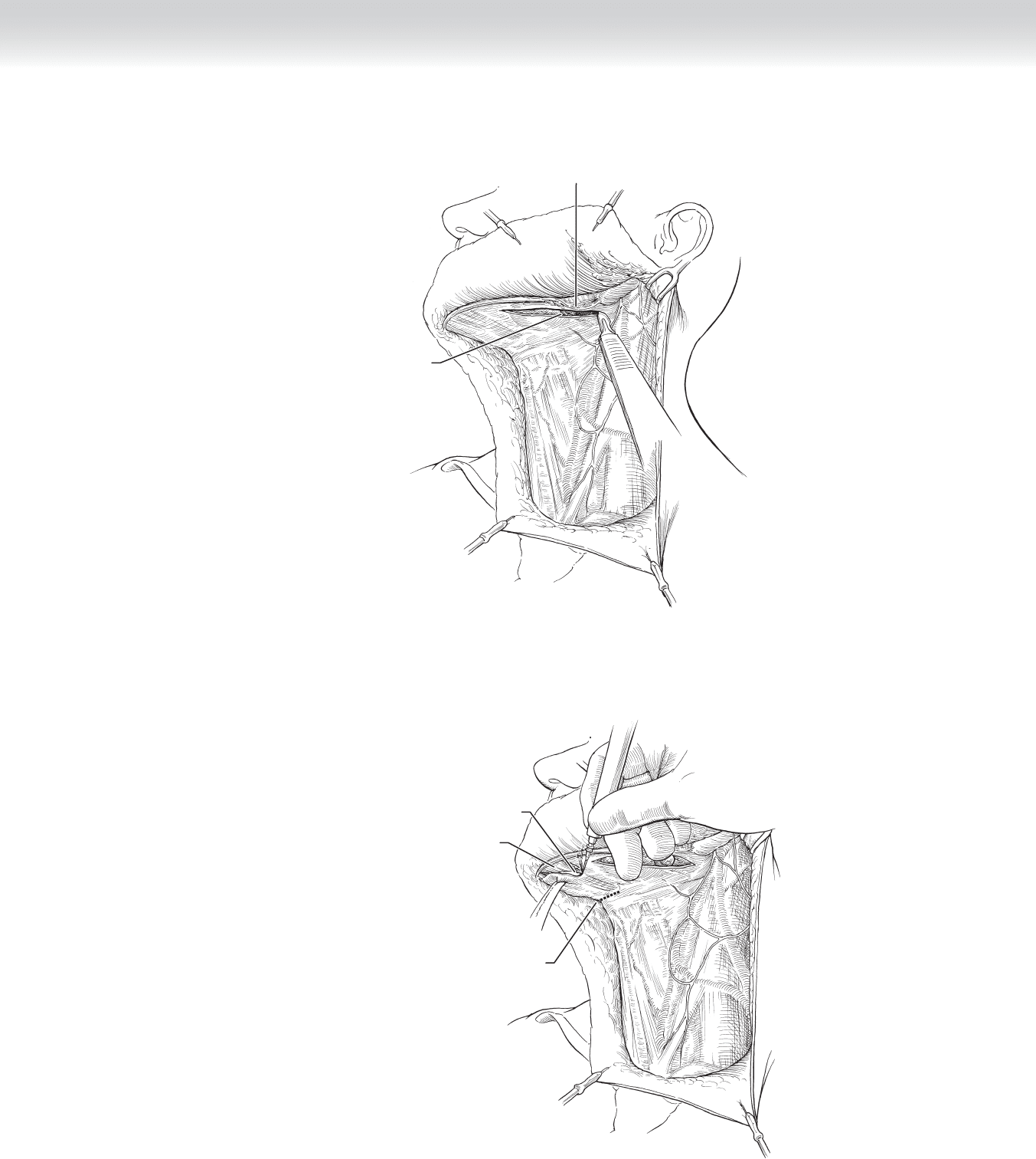
◆ The periosteum overlying the inferior border of the mandibular body is incised with
electrocautery, and the tissue in the submandibular triangle is retracted inferiorly. The facial
vessels are ligated at the lower border of the body of the mandible (Figure 2-5).
◆ The posterior border of the mylohyoid muscle is identifi ed during this dissection
(Figure 2-6).
Nerve to mylohyoid
Facial artery and vein
Marginal mandibular nerve
FIGURE 2 –5
Submandibular gland
and tissue
Mylohyoid muscle
FIGURE 2 –6
CHAPTER 2 • Modifi ed Radical Neck Dissection Preserving Spinal Accessory Nerve 27
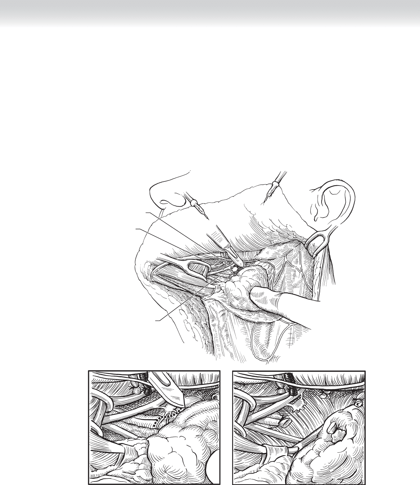
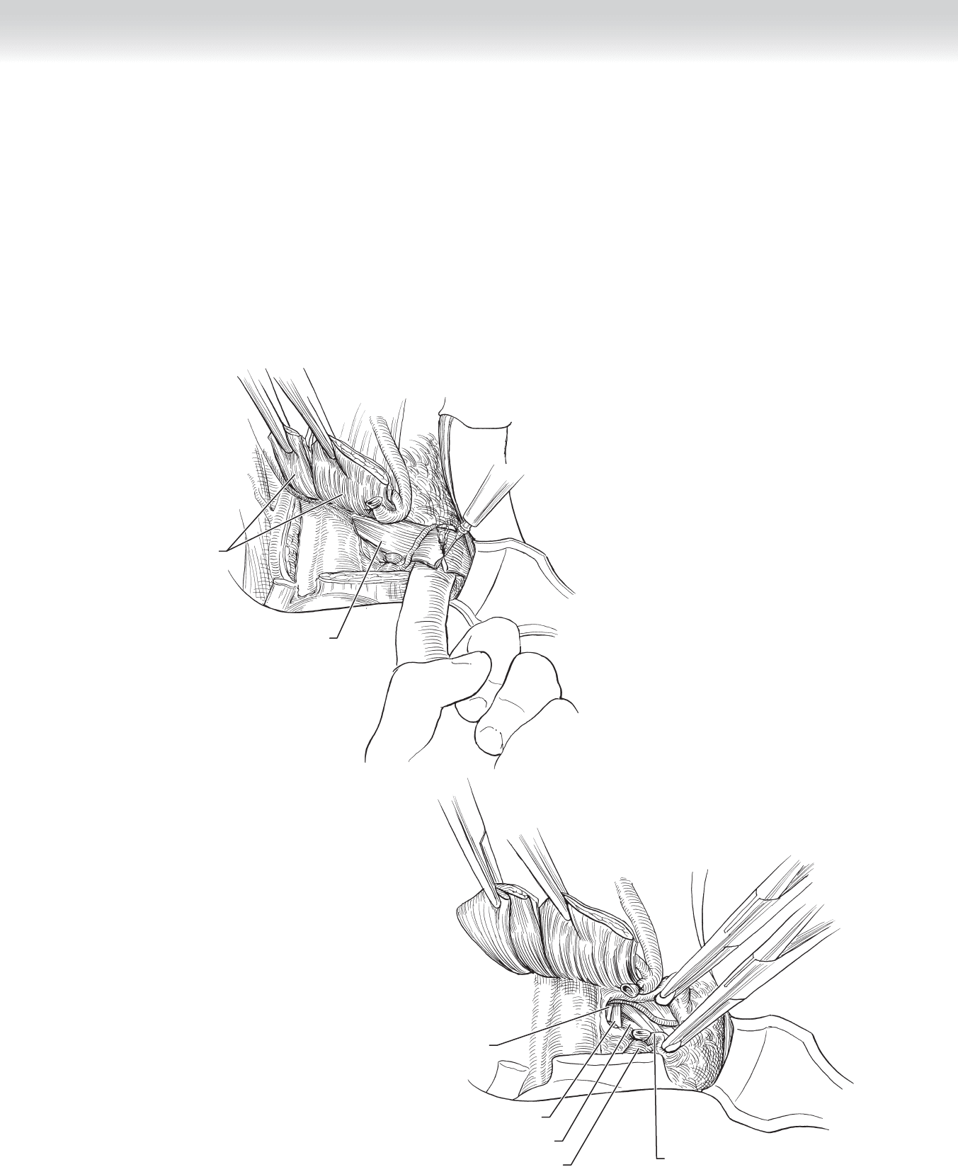
◆ An Army-Navy retractor is placed under the posterior aspect of the mylohyoid muscle, and
it is retracted cephalad. The lingual nerve, submandibular ganglion, and submandibular
duct are identifi ed (Figure 2-7, A).
◆ A clamp is placed below the submandibular ganglion, and the postganglionic fi bers are
transected and ligated. This releases the lingual nerve (Figure 2-7, B-C).
◆ The submandibular duct is located medial to the ganglion; it is transected and ligated
(Figure 2-7, B-C).
A
Submandibular ganglion
Submandibular duct
Lingual nerve
B
C
FIGURE 2 –7

28 Section I • Head and Neck and Endocrine Procedures
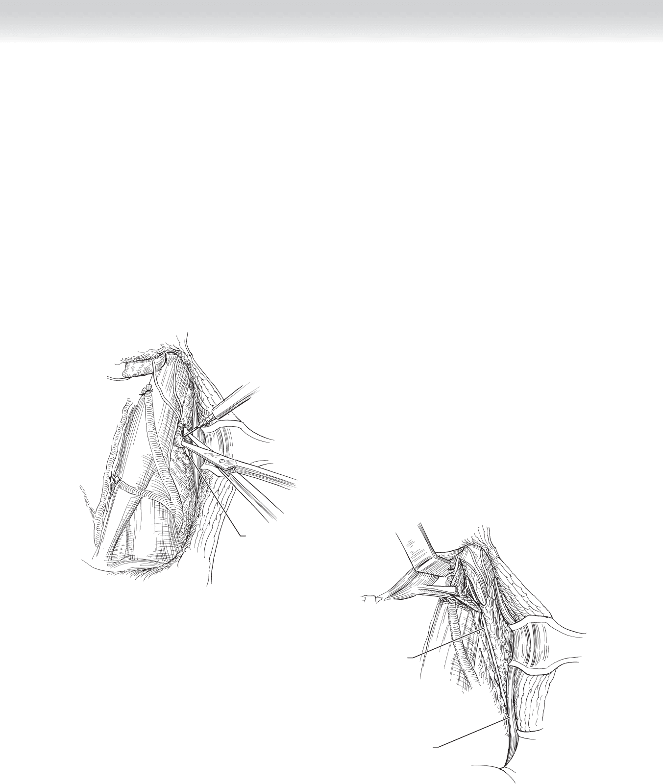
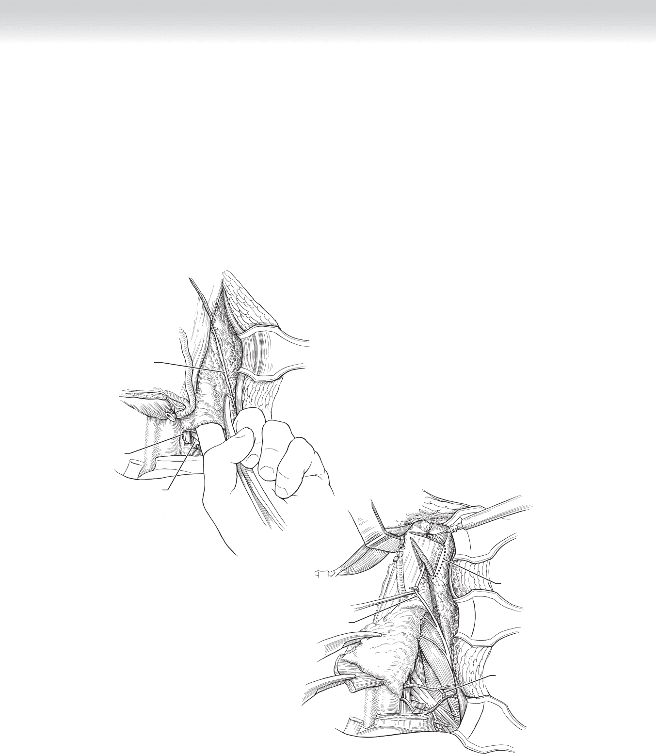
◆ Inferior retraction of the submandibular contents reveals the facial vessels as they cross the
superior aspect of the posterior belly of the digastric muscle. The vessels are clamped, tran-
sected, and ligated. The posterior belly of the digastric muscle is isolated in its entirety. This
muscle belly provides a landmark for levels I and II and the carotid sheath. The contents of
the submental and submandibular triangles, including the prevascular nodes, are pedicled
at the level of the hyoid bone (Figure 2-8).
◆ Attention is now directed to the posterior skin fl ap. Elevation of the fl ap proceeds in a sub-
cutaneous plane until the anterior border of the trapezius muscle is reached (Figure 2-9).
The platysma is defi cient in this area, and care must be taken to not “button hole” the skin
fl ap by dissecting too superfi cially or to injure the SAN by dissecting too deeply; the SAN
lies superfi cial in the posterior triangle. The use of electrocautery may stimulate the SAN
and cause the shoulder to “jump.”
Facial artery and vein
Submandibular gland
Marginal mandibular nerve
External jugular vein
Greater
auricular nerve
Tail of
parotid gland
FIGURE 2 –8
External jugular vein
Greater auricular nerve
Lesser occipital nerve
FIGURE 2 –9
CHAPTER 2 • Modifi ed Radical Neck Dissection Preserving Spinal Accessory Nerve 29
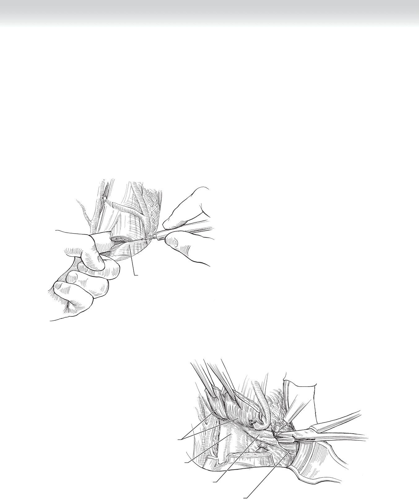
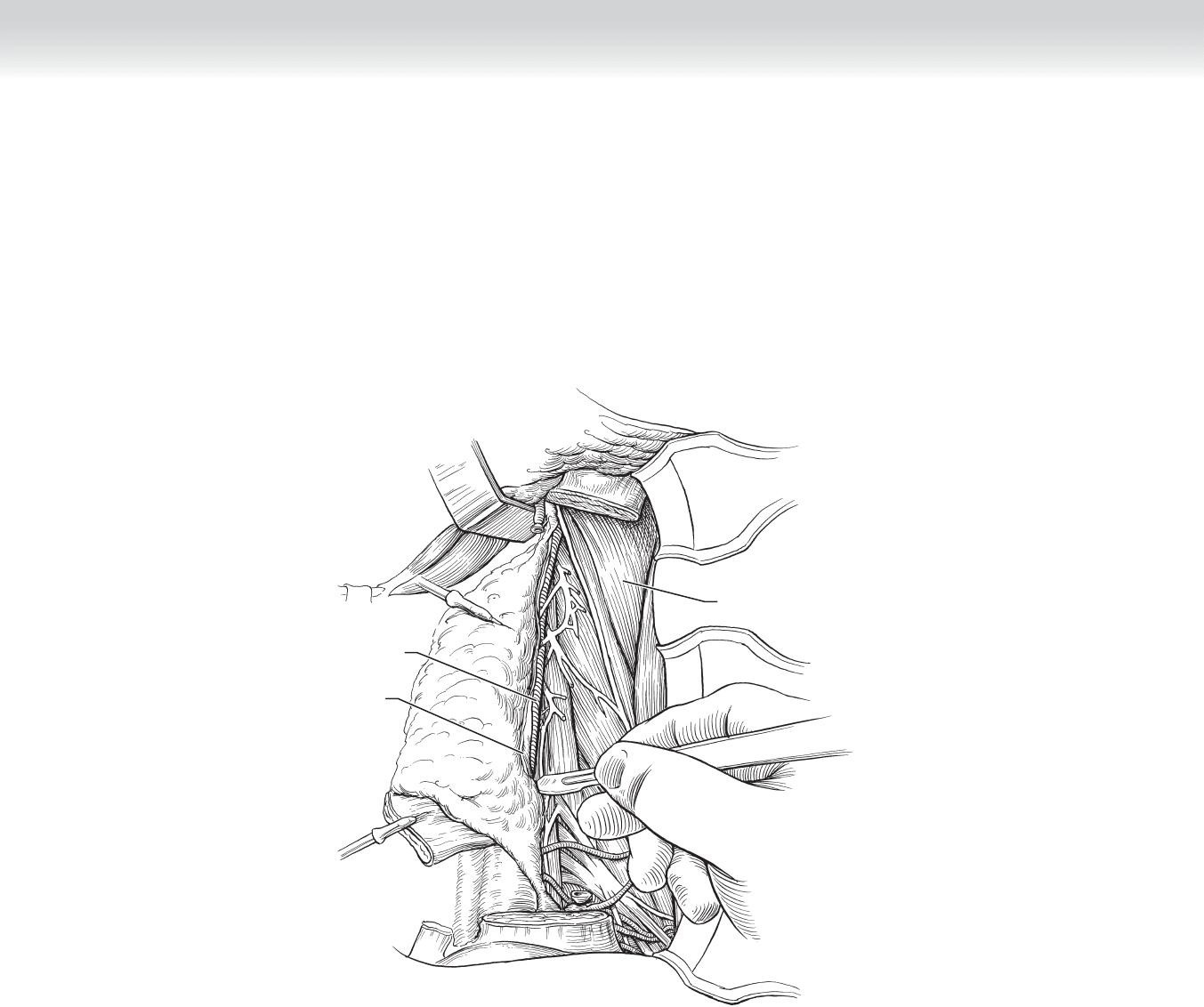
◆ The SAN is identifi ed in the posterior triangle as it enters the trapezius muscle. A spreading
technique using a fi ne hemostat or Metzenbaum scissors is used to dissect the soft tissue
and fascia overlying the nerve. The nerve is traced as it passes from the trapezius muscle to
the SCM muscle (Figure 2-10).
◆ The SAN exits the SCM muscle and dissection continues anteriorly and superiorly to the
skull base, transecting the overlying muscle with the nerve constantly in view. This divides
the SCM muscle in two (Figure 2-11). The posterior belly of the digastric muscle is
retracted superiorly for exposure of the nerve and the IJV at the skull base. The relationship
of the SAN to the IJV is noted during this dissection.
◆ The nerve is sharply dissected from the underlying tissue. The branch to the SCM muscle
must be divided to mobilize the nerve. A nerve hook or vein retractor can be used to
retract the nerve as it is being skeletonized to minimize trauma.
Accessory nerve
FIGURE 2 –10
Accessory nerve
Trapezius muscle
FIGURE 2 –11
30 Section I • Head and Neck and Endocrine Procedures
◆ The IJV at the skull base is isolated circumferentially from the surrounding tissue so that it
can be ligated at a later time.
◆ The sternal and clavicular heads of the SCM muscle are transected one fi ngerbreadth
above the clavicle (Figure 2-12). Upward traction is placed on the muscle with a sponge,
and the layers of the muscle are carefully transected so as not to injure the contents of the
carotid sheath that lie immediately deep to the muscle.
◆ Once the SCM muscle is divided inferiorly, the posterior belly of the omohyoid muscle is
visualized. The tissue overlying the muscle posteriorly is incised (Figure 2-13).
Sternocleidomastoid muscle
(clavicular head)
FIGURE 2 –12
Fascia covering
Omohyoid muscle
Omohyoid muscle
(posterior belly)
Sternocleidomastoid
muscle
Carotid sheath
FIGURE 2 –13
CHAPTER 2 • Modifi ed Radical Neck Dissection Preserving Spinal Accessory Nerve 31
◆ The muscle belly itself is transected near its origin at the scapula (Figure 2-14) and elevated
anteriorly to its attachment at the hyoid bone. The anterior jugular veins will be encountered
at this point and should be ligated. This defi nes the anterior limit of the neck dissection.
◆ The fascia underlying the posterior belly of the omohyoid muscle is incised horizontally. The
supraclavicular fat pad is then opened using blunt dissection exposing the brachial plexus
and phrenic nerve, which lies on the surface of the anterior scalene muscle (Figure 2-15). The
dissection should not continue until the brachial plexus and phrenic nerve are identifi ed,
because injury to these structures can be catastrophic. The transverse cervical vessels will also
be seen in this area. It is not always necessary to divide these vessels.
Omohyoid muscle
Sternocleidomastoid
muscle
FIGURE 2 –14
Brachial plexus
External jugular vein
Cut edge of fascia
Anterior scalene muscle
Phrenic nerve
FIGURE 2 –15
32 Section I • Head and Neck and Endocrine Procedures
◆ The fi bro-fatty tissue between the brachial plexus and the anterior border of the trapezius
muscle (supraclavicular fat pad) is clamped and ligated. The brachial plexus must be
directly visualized while the clamps are being placed. This tissue can be bluntly dissected
using a fi nger. This area is known as the “bloody gulch,” and bleeding will occur if the
tissue is not ligated (Figure 2-16).
◆ Dissection is then carried superiorly along the anterior border of the trapezius muscle until the
SAN is encountered. The SAN is retracted anteriorly to avoid injury during this dissection. The
SCM muscle is transected just inferior to the mastoid tip, and the fascia is incised at its poste-
rior aspect (Figure 2-17). This allows the specimen to be retracted medially.
Incise through
fascia
Transverse
cervical vessels
Accessory nerve
FIGURE 2 –17
Brachial plexus
Accessory nerve
Phrenic nerve
FIGURE 2 –16

CHAPTER 2 • Modifi ed Radical Neck Dissection Preserving Spinal Accessory Nerve 33
◆ The specimen, including the fi bro-fatty and lymphatic tissue in level V, as well as the
superior aspect of the SCM muscle, is dissected in a posterior to anterior direction. The
specimen is passed underneath the SAN, gently retracting the SAN laterally (Figure 2-18).
◆ The deep limit of dissection is the fascia of the deep cervical muscles; the dissection proceeds
along the medial aspect of the levator scapulae and the scalene muscles. The rootlets of the
cervical plexus are exposed. The cutaneous branches are transected and removed with the
specimen. Care must be taken to preserve the nerve supply to the posterior compartment
musculature and the contributions to the phrenic nerve. This is done by transecting the
cervical rootlets approximately 1 cm anterior to the takeoff of the phrenic nerve, that is, “high”
in the specimen. Vessels typically accompany the rootlets and should be controlled using
bipolar cautery or suture ligation. In addition, care must be taken to avoid direct injury to the
phrenic nerve by lifting it off the anterior scalene muscle with the specimen (Figure 2-19).
Accessory nerve
Fibro-fatty tissue
Phrenic nerve
FIGURE 2 –18
Cervical rootlets
Phrenic nerve
FIGURE 2 –19
34 Section I • Head and Neck and Endocrine Procedures
◆ Mobilization of the specimen continues until the IJV is exposed in its full length
(Figure 2-20).
Splenius muscle
Carotid sheath
Internal jugular vein
FIGURE 2 –20