Caballero B. (ed.) Encyclopaedia of Food Science, Food Technology and Nutrition. Ten-Volume Set
Подождите немного. Документ загружается.


disease. The ingestion of B. bifidum together with
lactulose was shown to assist in reestablishing the
balance of the gut flora, which is usually disturbed
in liver cirrhosis, and this was accompanied by a
decrease in fecal pH and by a reduction of ammonia
and free phenols in the blood. (See Liver: Nutritional
Management of Liver and Biliary Disorders.)
0029 The beneficial effects of ingested bifidobacteria
and/or lactobacilli may be obtained if: (1) large
numbers of viable cells (10
8
–10
9
cells day
1
) are
introduced; (2) they survive gastric transit and, pref-
erably, can adhere to epithelial surfaces and grow; (3)
a fermentable carbohydrate is available in the gut;
and (4) they have a strong antagonistic effect against
harmful microorganisms.
Improved Lactose Utilization
0030 It was shown that lactase-deficient persons can digest
lactose in yogurt better than the same amount of
lactose in unfermented milk. Yogurt bacteria,
Streptococcus salivarius subsp. thermophilus and
L. delbrueckii sp. bulgaricus, produce the enzyme,
lactose (beta-galactosidase), which hydrolyzes lactose
to glucose and galactose. It is possible that they
supply preformed lactase to the gut, thus allowing
digestion of lactose. L. acidophilus and L. casei also
allow digestion of lactose by producing lactase. These
organisms survive in the gut better than yogurt bac-
teria, thus suggesting that lactase from the bacterial
cells remains in the small intestine for a longer time.
Possible Anticarcinogenic Activity
0031 The potential anticarcinogenic activity of some lacto-
bacilli and bifidobacteria has been shown in
many studies. The effect of L. acidophilus on Ehrlich
ascites tumor was studied using mice as an animal
model. Feeding acidophilus milk resulted in a smaller
number of tumor cells in mice than in those not receiv-
ing L. acidophilus. Similar results were obtained
with mice, transplanted with Meth-A ascites tumor
cells, that received a suspension of B. infantis,
repeated four to six times. (See Cancer: Diet in Cancer
Prevention.)
0032 Other studies with mice indicated a possible role of
macrophages in suppressing the growth of tumor
cells. Intraperitoneal injection of L. casei in mice
was shown to increase the phagocytic activity of peri-
toneal macrophages and their acid phosphatase activ-
ity. In addition, the consumption by mice of milk
containing L. casei resulted in activation of the
macrophages, measured by increased levels of lactic
dehydrogenase activity.
0033 Some of these effects may be due to substances
produced by the organism during growth, and/or to
the stimulation of the host immune response. It is also
possible that some lactobacilli and bifidobacteria in-
hibit the growth of organisms which may convert
procarcinogens into carcinogens in the gut. The oral
administration of L. acidophilus (10
10
cells day
1
)to
meat-fed rats was shown to reduce the activity of
azoreductase, beta-glucuronidase, and nitroreduc-
tase. These fecal enzymes can catalyze procarcinogen
conversion to a proximal carcinogen. In a study
with human subjects it was found that feeding
500 ml of milk containing 2 10
6
cells of
L. acidophilus ml
1
day
1
for 4 weeks significantly
reduced beta-glucuronidase, azoreductase, and
nitroreductase activities. When feeding with lactoba-
cilli had ceased, fecal enzyme levels returned to
normal after 4 weeks.
0034Another mechanism for the anticarcinogenic effect
refers to nitrosamines (potent carcinogens) which are
synthesized in vivo from amines and nitrite. The syn-
thesis of nitrosamines may be reduced by enzymatic
degradation carried out by some strains of lactobacilli
and bifidobacteria. (See Nitrosamines.)
0035Apparently, various mechanisms may be involved
in the potentially anticarcinogenic effects of lactoba-
cilli and bifidobacteria.
Control of Serum Cholesterol
0036The influence of gut bacteria on sterol metabolism
may be of significance in human nutrition in prevent-
ing the accumulation of cholesterol. The feeding of a
milk formula supplemented with L. acidophilus to
infants was shown to result in lower levels of blood
cholesterol than those recorded in infants receiving
the milk without L. acidophilus. Similar results were
reported on rats supplementary-fed skim milk fer-
mented with L. acidophilus, compared to those fed
unfermented skim milk.
0037Laboratory studies have shown that strains of
L. acidophilus originating from humans actively
assimilate cholesterol, but there are considerable
differences in the activity of strains. These results
are supported by experiments with pigs using pig
isolates of L. acidophilus. It was shown that supple-
mentary-feeding L. acidophilus to pigs resulted in
lower blood cholesterol than in those not receiving
L. acidophilus.
0038Strains of L. casei and B. bifidum were also shown
in laboratory studies to assimilate cholesterol to vary-
ing degrees. In a study with human subjects it was
found that the ingestion of large numbers of
B. bifidum daily during 6 weeks, significantly reduced
the levels of serum cholesterol and serum triglycerides
in subjects suffering from elevated serum lipids. This
treatment was without effect in subjects with normal
values of serum lipids. (See Bile.)
3914 MICROFLORA OF THE INTESTINE/Probiotics

Stimulation of the Immune System
0039 There are indications that the ingestion of lactic acid
bacteria and/or bifidobacteria stimulates the immune
response of the host. A significant increase in the
number of B lymphocytes and T lymphocytes, was
observed, as well as an augmentation of some im-
munoglobulins in mice given yogurt or other lactic
acid bacteria. Also, phagocytic activity of macro-
phages was shown to be enhanced in mice supplemen-
tary-fed with lactic acid-producing bacteria (e.g.,
L. acidophilus, L. casei, B. longum).
0040 Studies with human subjects also showed the
immunogenical effects of lactic acid bacteria. For
example, (1) a significant increase of phagocytic ac-
tivity of leukocytes in peripheral blood, and an in-
crease of immunoglobulin A (IgA) were found in
subjects given fermented milk containing L. acido-
philus strain Lal for 3 weeks. This effect was observed
at least 6 weeks after termination of the consumption
fermented milk; (2) a significant increase in numbers
of B lymphocytes and natural killer cells, the augmen-
tation of IgG, and an increase of the serum levels of
gamma-interferon were observed in subjects given
large numbers of yogurt bacteria for 28 days.
0041 The immunogenic effects of lactic acid bacteria
and/or bifidobacteria may be influenced by: (1) the
bacterial strain characteristics; (2) the numbers of
culture bacteria ingested; and (3) a duration of
ingesting culture bacteria. Further research is needed.
Selection of Microbial Strains
0042 Development of improved microbial supplements in-
volves selection of proper strains of the organism. If
the ingested bacteria are to survive, they must be
resistant to all antimicrobial factors which exist in
the gut. Gastric acidity is an important barrier to
gut colonization, but some protection is afforded by
the buffering effect of the food and food boli will tend
to raise the pH of the stomach. Various strains of
lactobacilli and bifidobacteria were shown to differ
significantly in acid tolerance. Other factors affecting
bacterial survival in the gut include bile acids, lyso-
zyme, and organic acids. The ability of bacteria to
adhere to the gut epithelium is also an important
factor. Adhesion is host-specific, e.g., only lactobacilli
and bifidobacteria originating from the human gut
have the potential to attach to human epithelial
cells. The adhesion ability varies between bacterial
species, and even different strains of the same species
show variations. It was also shown that growth in
milk enhances the ability of lactobacilli to adhere to
epithelial cells.
0043 Various strains of lactobacilli and bifidobacteria
were shown to differ significantly in the antagonistic
effect against harmful bacteria. Other desirable prop-
erties include immunogenic effects, assimilation of
cholesterol, and hydrolysis of lactose.
0044The selection of probiotic strains may include the
following criteria: (1) not pathogenic; (2) of human
intestinal origin; (3) survival of intestinal passage;
(4) adhesion to epithelial cells; (5) clinically tested
for the specific therapeutic effect; and (6) applicable
for manufacturing food products.
Evidence of Influence of Ingested
Bacteria
0045The maximum effect of ingested probiotic bacteria is
obtained when the protective indigenous flora have
been changed by diet, antibiotic therapy, disease, or
some other factors, as the following examples illustrate.
1.
0046A disturbed balance of the gut flora in 28 patients
with leukemia was shown to be improved by
ingesting 2 10
9
bifidobacteria and 2 10
9
L. acidophilus in 200 ml of milk daily for 3
months. The counts of Klebsiella spp., Proteus
spp., Candida spp., and Pseudomonas spp. in the
feces decreased, and the levels of urine indican and
blood endotoxin reduced, compared to patients
not receiving supplements.
2.
0047Fifteen patients (mean 2.5 years) received antibi-
otics, such as cephema, penicillins, and aminogly-
cosides, for curing septicemia and respiratory tract
infections. During treatment, diarrhea occurred
and lasted for 1–10 weeks. The counts of bifido-
bacteria decreased in the feces, and those of Can-
dida and Enterococcus increased. The ingestion of
bifidobacteria and L. acidophilus improved the
stool frequency within 3–7 days, along with the
restoration of the gut flora.
0048The beneficial effects of probiotics may be en-
hanced by including special ingredients (prebiotics),
and thus improving host health.
0049Probiotic bacteria can affect the composition of the
gut flora and/or its metabolic activity during the
period of ingestion and for some time after. For
example:
1.
0050Milk fermented with L. acidophilus fed to human
volunteers reduced the E. coli count and increased
the lactobacillus count, but when the supplement
ceased, the bacterial counts returned to normal
after 9 days.
2.
0051Five healthy volunteers ingested 3 10
9
B. longum
daily for 5 weeks; during feeding, counts of bifido-
bacteria increased and those of clostridia decreased;
in addition, ammonia levels and beta-glucuronidase
activity decreased in the feces and serum.
MICROFLORA OF THE INTESTINE/Probiotics 3915

3.0052 As already mentioned, feeding L. acidophilus to
humans reduced beta-glucoronidase, azoreduc-
tase, and nitroreductase activities, but when the
supplement ceased, enzyme levels returned to
normal after 4 weeks.
0053 It is difficult to achieve a permanent colonization of
ingested probiotic bacteria. However, their ingestion
continuously or at regular intervals may insure the
desired benefits.
See also: Bile; Cancer: Diet in Cancer Prevention;
Carbohydrates: Classification and Properties;
Metabolism of Sugars; Cholesterol: Factors Determining
Blood Cholesterol Levels; Dietary Fiber: Properties and
Sources; Physiological Effects; Immunology of Food;
Infants: Breast- and Bottle-feeding; Liver: Nutritional
Management of Liver and Biliary Disorders;
Nitrosamines; Protein: Synthesis and Turnover
Further Reading
Alm L (1991) The therapeutic effects of various cultures –
an overview. In: Robinson RK (ed.) Therapeutic Proper-
ties of Fermented Milks, pp. 45–64. Essex: Elsevier Ap-
plied Science.
Fuller R (1992) Probiotics. The Scientific Basis. London:
Chapman & Hall.
Gilliland SE (1989) Acidophilus milk products: a review of
potential benefits to consumers. Journal of Dairy Science
72: 2483–2494.
Havenaar R and Huis in’t Veld JHJ (1992) Probiotcs: a
general view. In: Wood BJB (ed.) The Lactic Acid Bac-
teria: Vol. 1. The Lactic Acid Bacteria in Health and
Disease, pp. 151–170. Essex: Elsevier Applied Science.
Mitsuoka T (1989) Bifidobacterium microecology. In: Les
Laits Fermente
´
s, pp. 41–48. Paris: John Libbey.
Mollet B, Donnet A, Neeser JR et al. (1997) Effects of
probiotics on the immune system. In: Probiotics – Facts
and Opinions, pp. 35–40. Bern: Verlag Schweizerische
Milchkommission.
Playne MJ and Crittenden R (1996) Commercially available
oligosaccharides. In: Bulletin IDF 313, pp. 10–22. Brus-
sels: International Dairy Federation.
Ras
ˇ
ic
´
JLJ and Kurmann JA (1983) Bifidobacteria and their
Role, p. 87 Basel: Birkha
¨
user Verlag.
Savage DC (1977) Interaction between the host and its
microbes. In: Clarke BTJ, Bauchop T (eds) Microbial
Ecology of the Gut, pp. 277–310. London: Academic
Press.
Sellars RL (1991) Acidophilus products. In: Robinson RK
(ed) Therapeutic Properties of Fermented Milks, pp. 81–
116. Essex: Elsevier Applied Science.
Simone CD, Bianchi Salvatori B, Jirilo E, Baldinelli L, Fabio
SD and Vesely R (1989) Yogurt and the immune re-
sponse. In: Les Laits Fermente
´
s, pp. 63–67. Paris: John
Libbey.
Microorganisms See Aeromonas; Antibiotic-resistant Bacteria; Bacillus: Occurrence; Detection; Food
Poisoning; Bifidobacteria in Foods; Campylobacter: Properties and Occurrence; Detection; Campylobacteriosis;
Clostridium: Occurrence of Clostridium perfringens; Detection of Clostridium perfringens; Food Poisoning by
Clostridium perfringens; Occurrence of Clostridium botulinum; Botulism; Escherichia coli: Occurrence; Detection;
Food Poisoning; Occurrence and Epidemiology of Species other than Escherichia col i; Food Poisoning by Species
other than Escherichia coli; Lactic Acid Bacteria; Microbiology: Classification of Microorganisms; Detection of
Foodborne Pathogens and their Toxins; Mycobacteria; Salmonella: Properties and Occurrence; Detection;
Salmonellosis; Shigella; Staphylococcus: Properties and Occurrence; Detection; Food Poisoning; Vibrios: Vibrio
cholerae; V i brio parahaemolyticus; V i brio vu l nificus; Viruses; Yeasts; Yersinia enterocolitica: Properties and
Occurrence; Detection and Treatment; Zoonoses
3916 MICROFLORA OF THE INTESTINE/Probiotics

MICROSCOPY
Contents
Light Microscopy and Histochemical Methods
Scanning Electron Microscopy
Transmission Electron Microscopy
Image Analysis
Light Microscopy and
Histochemical Methods
J H Holgate and J Webb, Reading Scientific Services
Ltd, Whiteknights, Reading, UK
Copyright 2003, Elsevier Science Ltd. All Rights Reserved.
Background
0001 Light microscopy has been in use in the food industry
since the first instruments were introduced. In the
early years, its primary application was for detecting
adulteration of foodstuffs. Identification was depend-
ent primarily upon the recognition of structural
features combined with the reaction to selected
microchemical tests. (See Adulteration of Foods: De-
tection.) Today, a wide range of routine and research
microscopes and microscopy techniques are avail-
able, providing the food scientist with microstruc-
tural information on the nature and form of
individual food components and on their distribution
within a product. This allows the role and inter-
actions of food ingredients to be examined and pro-
vides information valuable to the understanding of
the properties of finished products, cooking and
manufacturing processes, and the influence of both
the type and form of ingredients. Electron microscopy
techniques complement this approach, allowing the
microstructure of the same types of food materials to
be examined at greater magnifications and with dif-
ferent types of preparation procedures. (See Analysis
of Food.) The light microscope is probably one of the
most versatile laboratory instruments available to the
food scientist. A basic instrument, available today for
the same price as a good-quality balance, provides a
wide range of magnifications and other facilities to
provide structural information on almost any form of
food sample.
Principles
0002 The light microscope is an instrument for visualizing
fine detail of an object. It does this by creating a magni-
fied image through the use of a series of glass lenses,
which first focus a beam of light onto or through
an object, and convex objective lenses to enlarge the
image formed. In the majority of light microscopes,
the image is viewed directly through binocular eye-
pieces that act as a secondary lens in the form of a
magnifying glass to observe the projected image. Such
instruments are termed ‘compound microscopes,’ and
the total magnification is the sum of the objective
magnification and the eyepiece magnification. The
magnification range extends from 10 to 1000,
with a resolving power of the order of 0.2 mm,
depending on the type and numerical aperture (area
available for passage of light) of the objective lenses.
A number of books are available, providing compre-
hensive details on the theory of the light microscope
and guidance to the practical use of the instrument,
including methods of image enhancement and instru-
ment care. The reader is referred to these, in par-
ticular an extensive series of handbooks published
by the Royal Microscopical Society, for further in-
formation.
Microscopy Techniques
0003Microscopy techniques for use in the food industry are
frequently a combination of those used in biological
and materials sciences. Samples are viewed using either
transmitted or incident (reflected) illumination. Most
frequently, transmitted light is used where a beam of
light is allowed to pass through a relatively thin object.
The microstructural detail becomes visible due to the
absorption of a portion of the light. Incident light is
used mainly for the examination of solid, opaque
objects, although it now forms the basis for all modern
fluorescent microscopes, referred to as ‘epifluores-
cence.’ (See Spectroscopy: Fluorescence.)
0004The light source is often a single or multicoil tung-
sten lamp or quartz halogen lamp. However, xenon
arc or mercury vapor lamps provide more intense
beams, as required for fluorescence microscopy.
Contrasting Techniques
0005Though many structures are visible by standard
(bright field) transmitted light, there are components
MICROSCOPY/Light Microscopy and Histochemical Methods 3917

that are nonabsorbent and appear transparent or lack
any contrast. This can be overcome, or additional
information may be gained by using one of several
contrasting techniques available that will introduce
or enhance image contrast, frequently with minimal
disruption of the specimen. The most frequently used
are phase contrast, differential interference contrast,
and polarized light.
0006 When using phase contrast or differential interfer-
ence contrast (Nomarski) optics, for example, the
phase of part of the light is altered, and this is then
recombined with light that has passed through the
specimen and thus yields improved differentiation
within a specimen. Phase contrast images are
characterized by enhanced contrast and visibility of
unstained materials and are used routinely for exam-
ination of microorganisms isolated from food. Inter-
ference contrast provides a distinct relief appearance,
with a shallow depth of focus often giving the appear-
ance of a three-dimensional image. It has proved
particularly valuable for examining small particles
and emulsion droplets (Figure 1).
0007 A different type of contrast is generated in certain
samples when they are viewed under crossed polars.
Plane polarized light (light vibrating in only a single
plane) is generated using a polarized filter and allowed
to impinge upon the specimen. The light then travels
through a second polarization filter (the analyzer),
which is orientated at 90
to the first, effectively
blocking the uninterrupted plane polarized light. Any
material that is anisotropic or birefringent, e.g., crys-
tals, is capable of rotating the light plane so that the
polarization of emerging light will be altered and par-
tially extinguished and will have an orientation that
will pass through the analyzer filter. The resultant
image is of bright features against a black background.
0008 The use of polarized light has many applications in
the study of food. Food starches and many crystalline
food ingredients, e.g., sugar, have characteristic sizes,
shapes, etc., that can be observed using polarized
light. Intact starch granules are strongly birefringent
but lose this property during gelatinization (e.g.,
cooking), and polarized light microscopy is fre-
quently used to follow this process during, for
example, the baking of bread doughs. (See Starch:
Structure, Properties, and Determination.)
Fluorescence
0009Irradiation of samples with light of specific short
wavelengths can result in reemission of this energy
as light of longer wavelengths. This is referred to as
‘fluorescence.’ The emitted light, different in color
from the excitation light, may be separated by the
use of specific filters. Some components of food, for
example collagen and lignin, fluoresce naturally after
excitation with light (autofluorescence). However,
such fluorescence is often very weak, and it is more
common for selective, strongly fluorescent dyes
(fluorochromes) to be used to produce a high contrast
within a sample (Figure 2). (See Lignin.)
0010Almost all modern fluorescence microscopes use
epifluorescence, in which the specimen is illuminated
by a high-density mercury vapor source (via a beam
splitter through the objective) to excite only the
surface layers of the sample.
0011The technique has particular value in food micro-
scopy as it can be used to detect substances at low
concentrations and for rapid sample screening at
low magnification where the particles of interest are
in localized regions within the sample. Dyes in
common use in food microscopy are Acridine orange
for staining bacteria and milk proteins and Nile blue
for fats. However, the availability of fluorescent dyes
10 µm
fig0002Figure 2 Bacteria isolated from milk. Preparation is stained
with Acridine orange and viewed using epifluorescence.
10µm
fig0001 Figure 1 Emulsified droplets of fat in a salad cream. Prepar-
ation viewed using Nomarski interference contrast.
3918 MICROSCOPY/Light Microscopy and Histochemical Methods

is steadily increasing, especially in combination with
highly specific labeling agents such as antibodies
(immunolabeling) and lectins, and is likely to be one
of the main growth areas within food light micro-
scopy over the coming years.
Sample Preparation
0012 The majority of food materials and their ingredients
require some kind of preparation before the micro-
structure can be examined by light microscopy. The
choice of sample preparation will depend on the ma-
terial being examined and the kind of information
required. It may range from simple whole mounts
and smears, to more complex, sectioned prepar-
ations.
0013 Often, food samples or their ingredients, e.g., milk
powders, can be examined simply by direct viewing
on a glass microscope slide; alternatively, further
valuable information can be gained after supporting
the sample in a transparent mounting medium. Such
media are frequently inert as well as transparent and
include water, liquid paraffin, glycerol, chloral hy-
drate (used to ‘clear’ plant cells), and many commer-
cial mountants. The choice will depend on the nature
of the material to ensure that little or no change, for
example dissolution or swelling, occurs prior to
examination.
0014 Liquid or semisolid foods, for example viscous
pastes, fats and emulsions, are often viewed as
‘smears’ across a slide. However, it is important to
be aware of the potential disruption of the micro-
structure during this type of preparation and the dif-
ferences in detail provided by samples of different
thickness. Contrast is often enhanced by phase or
interference contrast. The more traditional prepar-
ation for light microscopy, and one frequently used
for food samples, is the preparation of thin slices or
sections through the material. Such sections are
mounted on to slides, their contrast often enhanced
by multiple staining before examination by bright-
field illumination.
Sectioning
0015 Sectioning techniques for light microscopy range
from the relatively simple freehand sectioning with
a knife or razor blade to the precision cutting of
embedded material using sophisticated bench micro-
tomes or ultramicrotomes. A few foods can be
sectioned with little or no prepreparation, and it is
still commonplace in a food laboratory to see initial
freehand sectioning. A bench microtome, however,
enables ribbons of sections of more uniform size and
thickness (5–50 mm) to be cut. Stainless-steel knives
are used most frequently, although more resilient ma-
terials, such as tungsten carbide, are often used to cut
hard materials and provide longer-lasting blades. Al-
ternatively, freshly cleaved glass knives, used rou-
tinely for ultrathin sectioning for transmission
electron microscopy, are used to cut thinner sections
(< 5 mm) for light microscopy. Food plant material,
particularly dried tissue and some solid composite
foods, can be sectioned successfully with no prior
preparation or treatment. However, the majority of
foods and their ingredients require some preparation
prior to sectioning to ensure that they are rigid
enough to enable sections to be cut with minimum
distortion. Wax or resin embedding and low-
temperature ‘cryofixation’ are the most frequently
used preparative techniques and have been used in
food microscopy for many years.
Fixation/embedding
0016Whatever the embedding media used, the food mater-
ial needs to be chemically preserved or ‘fixed’ prior to
infiltration and embedding with the wax or polymer.
Successful fixation depends on the type of food ma-
terial being prepared, in particular the ease of pene-
tration of the fixative into the material. The use of
traditional aldehyde fixatives (e.g., formaldehyde or
glutaraldehyde) is common, as is the use of osmium
tetroxide, in liquid or vapor form, particularly with
fats present. However, many different fixation pro-
cedures have now been adapted and developed for
specific foods in order to achieve optimum preserva-
tion of individual components. These include a
number developed more specifically for electron
microscopy. After fixation, the specimen is usually
dehydrated with solvent prior to being infiltrated
and then embedded in a wax or polymer. Solidifica-
tion or polymerization of the embedding media pro-
duces a hard, rigid preparation from which sections
can be cut. Wax embedding remains one of the
cheapest preparation procedures for light micro-
scopy; the procedure can be readily automated for
routine use, and considerable improvements have
been made to the waxes available. However, it is
considered an awkward, somewhat time-consuming
and messy procedure and is increasingly being re-
placed with resins and other polymeric materials for
preparing food samples. A wide range of different
types of resins and polymers are now available, pro-
viding high flexibility in viscosity and conditions for
polymerization, e.g., temperature and time and in-
cluding some acrylic polymers that provide low-
temperature hardening (often using ultraviolet light
polymerization). The use of the newer resins for
embedding samples generally offers a significantly
MICROSCOPY/Light Microscopy and Histochemical Methods 3919

improved safety in the form of the reduced hazards of
the solvents used compared with wax embedding and
in the handling of the resins compared with the previ-
ously used epoxy resins, etc.
Cryostat Sectioning
0017 Cryofixation of samples, followed by ‘cryostat’ low-
temperature sectioning, has the significant advantage
of speed. Samples are supported (using a gel or
commercial preparation such as Tissue-Tek) on
metal blocks (often after an initial chemical fixation),
and then fast-frozen, generally in liquid nitrogen
(196
C) to preserve or ‘cryofix’ their structure.
The frozen block is sectioned at approximately 20
to 30
C using a conventional bench microtome
housed in a refrigerated unit (cryostat). The technique
obviously has particular application for frozen foods
such as icecream, but is also used frequently for foods
that have high water contents or that are difficult to
handle in any other way. However, some food prod-
ucts are unsuitable for sectioning when frozen, for
example, if they remain soft or become brittle. Also,
the initial capital expenditure can be high and, al-
though the sections can be preserved and permanently
mounted, the frozen block can only be kept if stored
under liquid nitrogen. Sections are collected on to
glass slides, sometimes aided by a thin coating/layer
of adhesive such as egg albumen, and allowed to
warm to room temperature. They are often further
chemically fixed at this stage prior to staining.
Staining Methods
0018 Other than using the optical contrasting techniques
described above, the use of dyes to stain components
specifically is the most common way of increasing
contrast for light microscopy. Staining techniques
rely on the interaction or absorption of the dyes with
the sample components. Many different stains are
now available, and procedures have been developed
for food and food ingredients in order to color spe-
cific individual components and thus produce en-
hanced contrast and enable identification of protein,
fat, starch, and sugar, for example, (See Protein:,
Determination and Characterization; Starch: Struc-
ture, Properties, and Determination.)
0019 Frequently, two or more stains can be used in a
single preparation. Within food microscopy, many
different stains are now readily available for the
differentiation of different types of protein, carbohy-
drate, and fats. Some of these are available in vapor
form, e.g., iodine for starch and osmium tetroxide for
fats, which has a particular advantage in causing little
disruption to the specimen. The selection of stains and
the procedures used will vary, depending not only on
the type of food material under examination but also
on the sample preparation technique employed and
the exact information required. However, stains are
still often classed according to the nature of their
binding to the specimen. Acidic or basic dyes are
used for attaching to positively or negatively charged
sites, respectively, and thus can be used, for example,
to distinguish between different types of proteins.
Solubility dyes rely on the dye being more soluble in
one component than another. The Sudan family of
stains use this property to stain fats in food.
0020Stains are described as ‘histochemical’ when a spe-
cific chemical reaction is used to attach the chromo-
phore to a particular chemical grouping in the
specimen. The most frequently used is the periodic
acid Schiff (PAS) reaction used to demonstrate the
presence of different polysaccharides by the forma-
tion of aldehyde groups following oxidation.
Applications of Light Microscopy
0021The versatility of the preparation procedures and tech-
niques for light microscopy makes them applicable to
a wide variety of different foods and ingredients.
Selected samples are presented below, although many
more are presented in other parts of the Encyclopedia
as well as numerous food microscopy publications.
Powders
0022A wide range of food materials, in particular raw
ingredients, exist in powdered form. Common
examples include ground spices, spray-dried flavors
and milk powders, and freeze-dried beverages and
proteins. Light microscopy provides a way of examin-
ing the size, shape, and often the internal structure of
individual powder particles. Liquid paraffin is fre-
quently used as a simple mountant, although glycerol
is more successful when fat is present. Examination
under polarized light shows the presence of crystalline
or other birefringent structures, enabling their size and
location to be determined. The presence of lactose
crystals, for example, is often demonstrated by their
characteristic tomahawk shape when viewed under
polarized light (Figure 3). In more complex powder
mixtures, individual components can also be discrim-
inated by selective staining. The use of an iodine/
potassium iodide solution or simply iodine vapor
will stain starch a distinctive blue/black color, whereas
fat appears pale brown, and proteins appear yellow.
Animal-based Products
0023Many meat and comminuted meat products are
readily prepared using cryostat sectioning. A variety
of staining procedures are available for identifying
3920 MICROSCOPY/Light Microscopy and Histochemical Methods
individual components including fats, muscle, con-
nective tissue, and bone. These range from single-
step stain mountants, for example, Toluidine blue,
to more complex sequential staining with three or
four different solutions (e.g., the Picro–Mallory tech-
nique). Polarized light can further complement the
use of stains in order to locate crystalline fatty regions
and connective tissues and to differentiate fresh and
processed muscle tissue.
Fruit and Vegetables
0024 Many cellular structures are visible using phase con-
trast, but more detailed information on changes to
cell wall and cell contents can be obtained following
staining for cellulose, pectins, etc. The understanding
of the effects of processing of fruit and vegetables by
brining, freezing, cooking, etc. has been greatly facili-
tated by microscopical studies on samples taken at
different stages of such processes (Figure 4).
Bakery Products
0025A combination of preparation and observation tech-
niques has been important in examining the complex
microstructure of many bakery products, e.g., bread,
cakes, and biscuits. Such techniques have enabled the
changes in ingredients, especially starch and protein,
to be followed and correlations to be made between
the microstructure and the physical and sensory (e.g.,
texture) properties of the finished products. One of
the key changes in baking is the gelatinization of
starch, which can be followed successfully through
examination of preparations using polarized light.
Photomicrography
0026The recording of images obtained by light microscopy
is an important area in its own right and thus,
unfortunately, beyond the scope of this article. The
advent of high-resolution digital cameras has facili-
tated image acquisition, enabling rapid recording of
images, and, where the investment has been made,
even short ‘videos’ can be made, e.g., for studying the
dissolution of particles. As always, the food micro-
scopist still needs to experiment to ensure that high-
quality images observed through binocular eyepieces
are permanently recorded for future reference.
Developing Areas within Light Microscopy
0027The two areas of most recent development in the field
of light microscopy have been the use of specific
labeling techniques, in particular immunolabeling,
and the availability of confocal instruments. In the
former, labeled antibodies are used as specific re-
agents for the detection/staining of specific specimen
components. Labeling is most frequently achieved by
fluorescent dyes. Although antibodies have been
raised for many biological and biomedical applica-
tions, they are currently limited for foods mainly
because the antigenic sites are modified as a result of
food processing and manufacture.
0028In a confocal microscope, a confocal image is built
up point by point by scanning a sample with a focused
beam of laser light. It is a noninvasive technique
enabling optical sectioning of a sample and producing
images with well-defined focal planes but without the
problems of out-of-focus blur. Combined with cur-
rent developments in image-processing techniques,
confocal microscopy is being applied widely in non-
food areas and is beginning to be applied to food, and
has considerable potential for the future.
0029Light microscopy offers versatility in the number of
optical techniques available for examining food
materials, combined with ease of preparation and
100 µm
fig0003 Figure 3 Characteristic tomahawk crystals of lactose viewed
under crossed polars.
100 µm
fig0004 Figure 4 Section through a fresh onion shoot; Toluidine blue
staining.
MICROSCOPY/Light Microscopy and Histochemical Methods 3921

operation at a relatively low cost. Although a decline
in its routine use was evident after the introduction of
electron microscopes, it has reemerged as an invalu-
able instrument for investigations into the science of
food and food processing.
See also: Adulteration of Foods: Detection; Analysis of
Food; Lignin; Protein: Determination and
Characterization; Spectroscopy: Fluorescence; Starch:
Structure, Properties, and Determination
Further Reading
Bradbury S (1982) An introduction to the optical micro-
scope. Royal Microscopical Society Microscopy Hand-
book No. 01. Oxford: Oxford University Press.
Cook HC (1974) Manual of Histochemical Demonstration
Techniques. Oxford: Butterworths.
Flint O (1994) Food microscopy. Royal Microscopical
Society Microscopy Handbook No. 30. Oxford: BIOS
Scientific Publishers.
Hartley WG (1979) Hartley’s Microscopy. Oxford: Senecio.
Heathcock JF (1988) The use of light microscopy and elec-
tron microscopy in the food industry. Microscopy and
Analysis 2: 81–90.
Heathcock JF and Holgate JH (1990) The Continued Im-
portance of Light Microscopy as a Complementary
Technique to EM in the Elucidation of Food Micro-
structure. Proceedings of RMS Conference Micro 90,
20, pp. 713–718.
Ploem JS and Tanke HJ (1986) Introduction to fluorescence
microscopy. Royal Microscopical Society Microscopy
Handbook No. 10. Oxford: Oxford University Press.
Vaughan JG (ed.) (1979) Food Microscopy. London:
Academic Press.
Winton AL and Winton KB (1935) The Structure and Com-
position of Foods, vols. 1 and 2. New York: John Wiley.
Scanning Electron Microscopy
J Webb and J H Holgate, Reading Scientific Services
Ltd, Whiteknights, Reading, UK
Copyright 2003, Elsevier Science Ltd. All Rights Reserved.
Introduction
0001 A scanning electron microscope uses a finely focused
beam of electrons to reveal the detailed surface char-
acteristics of a specimen and provide information
relating to its three-dimensional structure. It also
has a particular advantage of providing great depth
of field. With the introduction of the first commercial
instruments in the mid-1960s the scanning electron
microscope provided the link between the magnifica-
tion ranges offered by light microscopy and the higher
resolving capability of the transmission electron
microscope (TEM). Nowadays, following develop-
ment of electron guns, electromagnetic lens systems
and vacuum systems, scanning electron microscopes
can have a resolution of 1 nm. Therefore, there is now
considerable overlap between the resolving power
of the two forms of electron microscope available,
although the techniques are complementary and
different types of microstructural information are
obtained in each case.
0002Food and its ingredients have been examined by
scanning electron microscopy (SEM) ever since the
first instrument was available. In the early days the
main applications were in studies on the micro-
structural characterization of dry foods, in particular
powders. However, with the development of a range
of different preparation techniques able to handle
many different types of foods, almost every food
material has now been examined in some way by
SEM. (See Analysis of Food.)
Principles
0003When a fine beam of electrons is focused on to the
surface of a specimen, different interactions occur,
including the emission of secondary and back-
scattered primary electrons. If these are collected
and amplified they can be used to create an image
corresponding to the surface topography of the
specimen. The electron beam is scanned across the
specimen repeatedly in a raster pattern, which is syn-
chronized with the scan of a cathode ray tube such
that the image is presented in a digitized form built up
on a TV monitor. Magnification is achieved through
the electron beam scanning an increasing smaller area
of the same specimen and most modern scanning
electron microscopes have a magnification range
of 7 20, up to more than 300 000, and resolution
in the order of 2–4 nm or better. The great depth of
focus of the microscope (> 500 that of light micro-
scopy) is achieved by the convergence angle of the
primary electron beam and the relatively long
working distance between the final lens and the
specimen. This depth of focus enables production of
images that appear to be three-dimensional.
0004As with transmission electron microscopes, con-
ventional instruments must be operated under high
vacuum in order that the electron beam can travel to
the specimen surface without collision with gas
molecules. However, the introduction of specialized
vacuum systems with pressure-limiting apertures
brings a new perspective to SEM and enables samples
3922 MICROSCOPY/Scanning Electron Microscopy

to be examined at higher pressures. In conventional
SEMs, the source of electrons is generally a heated
tungsten filament, which provides ease of operation,
low cost, and simplicity. More intense sources include
lanthanum hexaboride (LaB6) tips and field emission
guns and these are used where greater brightness is
required. Accelerating voltages used typically range
from less than 1 kV up to 40 kV. The higher voltages
can provide greater resolving power, but at the risk
of excessive penetration into many specimens and
damage by the electron beam. Lower voltages are
used to image fine structure on sample surfaces.
0005 The generation of an image from secondary elec-
trons remains the most frequently used form of
imaging of biological samples within a conventional
SEM (CSEM). However, the interaction of a primary
electron beam with a specimen surface can also pro-
vide other information, for example, from back-
scattered or Auger electrons and X-rays.
Specimen Preparation
0006 In its simplest form, preparation for SEM involves
securing a specimen on to a metal support ‘stub’
and, if the sample material is nonconducting, coating
the surface with a conducting thin layer of metal. The
requirement for conductivity is to prevent the build-
up of electrical charge on the specimen surface once it
is bombarded with electrons within the instrument
and also to enhance the secondary image. Many dif-
ferent procedures have been developed for securing
dry food specimens on SEM stubs. These vary
depending on the size, shape, and composition of
the specimen and the type of information required.
One of the most frequently used support media
employs a colloidal silver cement (silver DAG),
which not only holds the specimen firmly but im-
proves conductivity. However, others include carbon
cement (carbon mixed with an acrylic carrier), glues,
gels, and adhesive tape.
Metal Coating
0007 In the early days of SEM, surface metal coats were
prepared by resistive evaporation in a high-vacuum
coating unit. However, this procedure often caused
localized heating of the specimen surface and, as the
metal was applied unidirectionally, it did not always
cover all the crevices in the specimen. It was soon
superseded by the introduction of diode sputter
coating, which is carried out under a lower vacuum.
The coating material forms the cathode and the spe-
cimen the anode. Sputtering is carried out in an argon
plasma generating a multidirectional spray of metal
particles that carry into crevices and around corners.
Relatively inexpensive sputter coaters are now avail-
able, which provide continuous, uniformly thin metal
coats of controllable thickness, often in automated
units. Such instruments deposit a metallic layer with
a thickness typically of 10 nm and a grain size of
approximately 2–5 nm to achieve a continuous film.
Thicker films, up to 20 nm, are used for specimens
with particularly irregular surfaces.
0008For conventional SEM, the most frequently used
metal has been gold, although others used include
silver, platinum, palladium, tungsten, chromium, or
mixtures of two of these. Chromium is now being
used increasingly with the requirement for thinner
and finer layers which do not mask the fine detail of
the specimen and provide ultrahigh-resolution images.
It is deposited under a higher vacuum to achieve a
finer grain size (0.5 nm) and a continuous, thin
(< 5 nm) layer.
Drying Techniques
0009Many food products and ingredients contain rela-
tively high levels of water. In the virgin state, such
products are unsuitable for examination in a conven-
tional SEM, as the high vacuum system causes rapid
desiccation and consequential damage to the fine
structure of the sample. For these types of specimens,
examination by conventional SEM has been carried
out successfully following some form of drying step.
In many cases, preparation has involved a brief chem-
ical fixation to stabilize the components of the sample
in situ (for example, with glutaraldehyde, which
cross-links proteins) before solvent dehydration
and drying using critical-point drying. Alternatively
freeze-drying has been used.
0010Water needs to be removed with great care in order
to minimize shrinkage or other artifacts and a
great deal of work has been undertaken to optimize
the procedures. Critical-point drying involves the
replacement of the organic dehydration fluid in the
specimen with liquid carbon dioxide and subsequent
conversion of the CO
2
into gas in a chamber pressur-
ized to the critical point and temperature where liquid
and gaseous CO
2
are in equilibrium. After gaseous
CO
2
is released, the specimen is dry without having
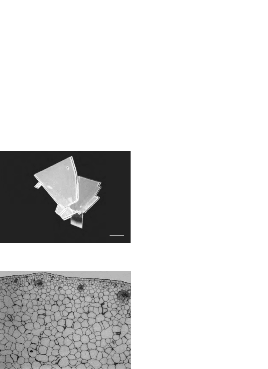
passed through any phase boundary. It has been
shown to be a particularly useful technique for
single-cell preparations (Figure 1) and food with
high tissue contents such as meat, fish, and vege-
tables. However, the technique has now been shown
to cause shrinkage of greater than 30% in some
samples and to generate many surface distortions
and is now losing favor as a routine SEM procedure.
Furthermore, any solvent-extractable components
are lost during the dehydration process.
MICROSCOPY/Scanning Electron Microscopy 3923