Cui Dongmei. Atlas of Histology: with functional and clinical correlations. 1st ed
Подождите немного. Документ загружается.

CHAPTER 11
■
Respiratory System
215
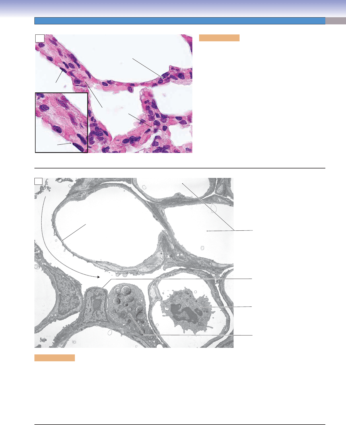
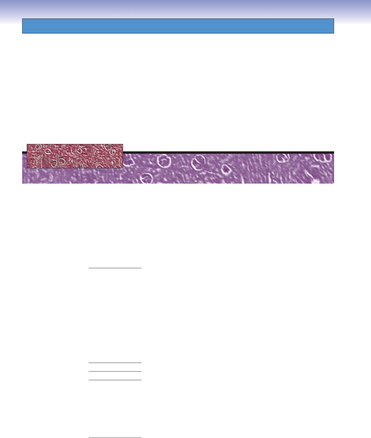
Figure 11-13A. Type I pneumocytes, lung. H&E,
725; inset, 1,145
Type I pneumocytes are squamous cells and make up
95% to 97% of the alveolar wall. A small percentage
of the alveolar wall is covered by type II pneumocytes
(Fig. 11-14A,B). Each type I pneumocyte has a fl at, dark
oval nucleus and very thin cytoplasm. These cells form
the blood-air barrier together with the endothelial cells
of the capillaries. They connect with each other by tight
junctions to prevent leakage of fl uid into the airspace.
Type I pneumocytes are not able to divide; if they are
damaged, type II pneumocytes will differentiate to replace
the damaged type I cells. There are delicate connective
tissues (including fi broblasts, elastic, and reticular fi bers)
and capillaries between the alveoli, forming the alveolar
septa (Fig. 11-12A). Alveolar septa contain a blood-air
barrier where gas exchange occurs. It is not easy to distin-
guish between type I pneumocytes and endothelial cells,
because they are both squamous cells.
Figure 11-13B. Type I pneumocytes and other constituents of alveoli. EM, 5,250
The open spaces in this view are a mixture of air-fi lled spaces (alveoli) and the lumens of capillaries that were emptied of blood in
the preparation of the specimen. The distinction between the air spaces and the blood spaces is not obvious because of the similar-
ity in ultrastructural appearance of the endothelial cells lining the capillaries and the type I pneumocytes lining most of the alveolar
surfaces. Both cell types are extremely fl attened to produce thin sheets of cytoplasm. The type I pneumocyte (squamous alveolar
cell) provides the covering of most (about 97%) of the surface of alveoli. Type II cells cover the remaining small fraction, and the
single type II cell provides an important clue in distinguishing the capillaries from the alveoli here. Adjacent to the type II cell is
the nucleus of a type I cell. This is the only part of the type I cell that is not extremely fl attened. The various cells and structures in
the fi eld provide some context for appreciating the thinness of the blood-air barrier.
Air space
(alveolus)
Type I
pneumocytes
Interalveolar
septa
Type I
pneumocyte
Type I
pneumocytes
A
Capillaries
Type I
pneumocyte
Type II
pneumocyte
Leukocyte in
capillary
Capillary
Capillary
Air
Air
Blood-air barrier
Capillary
Air
Air
Air
Air
B
CUI_Chap11.indd 215 6/16/2010 7:35:18 PM
216
UNIT 3
■
Organ Systems
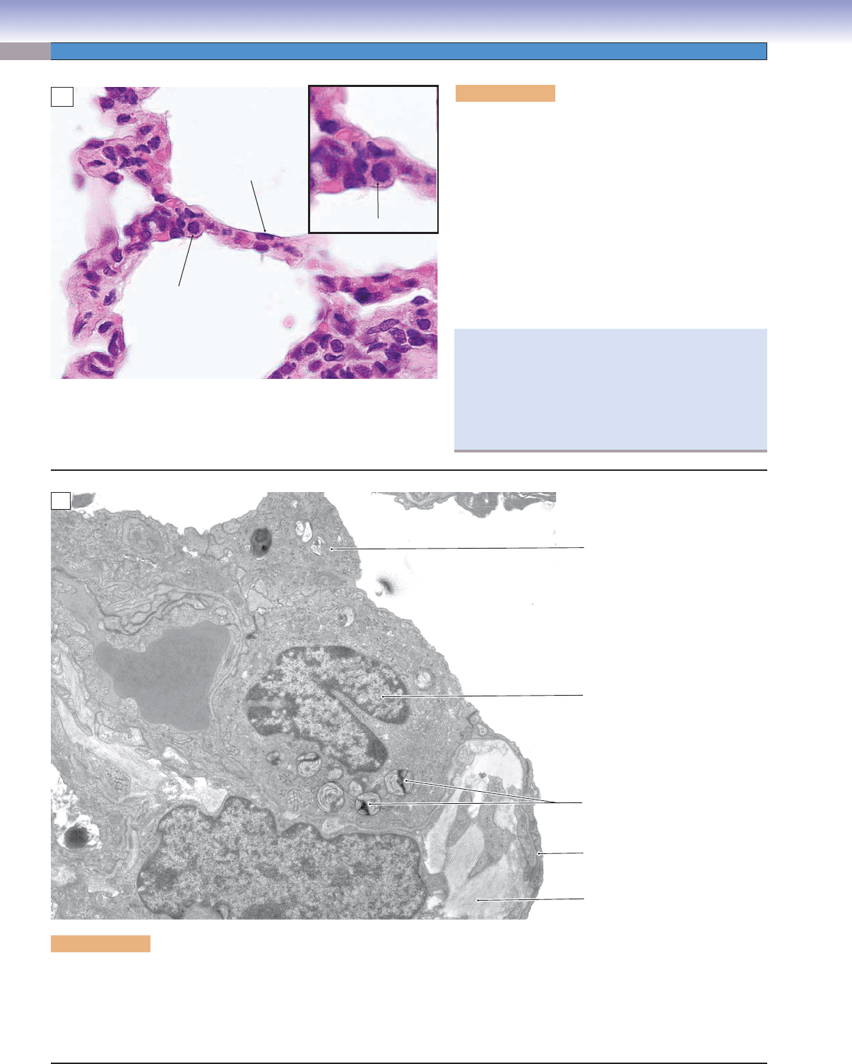
Figure 11-14B. Type II pneumocyte. EM, 10,900
Type II pneumocytes are easy to identify in transmission electron micrographs, owing to the presence in their cytoplasm of lamellar
bodies with their distinctive concentric lamellae. Unlike type I cells, the type II cells are compact in shape, either oval or cuboidal.
They have two main functions: They are precursors of type I cells and they secrete surfactant, an essential complex of lipids and
proteins that serves to reduce surface tension, thereby preventing collapse of the alveoli. Type II pneumocytes are connected to their
neighboring type I pneumocytes by junctional complexes. Part of an alveolar macrophage is visible in this fi eld.
Alveolar
macrophage
Type II
pneumocyte
Lamellar
bodies of type II
pneumocyte
Cytoplasm of
type I pneumocyte
Collagen
fibrils
Erythrocyte in
Erythrocyte in
capillary lumen
capillary lumen
Erythrocyte in
capillary lumen
B
Figure 11-14A. Type II pneumocytes, lung. H&E,
725; inset, 1,253
Type II pneumocytes are also called septal cells or type II
alveolar cells. These are large polygonal cells (or cuboi-
dal cells) with a large round nucleus. They bulge into the
air space, often sit at the corner of the alveoli (alveolar
septa), and make up 3% to 5% of the alveolar wall. Type
II pneumocytes have microvilli in their apical surfaces and
contain lamellar bodies in the cytoplasm (Fig. 11-14B).
Type II pneumocytes can divide and also regenerate both
type I and II pneumocytes. They produce a pulmonary
surfactant (phospholipids and proteins), which is impor-
tant in reducing the surface tension of the alveoli, thereby
preventing lung collapse.
Type I pneumocyte
Air space
(alveolus)
Type II pneumocyte
Type II pneumocyte
A
An example of lack of surfactant is respiratory distress
syndrome (RDS) in premature infants. These infants’
lungs are not suffi ciently well developed to produce
adequate surfactant. They have diffi culty breathing and
apnea (pauses in breathing). Treatment with surfactant
and the use of a mechanical respirator are necessary to
assure their survival.
CUI_Chap11.indd 216 6/16/2010 7:35:20 PM
CHAPTER 11
■
Respiratory System
217
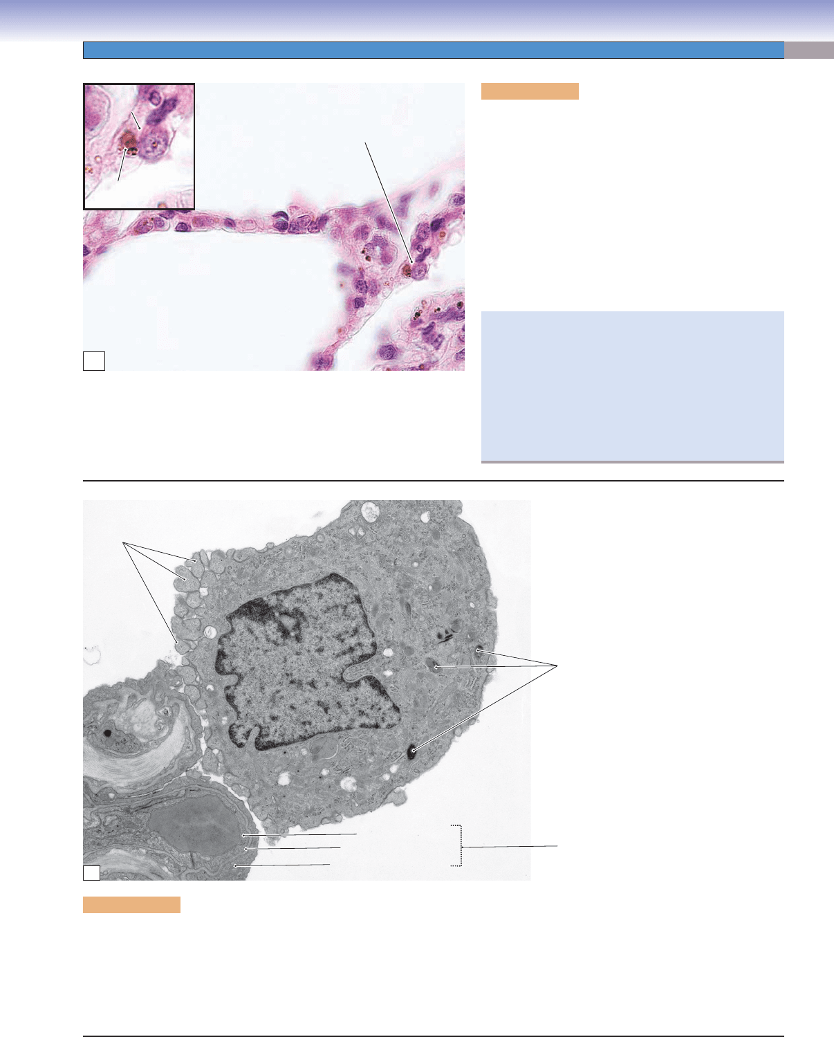
Figure 11-15A. Alveolar macrophages, lung. H&E,
725; inset 1,465
Alveolar macrophages are also called dust cells; they can
be found on the surface of the alveoli and in the connective
tissue of the septa. They are derived from blood monocytes
and migrate out of the capillaries to enter alveoli. Alveolar
macrophages are irregular in shape and have round nuclei;
they often contain phagocytized material (brown in color)
in the cytoplasm of active cells. Their function is to remove
dust particles, debris, and bacteria on the surface of the
alveoli, and they may also play an important role in initiat-
ing and maintaining chronic infl ammatory processes and
regulating tissue repair and remodeling in the lung.
Phagocytized
Phagocytized
material
material
Alveolar
Alveolar
macrophage
macrophage
Alveolar
macrophage
Phagocytized
material
Alveolar
macrophage
Air space
(alveolus)
A
Pseudopodia
Pseudopodia
Pseudopodia
Lysosomes
Blood-air barrie
r
Collagen
Collagen
Erythrocyte
Erythrocyte
Collagen
Erythrocyte
Endothelium of capillary
Endothelium of capillary
Fused basal lamina
Fused basal lamina
Type I pneumocyte
Type I pneumocyte
Endothelium of capillary
Fused basal laminae
Type I pneumocyte
B
Figure 11-15B. Alveolar macrophage. EM, 12,000
The surfaces of alveoli are continually swept by alveolar macrophages. In common with macrophages elsewhere, these cells are derived
from monocytes that have left the circulation, in this case through the walls of pulmonary capillaries. These cells phagocytose any
particles that have escaped capture in the conducting portion of the respiratory system. They also have a role in turnover of surfactant
produced by type II pneumocytes. As expected in a macrophage, the cytoplasm contains lysosomes, most of which appear to be primary
lysosomes in the cell shown here. Evidence of the cell’s motility is seen here as extensions of cytoplasmic processes (pseudopodia or
fi lopodia) extending from the surface of the cell that faces the surface of the alveolus. A view of the blood-air barrier can also be seen.
Clinically, alveolar macrophages may also be called
“heart failure cells.” During heart failure, the heart is
unable to pump blood at an adequate volume, and the
backup of blood causes increased pressure in the alveo-
lar capillaries. Red blood cells (erythrocytes) then leak
into the alveoli. Alveolar macrophages engulf these
erythrocytes. Pathologically, heart failure cells (alveolar
macrophages) are identifi ed by a positive stain for iron
pigment (hemosiderin).
CUI_Chap11.indd 217 6/16/2010 7:35:22 PM
218
UNIT 3
■
Organ Systems
CLINICAL CORRELATIONS
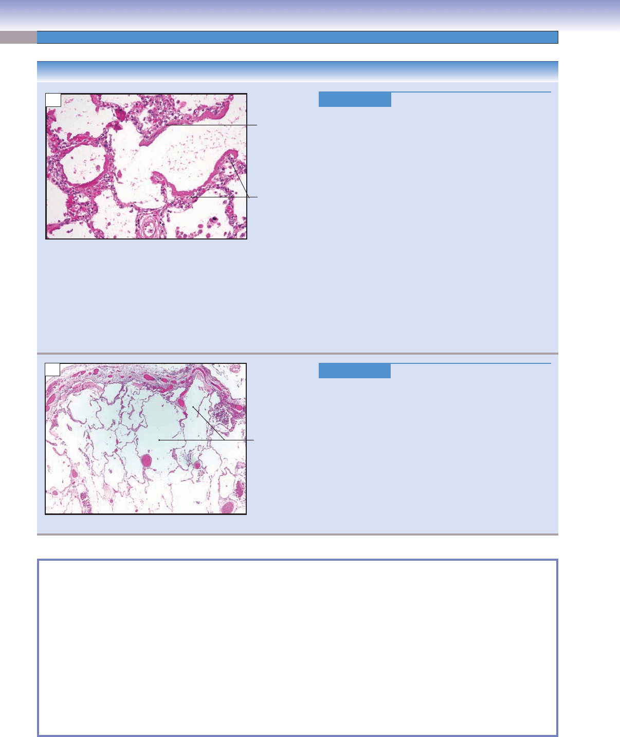
Figure 11-16A.
Acute Respiratory Distress Syndrome.
H&E, 1,079
Acute respiratory distress syndrome (ARDS) is a clinical
term describing acute lung injury, correlating with the
pathologic entity of diffuse alveolar damage. ARDS is a
respiratory emergency, characterized by an acute onset
of shortness of breath (developing in 4–48 hours), which
progresses to respiratory failure. It is caused by a broad
spectrum of diseases such as pneumonia, severe injury to
the lungs, severe trauma, burns, sepsis, medications, and
shock. ARDS is not a specifi c lung disease, but a spec-
trum of clinical and pathological changes due to acute
lung injury. Pathologic fi ndings depend on the stage of
the condition, and include (1) excess fl uid in the intersti-
tium and alveoli with rupture of the alveolar structures;
(2) proliferation of type II pneumocytes and squamous
metaplasia and myofi broblasts infi ltration; and (3) hya-
line membranes. Hyaline membranes consist of fi brin and
remnants of necrotic pneumocytes that line the alveo-
lar spaces, as seen in the photomicrograph. Treatment
includes using mechanical ventilation and treating the
underlying disease.
Figure 11-16B.
Emphysema. H&E, 27
Chronic obstructive pulmonary disease (COPD) includes
emphysema and chronic bronchitis. Emphysema is
characterized by the permanent destruction of alveolar
structures, enlargement of the alveolar airspaces distal
to the terminal bronchioles, and loss of elasticity of the
lung tissue without obvious fi
brosis. Cigarette smoking
is the primary cause of the disease. Signs and symptoms
include pursed-lip breathing, central cyanosis, fi nger club-
bing, and shortness of breath (dyspnea), hyperventila-
tion, “barrel chest,” and recurring respiratory infections.
Treatments include cessation of smoking, bronchodilating
agents, supplemental oxygen, and antibiotics for respira-
tory infections.
Hyaline
membrane
Inflammatory
cells
A
Enlargement
of the alveola
r
airspaces
B
SYNOPSIS 11-1 Pathological and Clinical Terms for the Respiratory System
Hyaline membrane ■ : A histological feature of diffuse alveolar damage in early ARDS. It is a proteinaceous alveolar exudate
at the periphery of the alveolar space; also seen in hyaline membrane disease (RDS) of neonates.
Dyspnea
■ : Shortness of breath; may be due to a myriad of causes including congestive heart failure (pulmonary edema),
pulmonary embolus, asthma, and COPD.
Finger clubbing
■ : Also called “hypertrophic osteoarthropathy,” it represents enlargement of the distal aspect of the digits
due to proliferation of connective tissue and bone changes caused by many conditions including pulmonary diseases such
as COPD, infection, and malignancy.
Asthma
■ : A chronic infl ammatory disease of airways that manifests as paroxysmal contraction of airway smooth muscle
which causes narrowing of the airway lumens in response to exposure to a variety of triggers including allergens, infection,
and exercise; airway narrowing results in shortness of breath.
Squamous metaplasia
■ : A reversible change from mature cell types to squamous epithelium, such as occurs in ciliated pseu-
dostratifi ed columnar respiratory mucosa when exposed to environmental changes such as cigarette smoke.
CUI_Chap11.indd 218 6/16/2010 7:35:24 PM

CHAPTER 11
■
Respiratory System
219
Tract Epithelium Glands Skeletal
Support
Muscle Special Features and Main
Functions
Upper Airway
Conducting portion
Nasal cavity
Nasal vestibule Stratifi ed squamous
epithelium
Sebaceous and sweat
glands
Hyaline
cartilage
None Vibrissae present; block large
particles and small insects
Nasal mucosa Respiratory epithelium Mixed mucoserous
glands
Bone and hya-
line cartilage
None Venous plexuses warm air to
body temperature
Olfactory
mucosa
Specialized olfactory
epithelium
Serous (Bowman)
glands
Bone None Olfactory receptor neurons in
epithelium detect smell and
odorants
Nasopharynx
and orophar-
ynx
Respiratory and
Stratifi ed squamous
epithelium
Seromucous (mixed)
glands
Bone Skeletal
muscle
Pharyngeal and palatine tonsils;
fi rst-line immunological defense
Larynx
Epiglottis and
Vocal cords
Stratifi ed squamous and
respiratory epithelium
Mostly mucous
glands and some
serous or mixed
glands
Hyaline and
elastic carti-
lage
Skeletal
(vocalis)
muscle
Vocal cords control airfl ow and
speaking; epiglottis prevents food
and fl uid from entering trachea
Lower Airway
Trachea Respiratory epithelium Mostly mucous
glands and some
serous or mixed
glands
C-shaped hya-
line cartilage
rings
Smooth
(trachealis)
muscle
Trachealis muscle bridges open-
ing ends of cartilage
Extrapulmonary
(primary) bronchi
Respiratory epithelium Mucous and serous
(mixed glands)
C-shaped
hyaline cartilage
rings
Smooth
muscle
Two primary bronchi outside
each lung bifurcate from trachea;
C-shaped cartilage
Intrapulmonary
bronchi
Respiratory epithelium Mucous and serous
(mixed glands)
Large and
small plates of
hyaline carti-
lage
Prominent
spiral band
of smooth
muscle
Secondary and tertiary bronchi
branch repeatedly; spiral smooth
muscle band lies between lamina
propria and submucosa
Bronchioles Simple ciliated columnar
to cuboidal; Clara cells
present
Occasional goblet
cells in large bron-
chioles, but not in
small bronchioles
No cartilage Smooth
muscle
Clara cells are present
Terminal bronchioles Simple ciliated cuboidal;
numerous Clara cells
No goblet cells in
normal cases
None Some
smooth
muscle
Numerous Clara cells are pres-
ent in these smallest conducting
airways
Respiratory portion
Respiratory
bronchioles
Simple cuboidal cells
with few cilia; a few
Clara cells; some type I
and II pneumocytes
No goblet cells None Few smooth
muscle cells
Alveoli interrupt simple cuboidal
epithelium; gas exchange begins
here
Alveolar ducts/
alveolar sacs
Rarifi ed simple cuboidal
epithelium between
alveoli; no Clara cells
No goblet cells None Few smooth
muscle cells
Air passes into alveoli for gas
exchange
Alveoli Type I and II
pneumocytes
No goblet cells None None Blood-air barrier is the primary
site for gas exchange
TABLE 11-1 Respiratory System
SYNOPSIS 11-2 Structural Differences (From Upper to Lower Airway) in the Respiratory System
Epithelium ■ changes from keratinized to nonkeratinized stratifi ed squamous and then to respiratory epithelium.
Respiratory epithelium
■ changes from simple columnar to cuboidal and then to simple squamous (alveolar cells).
Glands
■ gradually decrease in numbers and disappear in bronchioles as airways become smaller.
Skeletal support
■ changes from bone to cartilage to none at all.
Cartilage
■ changes from C-shaped rings to large irregular plates to small plates and disappears in bronchioles and levels below.
Muscle
■ changes from skeletal to smooth muscle; numbers of smooth muscle cells decrease.
CUI_Chap11.indd 219 6/16/2010 7:35:25 PM
220
12
Introduction and Key Concepts for the Urinary System
Figure 12-1 Overview of the Urinary System
Figure 12-2 Overview of the Kidney
Figure 12-3 Orientation of Detailed Urinary System Illustrations
Kidneys
Figure 12-4A Renal Cortex and Medulla, Kidney
Figure 12-4B Renal Cortex, Kidney
Figure 12-4C Clinical Correlation: Glomerular Disorders: Diabetic Nephropathy
Figure 12-5A Renal Corpuscle, Renal Cortex
Figure 12-5B Renal Corpuscle, Glomerulus and Bowman Capsule
Figure 12-6A Glomerulus, Renal Cortex
Figure 12-6B Glomerulus and Filtration Barrier
Figure 12-7 Glomerulus and Podocyte
Figure 12-8A Medullary Ray, Renal Cortex
Figure 12-8B The Nephron and Collecting System of the Kidney
Figure 12-9A,B Proximal Tubules
Figure 12-10A,B Distal Tubules
Figure 12-11A–C Medullary Tubules
Figure 12-11D Clinical Correlation: Renal Cell Carcinoma (Clear Cell Type)
Figure 12-12A Clinical Correlation: Renal Oncocytoma
Figure 12-12B Clinical Correlation: Hemodialysis
Synopsis 12-1 Clinical and Pathological Terms for the Urinary System
Table 12-1 Kidneys
Ureters
Figure 12-13A Ureter
Figure 12-13B Transitional Epithelium, Ureter
Figure 12-13C Clinical Correlation: Nephrolithiasis (Renal Stones)
Urinary System
CUI_Chap12.indd 220 6/2/2010 6:36:32 PM

CHAPTER 12
■
Urinary System
221
Urinary Bladder
Figure 12-14A Urinary Bladder, Bladder Wall
Figure 12-14B Urothelium, Bladder Wall
Figure 12-14C Clinical Correlation: Urothelial (Transitional) Carcinoma
Figure 12-15A,B Transitional Epithelium, Urinary Bladder
Urethra
Figure 12-16A Prostatic Urethra, Male Urethra
Figure 12-16B Penile (Spongy) Urethra, Male Urethra
Figure 12-16C Female Urethra
Introduction and Key Concepts for the
Urinary System
The urinary system is composed of two kidneys, two ureters,
the bladder, and the urethra. The kidneys produce urine, the
ureters transport urine to the bladder, and the bladder tempo-
rarily stores and empties urine through the urethra to outside of
the body. The urinary system functions to (1) fi lter blood and
reabsorb nutrients; (2) control the water, ion, and salt balance
of the body; (3) maintain the acid-base balance of the blood;
(4) excrete metabolic wastes (urea and uric acid), toxins, and
drug components; (5) secrete hormones, such as renin and eryth-
ropoietin; and (6) produce calcitriol (an active form of vitamin
D) to help the body absorb dietary calcium into the blood.
Kidneys
The kidneys are bean-shaped organs located in the posterior
abdominal region on each side of the vertebral column. The
kidney can be divided into the renal cortex, the renal medulla,
and the hilum. The renal cortex is composed of renal corpuscles
and various cortical tubules, which include the proximal con-
voluted tubules, the distal convoluted tubules, and the corti-
cal collecting tubules. The renal medulla is located deep to the
cortex, and its tubules extend as medullary rays into the cortex
region. The medulla comprises 10 to 18 renal pyramids; each
pyramid contains the loops of Henle, collecting ducts, and pap-
illary ducts. The apical projection of a renal pyramid is called
the renal papilla. The papillary ducts empty urine at the tip of a
renal papilla onto its surface, which is called the area cribrosa
(perforated area). Each renal papilla is surrounded by a space,
the minor calyx; several minor calices unite to form a major
calyx. There are two or three major calyces for each kidney.
The major calices unite to form the renal pelvis, which funnels
urine into the ureter. The hilum is the region in the medial por-
tion of the kidney where the renal artery, the renal vein, and
the ureter enter and exit the kidney (Fig. 12-2). Functionally
and structurally, the kidney can be divided into the nephron
and the collecting system (Fig. 12-8B). The nephron produces
urine. The collecting system adjusts the composition of urine
and transports urine to the calyces.
THE NEPHRON comprises a renal corpuscle, a proximal
convoluted tubule, a loop of Henele, and a distal convoluted
tubule.
A renal corpuscle is composed of a glomerulus and a
Bowman capsule. (1) A glomerulus consists of a spherical
knot of capillaries, which is fed by an afferent arteriole and
drained by an efferent arteriole at the vascular pole. (2) A Bow-
man capsule consists of a visceral layer and a parietal layer.
The visceral layer is composed of podocytes, which cover the
capillaries of a glomerulus. These cells have long, interdigitating
cellular processes and play an important role in blood fi ltration.
The interstitial tissues surrounding the glomerular capillaries
contain cells called intraglomerular mesangial cells. The pari-
etal layer of the Bowman capsule is a hollow spherical struc-
ture lined by simple squamous epithelium. The space between
the visceral and the parietal layers of the Bowman capsule is
called the Bowman space
. Blood fl
ows through the glomerular
capillaries, and its plasma passes through the glomerular fi ltra-
tion barrier (the fused basal laminae of the endothelial cells and
the podocytes); the fi ltrate is collected in the Bowman space
(Fig. 12-6A,B). Thus, the renal corpuscle, as a whole, forms a
blood-fi ltering unit, which allows water, metabolic wastes, ions,
and small molecules to pass through the capillary wall but pre-
vents circulating cells and large plasma proteins from leaving
the blood.
Proximal Convoluted Tubules are long tubes that follow
a serpentine course as they drain the fi ltrate from the renal
corpuscles into the loop of Henle. Each is lined by a simple
cuboidal epithelium with abundant long microvilli (brush bor-
der) bordering the lumen. Each proximal convoluted tubule
connects to a renal corpuscle at its urinary pole. The relatively
large epithelial cells of the proximal convoluted tubule contain
many mitochondria, which render their cytoplasm brightly
acidophilic (pink). The lateral boundaries between the cells
interdigitate, so that the boundaries between adjacent cells are
unclear in light microscopy. Their long microvilli appear to fi ll
much of the space within the lumen (Fig. 12-9A,B). The com-
bined structural features of the proximal convoluted tubules
contribute to their functions of actively transporting ions and
reabsorbing water, glucose, amino acids, proteins, and vitamins
from the fi ltrate.
The Loop of Henle is a continuation of the proximal
convoluted tubule. It is a U-shaped structure that includes a
descending limb and an ascending limb (Fig. 12-8B). The
descending limb consists of a thick descending limb (proximal
straight tubule) and a thin descending limb (descending thin
segment). The ascending limb contains a thin ascending limb
(ascending thin segment) and a thick ascending limb (distal
straight tubule). The loop of Henle plays a crucial role in gen-
erating a high sodium concentration gradient in the interstitium
of the renal medulla. This permits water to move passively
CUI_Chap12.indd 221 6/2/2010 6:36:39 PM

222
UNIT 3
■
Organ Systems
from collecting ducts into the interstitium. Some physiology
textbooks do not include the thick descending limb (proximal
straight tubule) as a part of the loop of Henle proper because
it does not signifi cantly contribute to its physiological function.
The proximal straight tubules are similar in structure to the
proximal convoluted tubules. The descending and ascending
thin segment tubules are lined by squamous cells and are struc-
turally similar to each other. The descending limb is perme-
able to water, Cl
-
, and Na
+
. The tubules of the descending limb
reabsorb water and salts and reduce the volume of the fi ltrate
that has passed through the proximal convoluted tubules. The
ascending limb is very active physiologically. It is impermeable
to water, and it actively pumps Cl
-
and Na
+
from the lumen into
the medullary interstitium.
Distal Convoluted Tubules are lined by small, simple
cuboidal epithelial cells, which have no brush border. They
may show a few short, irregular microvilli on their apical sur-
faces and plasma membrane infoldings on their basal region
at the EM level (Fig. 12-10A,B). Their lumens appear clearer
and wider than those of proximal tubules. The distal convo-
luted tubules are located in the cortex of the kidney and are
closely associated with the renal corpuscles. At the junction
between the distal straight and the convoluted tubules, there is
an important specialized sensory structure, the macula densa,
which senses and monitors ionic content and water volume
of the fi ltrate. The macula densa is composed of cells that are
taller and more tightly packed than other cells of the distal
tubule (see Fig. 12-5A,B). This portion of the distal tubule
is positioned between afferent and efferent arterioles at the
vascular pole of the renal corpuscle. The distal convoluted
tubules remove Na
+
and add K
+
to the fi ltrate if aldosterone
stimulation is present; they also reabsorb bicarbonate ions and
secrete ammonium to adjust the pH balance. The distal con-
voluted tubules connect distal straight tubules (thick ascend-
ing limb of the loop of Henle) to the collecting tubules. The
distal convoluted and straight tubules are structurally similar
to each other, differing mainly in their locations and courses.
THE COLLECTING SYSTEM consists of cortical collecting
tubules, collecting ducts, and papillary ducts. The collecting
tubules are small and lined by cuboidal cells. They are located
in the renal cortex, so they are also called cortical collecting
tubules. They drain the fi ltrate from distal convoluted tubules
into the collecting ducts of the medullary rays, which, in turn,
drain into larger collecting ducts in the medulla. Collecting
ducts have larger lumens than collecting tubules, and they
are lined by taller cuboidal or columnar cells. Both collecting
tubules and ducts have clear cytoplasm and distinct cell-to-cell
boundaries. These tubules become highly permeable to water
under the infl uence of antidiuretic hormone (ADH). Depending
on ADH levels, the tubules passively diffuse a variable volume
of water from their lumens into the medullary interstitium, thus
increasing the concentration of urine. The collecting ducts are
the last components of the kidney that process and determine
the fi nal urine composition. Papillary ducts, also called ducts
of Bellini, are continuations of the collecting ducts. They are
located in the papilla of the renal medulla. Several collecting
ducts merge into a single papillary duct, which empties urine
into the minor calyx at the tip of the renal papilla.
THE VASCULAR SUPPLY TO THE KIDNEY comes from the
renal artery, which enters the kidney at the hilum; segmental
branches of the renal artery give rise to the interlobar arteries.
These pass through the renal columns between the renal pyra-
mids and give rise to arcuate arteries. The arcuate arteries run
along the junction between the cortex and the medulla of the
kidney and give rise to the interlobular arteries, which extend
into the medulla to supply the afferent arterioles of renal cor-
puscles. Each afferent arteriole supplies a glomerulus of capil-
laries from which blood is drained by an efferent arteriole at the
vascular pole. The efferent arterioles of corpuscles in the outer
cortex feed into the peritubular capillary network, which sup-
plies the cortical tissue surrounding the cortical tubules. These
peritubular capillaries provide for gas and material exchange
and also receive renal interstitial fl uid, which is reabsorbed out
of the tubules and goes back into the vascular bed. Venules
carry blood to the interlobular veins and to the arcuate veins
in the renal corticomedullary junction. The efferent arterioles
of deeper (juxtamedullary) corpuscles extend into the medulla
where they give rise to capillaries called vasa recta, which
receive interstitial fl uid (reabsorbed from fi ltrate) in the medulla
and send it back to the circulation. The vasa rectae take a hair-
pin course in the medulla following the loop of Henle. They
return to the corticomedullary junction to join the interlobular
veins and then drain into the arcuate veins. The arcuate veins
drain blood into the interlobar veins, which then merge to form
the branches of the segmental renal veins, which in turn fi nally
merge into the renal vein (see Fig. 11-2).
Ureters
The two ureters lie in the extraperitoneal connective tissue,
laterally positioned on each side of the vertebral column. The
ureters are long, relatively small tubules lined by transitional
epithelium and surrounded by a thin layer of smooth muscle
and connective tissue. Superiorly, they drain the funnel-shaped
renal pelvis, and inferiorly, they empty into the bladder by pen-
etrating its posterior wall. The ureters have a much thinner wall
than the bladder. Like most tubular organs, the wall of the ure-
ter is composed of several layers of tissues: mucosa, muscularis,
and adventitia (Fig. 12-13A,B).
Urinary Bladder
The urinary bladder, a distensible sac-shaped organ located in
the pelvic cavity, temporarily stores urine. The wall of the blad-
der has three openings, two of them for ureters to enter and
one for emptying urine into the urethra. Like the ureter, the uri-
nary bladder wall consists of mucosal, muscularis, and adven-
titial layers, but the bladder wall is much thicker, having three
substantial layers of smooth muscle in the muscularis. (1) The
mucosa consists of a transitional epithelium lining and a layer
of connective tissue (lamina propria) containing blood vessels
and nerve fi bers. (2) The muscularis contains the three layers
of smooth muscle: inner longitudinal smooth muscle, middle
circular smooth muscle, and outer longitudinal smooth mus-
cle. The muscularis contracts in different directions to enable
the urinary bladder to empty urine. (3) The outer portion of
the bladder is protected by both a serosa and an adventitia
depending on whether it projects into the peritoneal cavity. The
superior surface of the bladder is covered by serosa, which is a
layer of connective tissue covered by mesothelium; the inferior
CUI_Chap12.indd 222 6/2/2010 6:36:39 PM

CHAPTER 12
■
Urinary System
223
surface of the bladder is covered by adventitia, which is a layer
of connective tissue without a mesothelial covering.
Urethra
The urethra is structurally different in the male and female. The
proximal end of the male urethra is surrounded by an internal
urethral sphincter (smooth muscle) that functions mainly to pre-
vent seminal fl uids from entering the bladder during ejaculation.
The male urethra is about 20 cm long, and it is composed of three
segments: prostatic, membranous, and penile (spongy) urethra.
The prostatic portion is surrounded by the prostate gland and is
lined by transitional epithelium. The membranous portion is a
short segment surrounded by the skeletal muscle of the external
sphincter (urogenital diaphragm) and is lined by pseudostratifi ed
columnar epithelium. The penile (spongy) urethra (also called the
cavernous urethra) is surrounded by the corpus spongiosum of
the penis, and its epithelial lining changes from pseudostratifi ed
columnar to stratifi ed squamous. In this region, there are many
small mucous glands called the glands of Littré, which secrete
mucus to coat and protect the lining of the urethra.
The female urethra is short, about 4 to 5 cm. It is lined
chiefl y by stratifi ed squamous epithelium and, in a few places,
may have patches of pseudostratifi ed columnar epithelium.
Glands of Littré are also present in the female urethra. The
proximal end of female urethra is surrounded by skeletal
muscle (external sphincter) where it penetrates the urogenital
diaphragm. The external sphincter muscle in both male and
female is innervated by the pudendal nerves; it functions to con-
trol retention or release of the urine from the urinary bladder
through the urethra and helps maintain urinary continence. A
female does not have an internal sphincter.
CUI_Chap12.indd 223 6/2/2010 6:36:40 PM
224
UNIT 3
■
Organ Systems
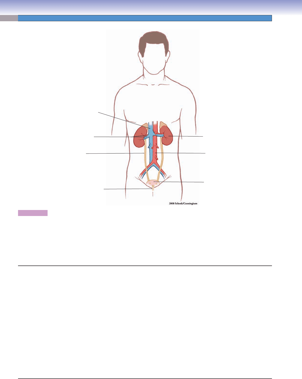
Figure 12-1. Overview of the urinary system.
The urinary system plays an important role in eliminating the body’s metabolic wastes and toxins; controlling water and ion balance
and regulating blood pressure; maintaining the acid-base (pH) balance of the blood; and in reabsorbing and conserving nutrients.
The urinary system achieves these goals by fi ltering the blood and producing urine. The complex tubule system in the kidney helps
to reabsorb and readjust water and ion content and to excrete urine. The urinary system consists of two kidneys, two ureters, the
urinary bladder, and the urethra. The kidneys are the organs that produce urine and accomplish the essential functions listed above.
After urine is produced, it passes through the ureters to the bladder for temporary storage, fi nally exiting the body through the
urethra.
Kidney
Urinar
y
b
l
adde
r
Abdominal
a
orta
U
reter
Renal artery
a
n
d
v
e
in
In
f
erior
v
ena ca
v
a
Urethra
General structure of the kidney:
I. Renal cortex
A. Renal corpuscles
B. Proximal convoluted tubules
C. Distal convoluted tubules
D. Cortical collecting tubules
II. Renal medulla (renal pyramids)
A. Outer medulla
B. Inner medulla
C. Renal papillae
III. Renal hilum
A. Minor calyx
B. Major calyx
C. Renal pelvis
Functional and histological unit of the kidney:
I. Nephron
A. Renal corpuscle
B. Proximal convoluted tubule
C. Loop of Henle
1. Descending limb
a. Thick descending limb (proximal straight tubule)
b. Thin descending limb (descending thin segment)
2. Ascending limb
a. Thin ascending limb (ascending thin segment)
b. Thick ascending limb (distal straight tubule)
D. Distal convoluted tubules
II. Collecting system
A. Cortical collecting tubule
B. Collecting ducts
C. Papillary ducts
Structures of the Kidney
CUI_Chap12.indd 224 6/2/2010 6:36:40 PM