Cui Dongmei. Atlas of Histology: with functional and clinical correlations. 1st ed
Подождите немного. Документ загружается.

CHAPTER 12
■
Urinary System
225
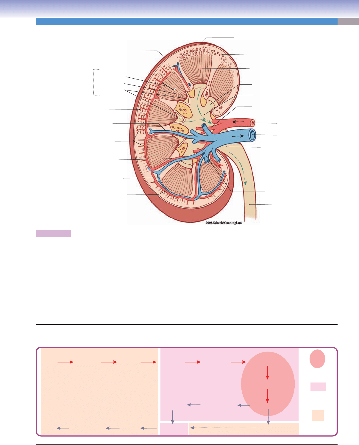
Figure 12-2. Overview of the kidney.
The kidney can be divided into three regions: the renal cortex, the renal medulla, and the hilum. The renal cortex is composed
of renal corpuscles, proximal and distal convoluted tubules, cortical collecting tubules, and the blood vessels supplying the renal
cortex. The renal medulla is made up of renal pyramids. The renal pyramids can be divided into three zones: the outer medulla, the
inner medulla, and the renal papillae. The renal medulla is composed of several types of tubules oriented parallel to one another: the
descending limb of the loop of Henle (thick descending and thin descending limbs), the ascending limb of the loop of Henle (thin
ascending and thick ascending limbs), the cortical collecting tubules, the collecting ducts, and the papillary ducts (see Fig. 12-8B).
The papillary ducts drain urine into the minor calices and then to the major calices; the major calices merge into the renal pelvis,
which drains urine into the renal ureter. The blood vessels supplying the kidneys include the renal artery and vein, branches of the
renal artery and vein (segmental arteries and veins), interlobar arteries and veins, arcuate arteries and veins, interlobular arteries
and veins, and afferent and efferent arteries. The microvasculature consists of the glomeruli of the renal corpuscle, the peritubular
capillaries, and the vasa recta.
Renal
pelvis
Renal
ureter
Renal vein
Branches (segmental)
of renal vein
Branches (segmental)
of renal artery
Arcuate artery
Arcuate vein
Interlobular artery
Interlobar artery
Interlobar vein
Renal
glomerulus
Renal artery
Renal medulla
Major calyx
Minor calyx
Renal papilla
Renal pyramid
Outer medulla
Inner medulla
Renal cortex
Renal corpuscle
Renal capsule
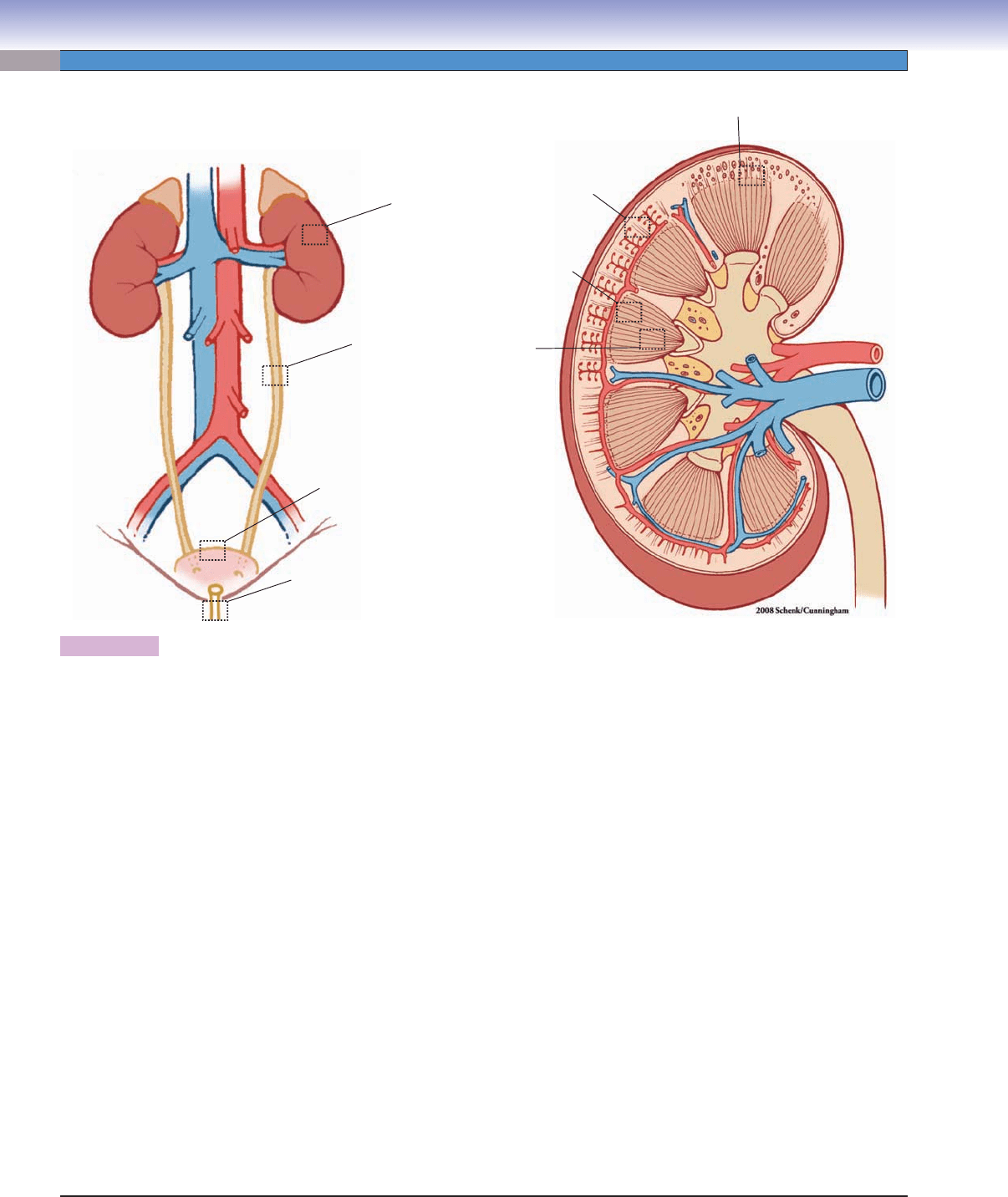
Vascular Supply of the Kidney
Renal
artery
Renal
vein
Arcuate veins
Interlobular
arteries
Peritubular
capillary network
to venules
Afferent arterioles
Renal
corpuscle
Renal
cortex
Renal medulla
and hilum
Efferent arterioles
Glomerulus
Vasa recta
Interlobar
arteries
Branches
of renal arteries
(segmental arteries)
Branches
of renal veins
(segmental veins)
Interlobar
veins
Interlobular
veins
Arcuate
arteries
CUI_Chap12.indd 225 6/2/2010 6:36:40 PM
226
UNIT 3
■
Organ Systems
Fig. 12-4A
Fig. 12-11A
Fig. 12-11B
Fig. 12-4B to
Fig. 12-10B
Fig. 12-13A,B
Fig. 12 4A to
Fig. 12-11C
Fig. 12-14A,B
Fig. 12-15A,B
Fig. 12-16A,B,C
Figure 12-3. Orientation of detailed urinary system illustrations.
Structures of the Urinary System with Figure Numbers
Kidney
Renal cortex and medulla
Figure 12-4A
Figure 12-4B
Figure 12-4C
Renal corpuscles
Figure 12-5A
Figure 12-5B
Glomerulus and fi ltration barrier
Figure 12-6A
Figure 12-6B
Figure 12-7
Medullary ray and urinary tubules
Figure 12-8A
Figure 12-8B
Proximal tubules
Figure 12-9A
Figure 12-9B
Distal tubules
Figure 12-10A
Figure 12-10B
Medullary tubules
Figure 12-11A
Figure 12-11B
Figure 12-11C
Figure 12-11D
Figure 12-12A
Ureter
Figure 12-13A
Figure 12-13B
Figure 12-13C
Urinary bladder
Figure 12-14A
Figure 12-14B
Figure 12-14C
Figure 12-15A
Figure 12-15B
Male and female urethrae
Figure 12-16A
Figure 12-16B
Figure 12-16C
CUI_Chap12.indd 226 6/2/2010 6:36:41 PM
CHAPTER 12
■
Urinary System
227
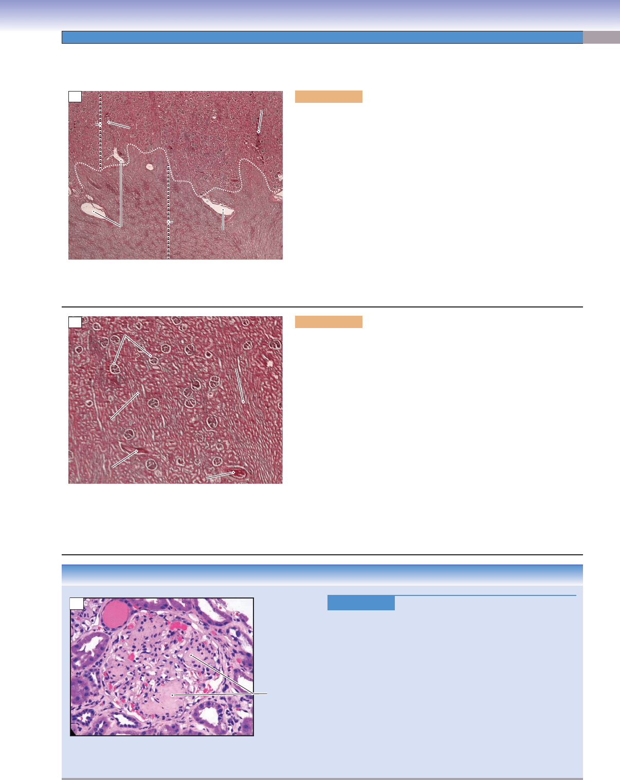
CLINICAL CORRELATION
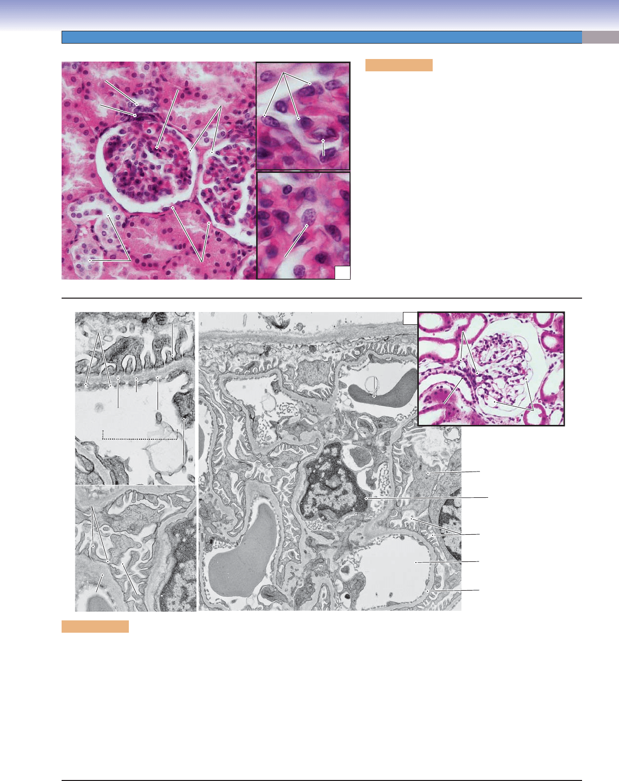
Figure 12-4C.
Glomerular Disorders: Diabetic Nephropathy.
H&E, 216
Diabetic nephropathy, a complication of both type 1 and type
2 diabetes mellitus, may result in chronic renal failure and is
the leading cause of end-stage renal disease in the United States
and other Western countries. Major histologic changes in the
glomeruli in diabetic nephropathy include thickening of the
glomerular basement membrane, diffuse glomerulosclerosis,
and nodular glomerulosclerosis, also called Kimmelstiel-Wilson
disease. As the disease progresses, edema (swelling), hyperten-
sion, foamy urine, fatigue, headache, and nausea and vomiting
may occur. Tight control of blood glucose levels tends to delay
the onset of development. Treatment includes dialysis and renal
transplantation. Shown here is a renal glomerulus with nodular
glomerulosclerosis, or Kimmelstiel-Wilson disease.
Kimmelstiel-
Wilson nodules
C
Figure 12-4A. Renal cortex and medulla, kidney. H&E, 11
This section shows the renal cortex and the medulla. The dashed white line
indicates the junction between the cortex and the medulla. The difference
in appearance between the cortex and the medulla is due to the arrange-
ment of the uriniferous tubules (nephrons and collecting ducts). The renal
cortex is stained darker than the renal medulla. There are numerous renal
corpuscles and various convoluted tubules in the cortex region. Both the
cortex and the medulla have a rich blood supply. The arcuate vessels (arter-
ies and veins) are visible at the border of the corticomedullary junction.
The interlobular vessels (arteries and veins) arise from arcuate vessels and
course upward (arteries) or downward (veins) in the renal cortex. The renal
medulla is composed of 10 to 18 renal pyramids. Each pyramid contains
numerous medullary tubules (loops of Henle, collecting ducts, and papil-
lary ducts). Each papillary duct opens at the surface of the renal papilla
(called the area cribrosa) where it empties urine into the minor calyx. The
renal medulla can be divided into inner and outer zones based on differ-
ences in the types of tubules residing in the two regions (Fig. 12-11A–C).
Cortex
Cortex
Cortex
Interlobular
Interlobular
vessel
vessel
Interlobular
vessel
Interlobular
Interlobular
vessel
vessel
Interlobular
vessel
Medulla
Medulla
Medulla
Arcuate
Arcuate
vessel
vessel
Arcuate
vessel
Arcuate
Arcuate
vessel
vessel
Arcuate
vessel
A
Renal
Renal
corpuscles
corpuscles
Renal
corpuscles
Medullary ray
Medullary ray
Medullary ray
Medullary
Medullary
ray
ray
Medullary
ray
Arcuate
Arcuate
vessel
vessel
Arcuate
vessel
Arcuate vessel
Arcuate vessel
Arcuate vessel
B
Figure 12-4B. Renal cortex, kidney. H&E, 32
The renal cortex is composed of the renal corpuscles, the proximal
convoluted tubules, the distal convoluted tubules, and the cortical
collecting tubules. The renal corpuscles look like small balls interspersed
among a tangle of tubules (cortical labyrinth) in the cortex region. The cor-
tical labyrinth (with its corpuscles) is subdivided into columns by groups
of parallel tubules called medullary rays. The medullary rays belong to
the renal medulla proper; however, they extend into the cortex region.
The renal cortex contains various convoluted tubules and is supplied by
interlobular arteries, which give rise to afferent arteries. The afferent arte-
rioles supply the glomeruli of renal corpuscles; blood exits the glomeruli
through efferent arterioles. The cortical tubules are supplied by a peritubu-
lar capillary network, which arises from efferent arterioles that exit renal
corpuscles located in the outer cortex. The renal medulla is supplied by the
vasa recta, which arise from efferent arteries that exit renal corpuscles in
the inner (juxtamedullary) cortex. The vasa recta follow the loop of Henle
downward into the medulla and loop back toward the cortex. Both the
peritubular capillaries and vasa recta converge into the interlobular vein
and then drain into the arcuate vein at the corticomedullary junction.
Kidneys
CUI_Chap12.indd 227 6/2/2010 6:36:43 PM
228
UNIT 3
■
Organ Systems
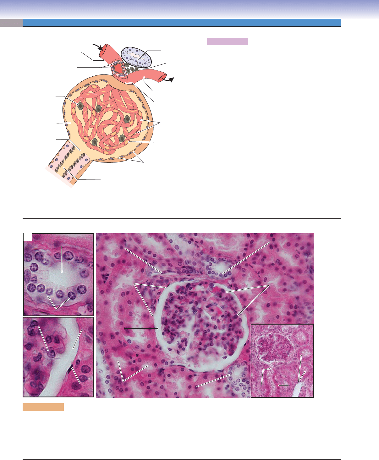
Figure 12-5A. Renal corpuscle, renal cortex.
The renal corpuscle is the site of blood fi ltration and
initial production of urine. The main components of a
renal corpuscle are a tuft of capillaries called the glom-
erulus and a surrounding sac, the Bowman capsule. The
area of the renal corpuscle through which the arterioles
pass into and out of the glomerulus is called the vascular
pole. The surfaces of the capillaries of the glomerulus are
covered by podocytes, which make up the visceral layer
of the Bowman capsule. The outer wall of the renal cor-
puscle is a simple squamous epithelium called the parietal
layer of the Bowman capsule. Fluid is fi ltered from the
blood in glomerular capillaries into the space between
the two layers, the Bowman space. Fluid exits through
the urinary pole to enter the proximal convoluted
tubule. There are phagocytic cells called mesangial cells
(or intraglomerular mesangial cells) in the interstitial tis-
sue between the glomerular capillaries. Similar cells are
called extraglomerular mesangial cells when located at
the vascular pole of the corpuscle. Juxtaglomerular cells
in the walls of afferent arteriole are modifi ed smooth
muscle cells that secrete renin in order to regulate blood
pressure. The macula densa of the distal tubule is located
between the afferent and the efferent arteries.
T. Yang & D. Cui
Juxtaglomerular cells
Afferent arteriole
Intraglomerular
mesangial cell
Bowman space
(urinary space)
Urinary pole
Proximal convoluted tubule
Parietal layer of
Bowman capsule
Vascular pole
Nucleus of
podocyte cell
Efferent arteriole
Visceral layer
(podocytes) of
Bowman capsule
Macula densa
of the distal tubule
Extraglomerular
mesangial cells
A
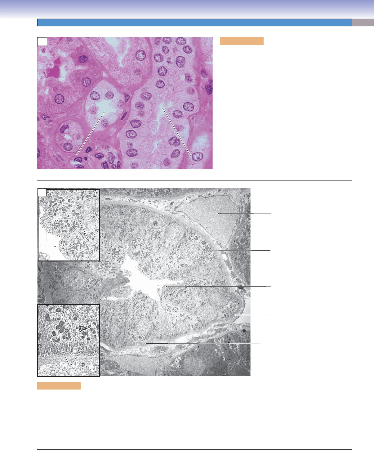
Figure 12-5B. Renal corpuscle, glomerulus and Bowman capsule. H&E, 402; insets (left) 921; insets (lower right) 183
A glomerulus housed within the Bowman capsule is shown here. The lighter space between these two structures is the Bowman space.
The upper left small inset shows the macula densa, a row of columnar cells that are densely packed together. This is a special sensory
structure of the distal tubule as it passes close to the afferent and efferent arterioles at the vascular pole (Fig. 12-5A). The macula
densa plays a role in monitoring ionic content and volume of the fi ltrate. The lower left small inset shows the Bowman space between
the glomerulus and the parietal layer of the Bowman capsule. The lower right inset shows the urinary pole.
Squamous cell
Squamous cell
of the parietal layer
of the parietal layer
Podocyte
Podocyte
Squamous cell
of the parietal layer
Podocyte
Parietal layer of
Parietal layer of
Bowman capsule
Bowman capsule
Parietal layer of
Bowman capsule
Urinary
Urinary
pole
pole
Urinary
pole
Afferent/efferent
Afferent/efferent
arteriole
arteriole
Afferent/efferent
arteriole
Macula densa
Macula densa
of distal tubule
of distal tubule
Macula densa
of distal tubule
Distal
Distal
convoluted tubules
convoluted tubules
Distal
convoluted tubules
Glomerulus
Glomerulus
Glomerulus
Bowman
Bowman
space
space
Bowman
Bowman
space
space
Bowman
space
Bowman
space
Proximal
Proximal
convoluted tubules
convoluted tubules
Proximal
convoluted tubules
Proximal
Proximal
convoluted
convoluted
tubules
tubules
Proximal
convoluted
tubules
Columnar cells of macular densa
Columnar cells of macular densa
Columnar cells of macular densa
Luman of the
Luman of the
distal tubule
distal tubule
Lumen of the
distal tubule
Glomerular
Glomerular
capillaries
capillaries
Glomerular
capillaries
Visceral layer of
Visceral layer of
Bowman capsule
Bowman capsule
Visceral layer of
Bowman capsule
B
CUI_Chap12.indd 228 6/2/2010 6:36:47 PM
CHAPTER 12
■
Urinary System
229
Figure 12-6A. Glomerulus, renal cortex. H&E,
310; insets 841
Each glomerulus is formed by a tuft of capillaries that is
fed by the afferent arteriole and drains into the efferent
arteriole. Pressure in the glomerulus due to resistance
of the efferent artery provides the force for fi ltration
into the Bowman space. The glomerular capillaries are
fenestrated capillaries lined by endothelial cells with
gaps (fenestrae) that lack the usual diaphragms (see
Fig. 9-13A). These capillaries are covered by processes
of podocytes. The endothelial cells, basal lamina, and
podocytes combine to form a glomerular fi ltration bar-
rier. Intraglomerular mesangial cells within the glom-
erulus provide structural support as well as phagocy-
tosis of debris and large molecules, thereby preventing
material from accumulating on the fi ltration barrier.
They also have a contractile capability, which may
function in regulating glomerular blood fl ow.
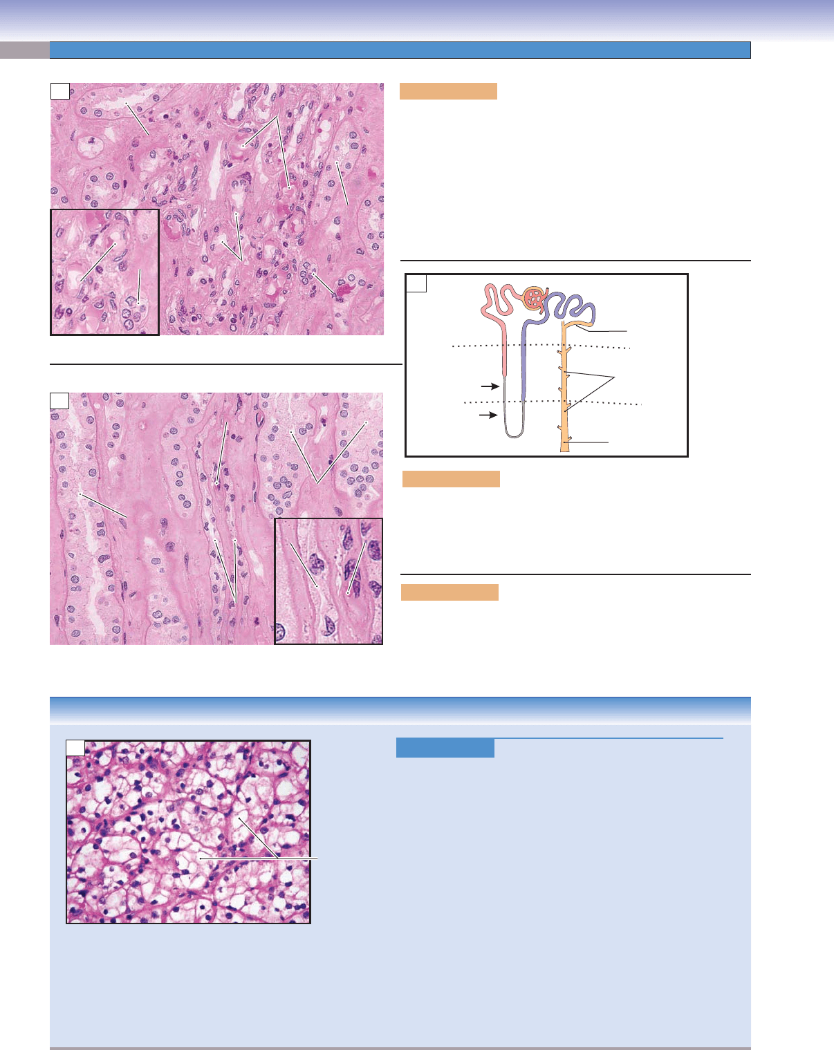
Figure 12-6B. Glomerulus and fi ltration barrier. EM, 16,667; insets, lower left 29,206; upper left 41,818; upper right (color),
H&E 219
The central part of a renal corpuscle is composed of a bed of capillaries, the glomerulus, and the cells and structures associated with the
glomerulus. In addition to the endothelial cells of the capillaries are two other cell types, podocytes and the intraglomerular mesangial
cells. The endothelial cells of the fenestrated glomerular capillaries coproduce and share a common basal lamina with the terminal
podocyte processes (pedicles, foot processes) that cover them. As blood fl ows through the capillaries, a fi ltrate of plasma is formed
as it passes through several layers (fenestrations of the capillary, trilayered basal lamina, and fi ltration slits between podocytes) to
enter the Bowman space (urinary space). Intraglomerular mesangial cells, lodged among the podocytes and endothelial cells, serve an
incompletely understood maintenance function. The upper left inset shows the basement membrane of the glomerulus, pedicles (small
podocyte processes), and cytoplasm of the endothelial cell, which together form a fi ltration barrier that selectively allows water, ions,
and small molecules to pass through but not large molecules and blood cells. The lower left inset shows foot processes of the podocyte
resting on the basement membrane of the glomerulus. The upper right color inset indicates the afferent and efferent arterioles.
Macula densa
Macula densa
of distal tubule
of distal tubule
Macula densa
of distal tubule
Fenestrated
Fenestrated
capillary wall
capillary wall
Fenestrated
capillary wall
Filtration slit
Filtration slit
Filtration slit
Lamina
Lamina
rara
rara
interna
interna
Lamina
rara
interna
Lamina
Lamina
rara
rara
externa
externa
Lamina
rara
externa
Lamina
Lamina
densa
densa
Lamina
densa
Endothelial
Endothelial
cell
cell
Endothelial
cell
Basement
Basement
membrane
membrane
Basement
membrane
Podocyte
Podocyte
Pedicles
Pedicles
Pedicles
Podocyte
Basement
Basement
membrane
membrane
Bowman
Bowman
space
space
Bowman
space
Basement
membrane
Erythrocyte
Erythrocyte
Erythrocyte
Erythrocyte
Erythrocyte
Erythrocyte
Luman of
Luman of
Glomerular
Glomerular
capillary
capillary
Lumen of
Glomerular
capillary
Basement
Basement
membrane
membrane
Lumen of
glomerular
capillary
Basement
membrane
Bowman space
Podocytes
Intraglomerular
mesangial cell
Capillaries
Capillaries
Capillaries
Arterioles
Arterioles
Arterioles
B
Macula densa
Macula densa
Macula densa
Arteriole
Arteriole
Arteriole
Glomerulus
Glomerulus
Bowman space
Bowman space
Glomerulus
Bowman space
Bowman
Bowman
capsule
capsule
Distal
Distal
convoluted tubules
convoluted tubules
Distal
convoluted tubules
Bowman
capsule
Mesangial
Mesangial
cell
cell
Mesangial
cell
Endothelial cell
Endothelial cell
Endothelial cell
Podocytes
Podocytes
Podocytes
B
CUI_Chap12.indd 229 6/2/2010 6:36:52 PM
230
UNIT 3
■
Organ Systems
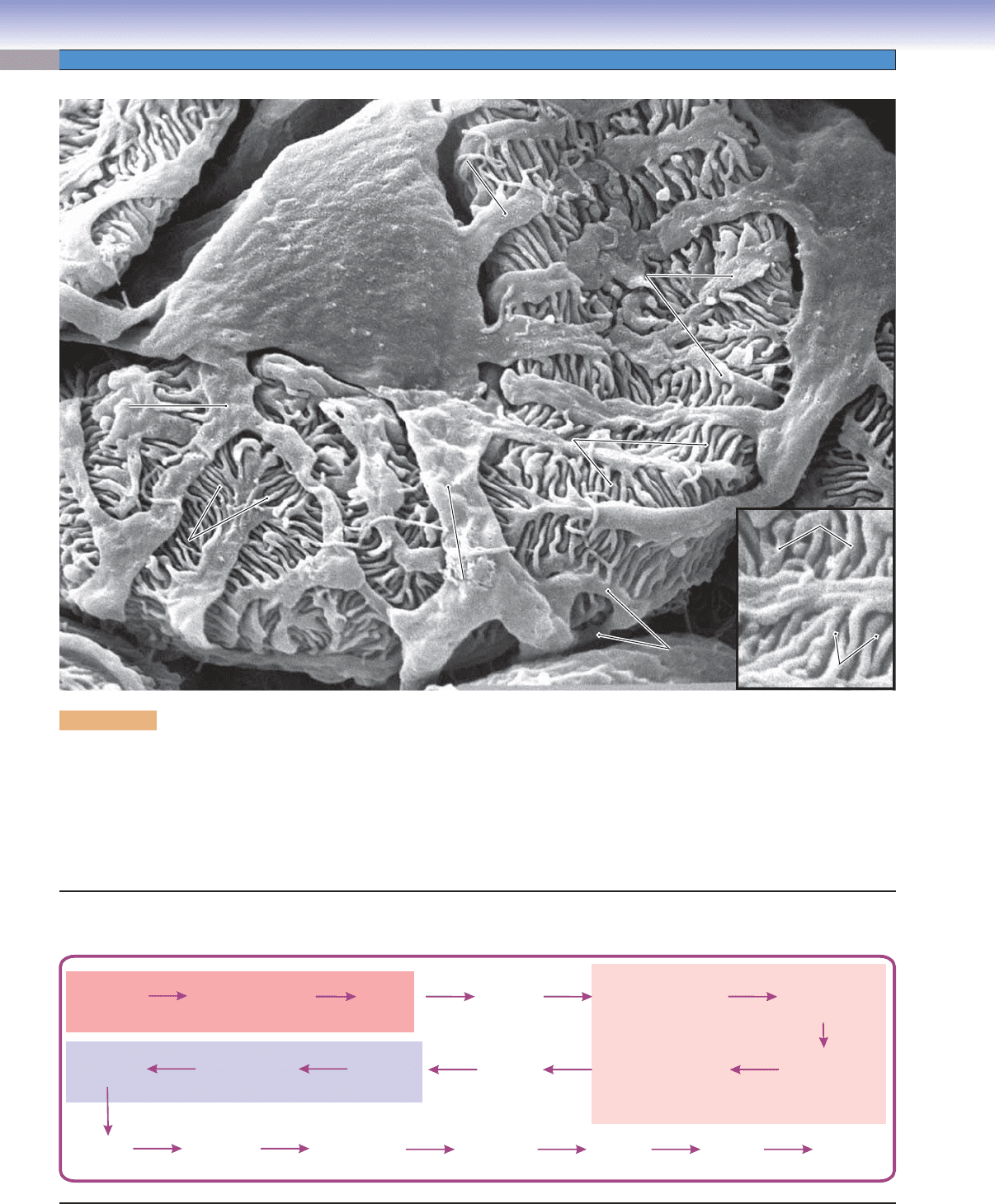
Figure 12-7. Glomerulus and podocyte. SEM, 9,677
This scanning electron microscopy (SEM) image shows that the surface of the glomerulus is entirely covered by podocytes and their
processes. Each podocyte is composed of a cell body (with nucleus) and several branching processes. The small terminal branches
that cover the capillaries are called foot processes or pedicles. The pedicles from two podocytes interdigitate with each other. The
gaps between adjacent pedicles are referred to as fi ltration slits, which are bridged by fi ltration slit diaphragms. The inner core of
the pedicles is supported by actin fi laments. The inner core of the primary processes is supported mainly by microtubules and inter-
mediate fi laments. The podocytes and their unique arrangement are important components in establishing the glomerular fi ltration
barrier.
Podocyte
Podocyte
Podocyte
Pedicles
Pedicles
Pedicles
Filtration slits
Filtration slits
Filtration slits
Branches of
Branches of
primary processes
primary processes
Branches of
primary processes
Primary
Primary
process
process
Primary
process
Pedicles
Pedicles
Pedicles
Primary
Primary
process
process
Primary
process
Podocyte
Podocyte
Podocyte
Primary
Primary
process
process
Primary
process
Branches of
Branches of
primary processes
primary processes
Branches of
primary processes
Filtration slits
Filtration slits
Filtration slits
Bowman space
(filtrate into the space)
Glomerulus
(filters blood)
Urinary
pole
Proximal
convoluted
tubule
Thick descending limb
(proximal straight
tubule)
Thin descending limb
(descending
thin segment)
Ascending thin limb
(ascending
thin segment)
Thick ascending limb
(distal straight
tubule)
Distal
convoluted
tubule
cortical
collecting
tubule
Collecting
duct
Papillary
duct
Minor
calyx
Area
cribrosa
Major
calyx
Renal
pelvis
Ureter
Urinary
bladder
(store)
Urethra
(to outside)
Loop of Henle
Renal corpuscle
Collecting system
Production and Drainage of Urine
CUI_Chap12.indd 230 6/2/2010 6:36:57 PM
CHAPTER 12
■
Urinary System
231
Figure 12-8B. The nephron and collecting system of the kidney.
The kidney is composed of nephrons and a collecting system. Each nephron comprises a renal corpuscle, a proximal convoluted tubule, a
loop of Henle, and a distal convoluted tubule. The U-shaped loop of Henle connects the proximal convoluted tubules to the distal convo-
luted tubules. The loop of Henle creates a high concentration of solutes in the interstitium of the medulla, which is essential in controlling
the concentration of urine. The collecting system (yellow) includes cortical collecting tubules, collecting ducts, and papillary ducts. The
dashed lines indicate the junction between the cortex and the medulla and the medullary ray region in the cortex. When the blood pressure
in the glomerular capillaries is within certain limits, water and some solutes of the blood plasma are forced through the fi ltration barrier
into the Bowman space and then into the proximal convoluted tubules. Most glucose and amino acids and a large volume of water and
salt are reabsorbed by the proximal convoluted and straight tubules before the fi ltrate enters the descending thin segment of the loop
of Henle. The thin descending segment is highly permeable to water and less permeable to salt, so water passes from the lumen to the
interstitium of the medulla and returns back to the blood circulation via the vasa recta. The thin ascending limb is impermeable to water
but permeable to salt, and the thick ascending segment, which is also impermeable to water, actively pumps salt into the interstitium. As
a result, the concentration of solutes increases to about four times the normal amount in the interstitium of the deep medulla. This hyper-
osmotic environment drives the movement of water from the lumens of the collecting ducts, therefore increasing the concentration of the
urine. The permeability to water in the cortical collecting tubules and collecting ducts (and, therefore, the fi nal concentration of urine) is
controlled by the level of ADH released by pituitary glands. The reabsorption of various ions by the distal convoluted tubule is controlled
by hormones, primarily aldosterone. The fi nal urine is collected by papillary ducts and emptied at the area cribrosa into the minor calyx.
Collecting
Collecting
duct
duct
Collecting
duct
Collecting
Collecting
duct
duct
Collecting
duct
Collecting
Collecting
duct
duct
Collecting
duct
Glomerulus
Glomerulus
Glomerulus
Proximal
Proximal
straight tubule
straight tubule
Proximal
straight tubule
Distal straight
Distal straight
tubule
tubule
Distal straight
tubule
Proximal
Proximal
straight
straight
tubule
tubule
Proximal
straight
tubule
Distal
Distal
straight
straight
tubule
tubule
Distal
straight
tubule
Medullary ray
Medullary ray
Medullary ray
A
D. Cui &T. Yang
Area cribrosa
Papillary duct
(duct of Bellini)
Papillary duct
(duct of Bellini)
Collecting duct
Medullary
ray
Thick descending limb
(proximal straight tubule)
Loop of
Henle
Thick ascending limb
(distal straight tubule)
Thin descending limb
(descending thin segment)
Thin ascending limb
(ascending thin segment)
Distal
convoluted tubule
Macula densa (region)
Glomerulus
Proximal
convoluted tubule
Cortical
collecting tubule
Cortex
Medulla
Renal
medulla
Renal
cortex
H O (+ ADH)
2
HO
2
Cl
–
,Na
+
B
Cl
–
,Na
+
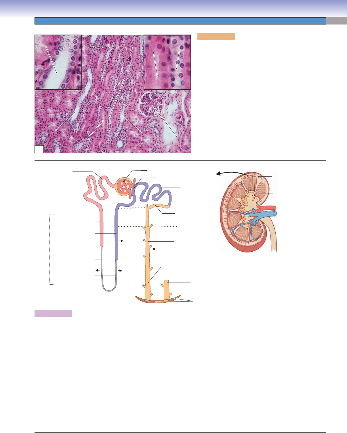
Figure 12-8A. Medullary ray, renal cortex. H&E,
142; insets 448
Each medullary ray is composed of proximal straight
tubules, distal straight tubules, and collecting ducts. These
tubules run parallel to each other within a medullary ray,
which separates the glomeruli into groups. Although med-
ullary rays are located in the renal cortex region, they are
an extension of the renal medulla. The proximal straight
tubules are lined by cuboidal cells with acidophilic cyto-
plasm and long microvilli. The distal straight tubules and
collecting ducts are lined by cuboidal cells with clear cyto-
plasm. However, the collecting ducts have a larger lumen
and more distinct cell-to-cell borders than do distal straight
tubules. The proximal straight tubules convey fi ltrate from
the proximal convoluted tubules into the thin segment
tubules. The distal straight tubules convey the fi ltrate into
the distal convoluted tubules, from which it drains into the
collecting tubules and ducts (see below).
CUI_Chap12.indd 231 6/2/2010 6:36:59 PM
232
UNIT 3
■
Organ Systems
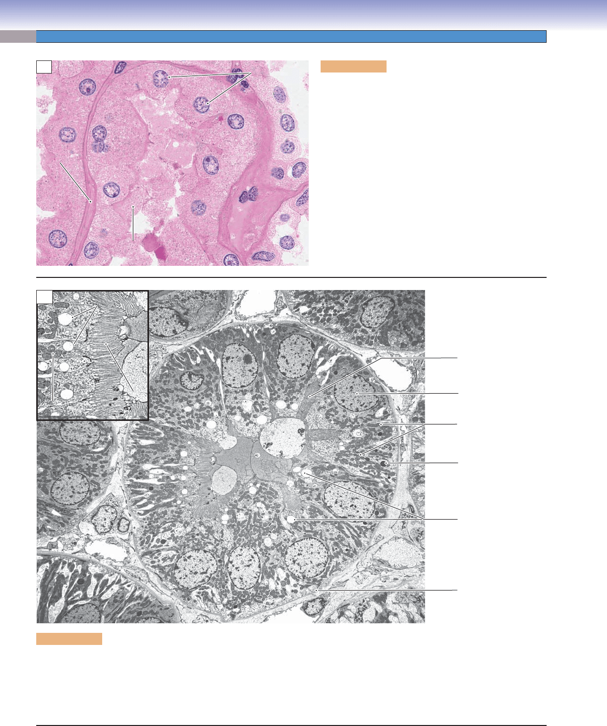
Figure 12-9B. Proximal tubule. EM, 4,606; inset 9,208
This is a cross section of a proximal tubule. It is lined by cuboidal and low columnar cells with apical microvilli, which form a brush
border, a feature that is associated with reabsorption function. The numerous mitochondria are more concentrated at the basolateral
surface where they support the energy requirements of sodium pumps located in the expanded plasmalemma. The apical regions of
the cells contain pinocytotic vesicles, which refl ect the uptake of proteins that evaded the fi ltration barrier in the renal corpuscle and
entered the fi ltrate. The inset shows long microvilli and pinocytotic vesicles in the apical surface of the cells.
Basement
Basement
membrane
membrane
Basement
membrane
Brush border
Brush border
with glycocalyx
with glycocalyx
Brush border
with glycocalyx
Nuclei of the
Nuclei of the
cuboidal cells
cuboidal cells
Nuclei of the
cuboidal cells
Lumen
Lumen
Lumen
A
Pinocytotic
Pinocytotic
vescle
vescle
Pinocytotic
vesicle
Mitochondria
Mitochondria
Mitochondria
Microvilli
Microvilli
Microvilli
Nucleus
Nucleus
Nucleus
Lysosome
Lysosome
Lysosome
Pinocytotic
Pinocytotic
vesicles
vesicles
Pinocytotic
vesicles
Basement
Basement
membrane
membrane
Basement
membrane
Microvilli
Microvilli
Microvilli
Lysosome
Lysosome
Mitochondria
Lumen
Lumen
Lumen
B
Figure 12-9A. Proximal tubules, renal cortex. H&E,
754
Both proximal convoluted and straight tubules have a
similar structure and function. They are lined by large
acidophilic cuboidal cells with brush borders formed
by numerous long microvilli. The brush border extends
into the lumen, which, in conjunction with postmortem
changes, makes the lumen appear smaller and fi lled with
acidophilic material (brush border and glycocalyx). The
proximal tubules have a substantial reabsorption function.
About 65% of the water and sodium and more than 90%
of glucose, amino acids, and bicarbonate are reabsorbed
by the proximal convoluted tubules. The proximal convo-
luted tubules are located in the cortical labyrinth and are
connected to the renal corpuscle at the urinary pole. The
proximal straight tubules are part of the loop of Henle
(Fig. 12-8B).
CUI_Chap12.indd 232 6/2/2010 6:37:03 PM
CHAPTER 12
■
Urinary System
233
Figure 12-10A. Distal tubules. H&E, 739
The distal tubules are lined by small cuboidal cells
with faintly eosinophilic or clear cytoplasm. The lat-
eral boundaries between cells are not as distinguishable
as those of the collecting duct. The distal convoluted
and straight tubules are similar in structure. The distal
straight tubule exits the medullary ray and approaches
the vascular pole of the renal corpuscle of the same
nephron to which the distal tubule belongs. As the distal
tubule passes adjacent to the afferent and efferent arteri-
oles, part of its wall becomes modifi ed as a sensory struc-
ture, macula densa, which monitors ionic content and
water volume of the fi ltrate (Fig. 12-5A,B). The macula
densa is considered to mark the transition from the dis-
tal straight to the distal convoluted tubule. This fi gure
shows the distal convoluted tubules in the renal cortex.
Distal tubules function mainly to remove sodium and
add potassium ions to the fi ltrate when stimulated by
aldosterone, a hormone produced by the adrenal gland.
Proximal
Proximal
tubule
tubule
Proximal
tubule
Proximal
Proximal
tubule
tubule
Proximal
tubule
Lumen of
Lumen of
distal tubule
distal tubule
Lumen of
distal tubule
Lumen of
Lumen of
collecting duct
collecting duct
Lumen of
collecting duct
A
Figure 12-10B. Distal tubule. EM, 4,441; insets 7,377
A cross section of a distal tubule, which is lined by cuboidal and columnar cells, is shown. In contrast to the extravagant brush
border of cells lining the proximal tubule, these cells have just a few, short microvilli. These cells have basally located nuclei and
tightly interdigitated lateral walls. In distal tubules, there are many mitochondria in the cytoplasm as there are in the proximal
tubules. The upper left inset shows short and irregular microvilli bulging into the lumen and many mitochondria beneath them. The
lower left inset shows a nucleus and basal enfolding (basal plasma membrane enfolding) of the cell. The basal enfolding is due to
corrugation of the cell membrane in the basal region of the cell. This increases the surface area of the cell and is closely associated
with mitochondria, which produce adenosine triphosphate for active transport of ions.
Nucleus
Nucleus
Nucleus
Blood vessel
Blood vessel
Blood vessel
Microvilli
Microvilli
Microvilli
Basement
membrane
Basal enfolding
Basal enfolding
Basal enfolding
Lumen
Lumen
Lumen
Microvilli
Microvilli
Microvilli
Mitochondria
Mitochondria
Mitochondria
Basal enfolding
Basal enfolding
Basal enfolding
B
CUI_Chap12.indd 233 6/2/2010 6:37:06 PM
234
UNIT 3
■
Organ Systems
Figure 12-11A. Medullary tubules, outer zone of the medulla.
H&E, 296; inset 435
The renal medulla is composed of the loop of Henle, the collecting
ducts, and the papillary ducts (ducts of Bellini). It can be divided
into an outer zone and an inner zone. The thin segment tubules
and distal straight tubules of the loop of Henle and collecting
ducts of the outer zone are shown here. The inner zone contains
only thin segments and collecting ducts, along with the vasa recta.
Blood vessels (vasa recta) are found throughout the medulla.
Vasa recta
Vasa recta
(blood vessels)
(blood vessels)
Vasa recta
(blood vessels)
Distal straight
Distal straight
tubule
tubule
Distal straight
tubule
Thin
Thin
segment
segment
Thin
segment
Thin
Thin
segment
segment
Thin
segment
Vasa recta
Vasa recta
Vasa recta
Thin
Thin
segment
segment
Thin
segment
Collecting
Collecting
duct
duct
Collecting
duct
A
D. Cui &T. Yang
Loop of Henle
Papillary duct
Collecting
duct
Collecting
tubule
Cortex
Outer
medulla
(Fig. 12-11A)
(Fig. 12-11B)
Inner
medulla
B
Collecting
Collecting
duct
duct
Collecting
duct
Vasa
Vasa
recta
recta
Vasa
recta
Thin
Thin
segment
segment
Thin
segment
Vasa
Vasa
recta
recta
Vasa
recta
Collecting
Collecting
duct
duct
Collecting
duct
Thin
Thin
segment
segment
Thin
segment
C
CLINICAL CORRELATION
Figure 12-11D.
Renal Cell Carcinoma (Clear Cell Type).
H&E, 216
Renal cell carcinoma, which arises from the renal tubular
epithelium, is the most common renal cancer in adults. Risk
factors for development of renal cell carcinoma include smok-
ing, exposure to toxic substances, chronic renal failure, and
acquired cystic disease of the kidney as well as genetic predis-
position in various familial syndromes. Renal carcinoma pres-
ents clinically with hematuria, abdominal mass, or fl ank
pain.
Renal cell carcinoma tends to metastasize early, especially to the
lungs and bones. On gross examination, renal cell carcinoma is
well circumscribed, lobulated, and yellow, with areas of hemor-
rhage and necrosis. The most common renal cell carcinoma is
the clear cell type, the cells of which may be arranged in cords,
nests, or tubules. The cells are large and polygonal with clear or
granular cytoplasm. Other types of renal cell carcinoma include
papillary carcinoma, chromophobe carcinoma, and collecting
duct carcinoma. Treatment is primarily surgical removal, with
lesser roles for immunotherapy and chemotherapy.
Clear cells
(tumor cells)
D
Figure 12-11B. Orientation of the kidney tubules in the cortex
and medulla.
This illustration shows the orientation of the kidney tubules in
the cortex and medulla. The outer zone and inner zone of the
medulla indicate the levels of Figures 12-11A and 12-11B.
Figure 12-11C. Medullary tubules, inner zone of the medulla.
H&E, 296; inset 726
This fi gure shows the inner zone of the medulla. The collecting
ducts gradually increase in size. The thin segment tubules and the
vasa recta (blood vessels) are seen here.
CUI_Chap12.indd 234 6/2/2010 6:37:10 PM