Feny? D. (Ed.) Computational Biology
Подождите немного. Документ загружается.

171
3-D Structures of Macromolecules Using Single-Particle Analysis in EMAN
tomographic series of orientations, i.e., a series of particle
orientations covering a 180° rotation of the object from any
one arbitrary orientation.
10. The “boxes” menu in boxer also has an “autobox from refer-
ences” option. Better particle picking may be achieved by
preparing a set of references from the refine2d.py results, and
using these to rebox the micrographs. After an initial 3-D
refinement, projections of the model can also be used in this
process with makeboxref.py. Note 7 should also be consid-
ered, however.
11. It is theoretically impossible to determine absolute handed-
ness from untilted single-particle data, since the recorded
images represent near ideal projections of the object. To deter-
mine absolute handedness experimentally, some other protocol,
such as tomography or random conical tilt, must be followed.
At sufficiently high resolutions, however, it may be possible
to determine handedness by comparison of fragments to
X-ray crystal structures or homology models. At even higher
resolutions (~4 Å), the pitch of the alpha-helices may become
visible, also solving the handedness problem.
12. For particles with greater than threefold rotational symmetry,
the program startcsym can be used to generate initial models
from raw particle data. Prior to use, the particles should first
be centered with cenalignint. In some cases the “fixrot=90”
option will need to be specified to get an accurate model.
Also note that for Dn symmetries, the symmetry must still be
specified as Cn.
13. Generally the class-averages produced by refine2d.py will give
some idea of what symmetry is present, but in general, when
dealing with objects of unknown symmetry, the best approach
is to try refining with the suspected symmetry, assess the
results, then relax the symmetry for a few refinement itera-
tions to see how the model changes. The process can then be
repeated for another symmetry choice and compared.
14. The threshold value to use for resolution assessment has been
hotly debated over the years. Other values that have been
used include 0.33 (20), 0.143 (21), and use of a sigma curve
(22). It is generally agreed among reviewers, however, that
FSC curves should be included in the supplementary data
when publishing. This serves the dual purpose of allowing the
reviewer to assess the shape of the FSC curve in addition to
applying a threshold of their choice.
15. Note that in many cases EMAN1 will use the IMAGIC file
format by default. IMAGIC files separate images into two
parts, a “.hed” file containing image header information, and
a “.img” file containing the actual image data. These two files
172 Ludtke
exist as a pair, and one file must never be renamed, moved, or
deleted without also removing its companion. When issuing
EMAN commands, either of these files may be specified.
EMAN also supports most other TEM formats such as MRC,
SPIDER, TIFF, DM3, PIF, etc. The Gatan .DM3 format is
supported, but is read-only.
16. Image sampling can be quite important. Generally speaking,
interpretable images can only be obtained up to resolutions
~3× the pixel size. That is, a 3 Å/pixel image can at best pro-
duce a 9 Å resolution reconstruction. However, sampling too
finely can also lead to a number of problems. In addition to
the obvious issue of computational inefficiency, certain algo-
rithms make assumptions about the sampling level. While
some additional oversampling, perhaps as much as 5×, should
be fine, beyond this point reconstructions may actually
become worse, not better.
17. EMAN2 has a program called e2initialmodel.py, which can
automate the entire task of initial model determination from
class-averages, but using it will require some familiarity with
EMAN2.
References
1. Frank J. Three-dimensional electron micros-
copy of macromolecular assemblies: visualiza-
tion of biological molecules in their native state.
New York: Oxford University Press, 2006.
2. Ludtke SJ, Baker ML, Chen DH, Song JL,
Chuang DT & Chiu W. De novo backbone
trace of GroEL from single particle electron
cryomicroscopy. Structure 2008;16:441–8.
3. Jiang W, Baker ML, Jakana J, Weigele PR, King
J & Chiu W. Backbone structure of the infec-
tious epsilon15 virus capsid revealed by electron
cryomicroscopy. Nature 2008;451:1130–4.
4. Zhang X, Settembre E, Xu C, et al. Near-
atomic resolution using electron cryomicros-
copy and single-particle reconstruction. Proc
Natl Acad Sci USA 2008;105:1867–72.
5. Yu X, Jin L & Zhou ZH. 3.88 Å structure of
cytoplasmic polyhedrosis virus by cryo-electron
microscopy. Nature 2008;453:415–9.
6. Glaeser RM. Electron crystallography of bio-
logical macromolecules. New York: Oxford
University Press, 2007.
7. Chen DH, Song JL, Chuang DT, Chiu W &
Ludtke SJ. An expanded conformation of single-
ring GroEL–GroES complex encapsulates an
86 kDa substrate. Structure 2006;14:1711–22.
8. Brink J, Ludtke SJ, Kong Y, Wakil SJ, Ma J
& Chiu W. Experimental verification of
conformational variation of human fatty acid
synthase as predicted by normal mode analysis.
Structure 2004;12:185–91.
9. Leschziner AE & Nogales E. Visualizing flex-
ibility at molecular resolution: analysis of het-
erogeneity in single-particle electron
microscopy reconstructions. Annu Rev
Biophys Biomol Struct 2007;36:43–62.
10. Ludtke SJ, Baldwin PR & Chiu W. EMAN:
semiautomated software for high-resolution
single-particle reconstructions. J Struct Biol
1999;128:82–97.
11. Tang G, Peng L, Baldwin PR, et al. EMAN2: an
extensible image processing suite for electron
microscopy. J Struct Biol 2007;157:38–46.
12. Ludtke SJ, Jakana J, Song JL, Chuang DT &
Chiu W. A 11.5 A single particle reconstruc-
tion of GroEL using EMAN. J Mol Biol
2001;314:253–62.
13. Frank J, Radermacher M, Penczek P, et al.
SPIDER and WEB: processing and visualiza-
tion of images in 3D electron microscopy and
related fields. J Struct Biol 1996;116:190–9.
14. van Heel M, Harauz G, Orlova EV, Schmidt
R & Schatz M. A new generation of the
IMAGIC image processing system. J Struct
Biol 1996;116:17–24.
15. Grigorieff N. FREALIGN: high-resolution
refinement of single particle structures. J Struct
Biol 2007;157:117–25.
173
3-D Structures of Macromolecules Using Single-Particle Analysis in EMAN
16. Stewart A & Grigorieff N. Noise bias in the
refinement of structures derived from single
particles. Ultramicroscopy 2004;102:67–84.
17. Ludtke SJ, Chen DH, Song JL, Chuang DT
& Chiu W. Seeing GroEL at 6 A resolution by
single particle electron cryomicroscopy.
Structure 2004;12:1129–36.
18. Saad A, Ludtke SJ, Jakana J, Rixon FJ, Tsuruta
H & Chiu W. Fourier amplitude decay of
electron cryomicroscopic images of single
particles and effects on structure determina-
tion. J Struct Biol 2001;133:32–42.
19. Zhu Y, Carragher B, Glaeser RM, et al.
Automatic particle selection: results of a
comparative study. J Struct Biol 2004;145:
3–14.
20. Penczek PA. Three-dimensional spectral
signal-to-noise ratio for a class of recon-
struction algorithms. J Struct Biol 2002;138:
34–46.
21. Rosenthal PB & Henderson R. Optimal
determination of particle orientation, abso-
lute hand, and contrast loss in single-parti-
cle electron cryomicroscopy. J Mol Biol
2003;333:721–45.
22. van Heel M & Schatz M. Fourier shell corre-
lation threshold criteria. J Struct Biol 2005;
151:250–62.

175
Chapter 10
Computational Design of Chimeric Protein Libraries
for Directed Evolution
Jonathan J. Silberg, Peter Q. Nguyen, and Taylor Stevenson
Abstract
The best approach for creating libraries of functional proteins with large numbers of nondisruptive amino
acid substitutions is protein recombination, in which structurally related polypeptides are swapped among
homologous proteins. Unfortunately, as more distantly related proteins are recombined, the fraction of
variants having a disrupted structure increases. One way to enrich the fraction of folded and potentially
interesting chimeras in these libraries is to use computational algorithms to anticipate which structural
elements can be swapped without disturbing the integrity of a protein’s structure. Herein, we describe
how the algorithm Schema uses the sequences and structures of the parent proteins recombined to
predict the structural disruption of chimeras, and we outline how dynamic programming can be used to
find libraries with a range of amino acid substitution levels that are enriched in variants with low Schema
disruption.
Key words: Chimera, Directed evolution, Dynamic programming, Optimization, Protein design,
Recombination
Proteins are widely used for synthetic biology applications,
but they often do not exhibit the functional properties desired
for engineered biological systems. However, protein variants are
thought to exist in protein sequence space that meet the specifi-
cations of almost any artificially engineered biological system
imaginable. Evidence for this comes from studies using knowl-
edge-based protein design, which have identified proteins with
structures and functions distinct from those observed in nature
(1, 2). Unfortunately, our understanding of protein sequence-
structure–function relationships is not yet sophisticated enough
to consistently alter protein functions rationally, especially when
the design goal is to optimize a preexisting property. Directed
1. Introduction
David Fenyö (ed.), Computational Biology, Methods in Molecular Biology, vol. 673,
DOI 10.1007/978-1-60761-842-3_10, © Springer Science+Business Media, LLC 2010

176 Silberg, Nguyen, and Stevenson
evolution, in contrast, has repeatedly proven effective at protein
optimization when applied alone and when used with knowledge-
based mutagenesis (3). In this approach, a selection (or screen) is
used to sieve through libraries of artificial protein variants to find
those rare mutations that lead to desired changes in function.
Directed evolution has several limitations that must be consid-
ered when engineering a protein. Typically, functional proteins
cannot be fished out of libraries encoding random protein
sequences. The frequency with which functional proteins occur in
protein sequence space is thought to be miniscule compared with
the maximum number of protein variants that can be evaluated in
a given experiment (4). One way to improve your chances of dis-
covering proteins with a desired function is to increase the fraction
of folded variants in your combinatorial library. This can be achieved
by infusing into your library design some knowledge about the
protein(s) used as parents for directed evolution. This information
can draw from our understanding of protein stability (5), family
sequences (6), structure–function relationships (7), and laboratory
evolution experiments (8). In this chapter, we describe how one
can use sequence, structural, and thermodynamic information to
enrich the fraction of functional protein variants in a library created
using protein recombination. A protocol is outlined for using the
Schema algorithm to identify libraries with a user-defined level of
amino acid substitutions that minimize structure disruption
(9–11).
With Schema, the three-dimensional structural coordinates from
one of the parent proteins being recombined are required to esti-
mate chimera disruption (see Note 1). Only those sequence posi-
tions with defined structural coordinates are considered in the
calculation of structural disruption (see Note 2). In cases where
there are no structural reports for the proteins being recombined,
structural coordinates can be generated using algorithms that
generate homology models of proteins (see Note 3), such as
Swiss-Model (12).
The primary amino acid sequences of the proteins being recom-
bined must be aligned before performing calculations of struc-
tural disruption. If PDB coordinates are available for all of the
proteins being recombined, the sequence alignment should be
generated using the SwissProt or Combinatorial-Extension
algorithms, which use structural information to guide the creation
of a sequence alignment (13, 14). In all other cases, multiple
2. Materials
2.1. Protein Structural
Coordinates
2.2. Protein Sequence
Alignment

177
Computational Design of Chimeric Protein Libraries for Directed Evolution
sequence alignments should be created using algorithms that only
consider sequence information, such as the BLAST (15) and
ClustalW2 (16) algorithms.
Schema posits that the best way to conserve a protein’s structure
upon recombination of homologous proteins is to minimize the
number of residue–residue interactions in the parental structures
that are altered by recombination (see Fig. 1). The physiochemi-
cal characteristics of residues incorporated in chimeras at each
position are ignored because they have been preselected to be
compatible with the parental structure (17). Interactions are sim-
ply defined as any pair of residues whose side chains are within a
defined cutoff distance d
c
(see Note 4). The major advantages of
Schema are its simplicity (11), proven effectiveness (10), and abil-
ity to be used with the library design algorithm Recombination
As a Shortest-Path Problem (RASPP) (9). For any set of parent
proteins being recombined, RASPP uses dynamic programming
to identify the optimal tradeoff surface for mutation and struc-
tural disruption in library space. For a library of a user-defined
size (e.g., n crossovers between two parents), RASPP identifies
libraries with a range of average amino acid substitution levels ámñ
that have lower than average Schema disruption áE ñ (9).
Chimeric libraries optimized using Schema and RASPP are cre-
ated using Sequence-Independent Site-Directed Chimeragenesis
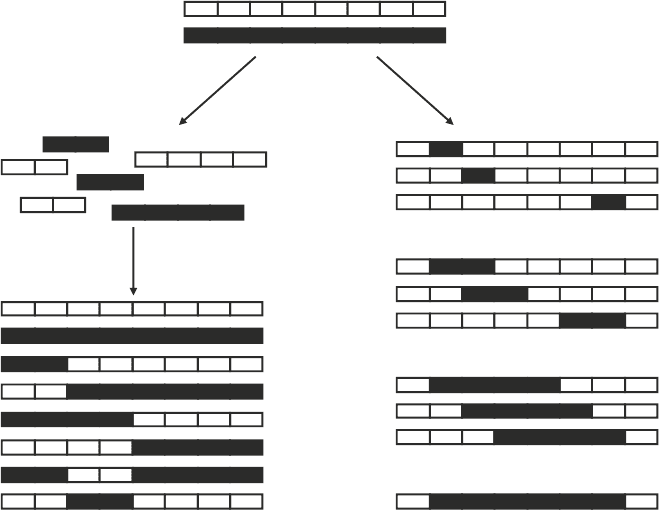
(SISDC) (8, 18, 19). With SISDC, the number of parents and
crossover sites controls library size (see Fig. 2a). Upon creating a
n-crossover library using p parents, the number of possible chi-
meric variants is p
n+1
(see Note 5). The amino acid substitution
level accessible in your chimeras can also be adjusted through
your parental choice (see Note 6). The number of amino acid
substitutions that can be incorporated into a chimera increases as
the sequence identity among the parents used for recombination
decreases (17). The accessible substitution level also increases as
the number of parents recombined increases.
When recombining homologous proteins, it is thought to be best
to recombine the most closely related proteins that will yield your
desired level of amino acid substitution (17). Libraries created in
this way are predicted to contain a higher fraction of folded (and
functional) variants than libraries created using more distantly
related parents. In addition, the thermostability of the proteins
being recombined should be considered when using SISDC.
3. Methods
3.1. Library Diversity
3.2. Choosing Parental
Proteins
178 Silberg, Nguyen, and Stevenson
Among protein homologs, those with higher stability have been
shown to yield libraries that are enriched in the number of unique,
functional, and potentially interesting proteins in both random
mutation (20) and recombination (21) experiments. Thus, when
you have a choice of multiple proteins for SISDC, you should
00000000
1
0
1
0000
1
0
00000
1
000
1
0
1
000
1
0
0
1
00
1
00
00
0
00000000
0
0
0
0000
0
0
00000
0
000
1
0
1
000
1
0
0
1
00
1
00
00
0
0000000 0
0
0
0
0000
0
0
00000
01
000
1
0
1
000
1
0
0
1
00
1
00
00
0
1. Identify all residue-residue interactions (solid lines &
ij
pairs = 1) using
structural coordinates from one of the parent proteins recombined.
1234
5678910
1234
5678910
123
910
Chimera
2. Remove interactions that cannot be broken by recombination using the
sequence alignment of the parent proteins.
3. Count the interactions broken (left = dashed lines; right = black boxes) when
a chimera inherits peptides from different parents (left = gray & white boxes).
7
Residue
Residue
4
56 8
Residue
j
Residue
i
=
=
=
1
1
Fig. 1. Protocol for calculating the structural disruption of a chimera. When recombining two structurally related proteins,
you first generate a contact matrix that accounts for all pairwise residue–residue interactions in the parent structures.
Interactions involving residues that are identical in the parents are removed from the matrix, since they cannot be broken
by recombination. The structural disruption E is simply the number of residue–residue contacts broken by recombination
(11). The chimera shown, which inherits residues 4–8 from parent X and all other residues from parent Y (1–3 and 9–10),
has an E = 2.
179
Computational Design of Chimeric Protein Libraries for Directed Evolution
recombine proteins that exhibit the greatest thermostability
available, provided that there are no other functional differences
in the enzymes.
When creating a chimera by recombining homologous proteins,
a matrix s
i
is created to indicate which parent is incorporated at
each position i in the chimeric sequence. For example, if the first
residue in a chimera (i = 1) is inherited from the first parent, then
s
1
= 1, but if that residue is inherited from the second parent,
then s
1
= 2. In addition, the structural coordinates of one parent
protein and a sequence alignment of all parents proteins recombined
are read into the matrices pdb and align, respectively (see Note 7).
These three matrices are used to calculate the disruption of each
chimera, which is defined as
3.3. Calculating
the Structural
Disruption
of a Single Chimera
11
NN
ij ij
i ji
ECD
= =+
=
∑∑
Residue number
12345678
Library assembly
strategy
Double-
crossover chimeras
ab
Fig. 2. Sequence-independent site-directed chimeragenesis. (a) When constructing a n-crossover library, n recombination
sites are chosen that define the possible polypeptide inheritance in the chimeras. In the example shown, two
crossovers between two natural proteins yield 2
3
sequences, two of which are the original parent proteins. (b) A simple
way to find chimeras for calibration experiments is to calculate E for all chimeras in which a single contiguous poly-
peptide is exchanged among the parent proteins (22). By plotting the E vs. m for all such chimeras, one can rapidly
identify chimeras with a range of amino acid substitution and disruption levels that are easy to build for calibration
studies (see Fig. 3).
180 Silberg, Nguyen, and Stevenson
where N specifies the number of residues in the structure used for
calculations, C
ij
indicates whether residues i and j are sufficiently
close in the parental structure to represent an important interac-
tion that should not be broken, and D
ij
designates whether the
interaction between residues i and j in the chimera is present in
either of the parents recombined. C
ij
is given a value of 1 when
residues i and j are inherited from different parent proteins (s
i
¹ s
j
)
and when these residues are interacting, i.e., within a user-defined
cutoff distance d
c
in the parental structure. C
ij
is given a value of
0 in all other cases (see Notes 8 and 9). Because some exchanged
polypeptides do not effectively disrupt residue–residue interac-
tions observed in the parents, the delta function D
ij
uses the
sequence alignment of the proteins recombined to determine
which of the residue–residue interactions in a chimera are distinct
from those present in either of the parents. In cases where an
interaction between residues i and j in a chimera involves amino
acids distinct from those at structurally related positions in all of
the parent proteins, D
ij
is given a value of 1 to indicate that this
new pairwise interaction is capable of disrupting the chimeras’
structure (otherwise D
ij
= 0).
Because of its simplicity, Schema cannot differentiate the
structural disruption of chimeras that have opposite polypeptide
inheritance (see Note 10). In addition, this algorithm does not
account for intersubunit residue–residue contacts that are broken
by recombination, although these could be easily considered
(see Note 11).
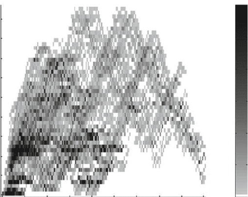
Functional analysis of chimeric libraries is time consuming and
expensive, so you should calibrate the disruption nature of E for
the proteins that you are recombining before selecting a library to
construct for directed evolution (22). The easiest way to do this
is to evaluate the folding (and function) of a small number of
chimeras created by swapping single contiguous polypeptides (see
Fig. 2b). To do this, you first calculate E for all possible double
crossover chimeras and their amino acid substitution level m (see
Fig. 3). From this library, you select a handful of chimeras with a
broad range of E and m values (e.g., 20), you build the genes
encoding these chimeras using splicing by overlap extension in
the laboratory (22), and you characterize which of these proteins
exhibit parent-like structure (see Note 12). The results from these
measurements are then used to identify a threshold level of dis-
ruption below which a majority of chimeras retain parent-like
structure. This threshold is used to estimate the fraction of vari-
ants that are folded in chimeric libraries identified by RASPP and
to guide the selection of a chimeric library to construct (8).
The structural disruption of individual chimeras can be rapidly
calculated using Schema as described above (11). However,
3.4. Calibrating
the Disruptive Nature
of Substitutions
3.5. Using RASPP
for Library Design
181
Computational Design of Chimeric Protein Libraries for Directed Evolution
this approach is not practical for finding libraries that minimize
the average disruption áE ñ of chimeras subject to constraints on the
average amino acid substitution level ámñ, i.e., libraries with the
best energy-diversity tradeoff (see Note 13). While no optimi-
zation protocol has been described for minimizing the áE ñ of a
library subject to constraints on the ámñ, one approach that has
been applied to this problem identifies libraries that minimize
the áE ñ of a library subject to constraints on the length of the
polypeptides recombined (8, 23). By posing this surrogate opti-
mization goal, RASPP uses dynamic programming to identify
libraries over a range of ámñ that minimize áE ñ (9).
RASPP uses graph theory to establish the global optimization
problem as an all-pairs shortest path problem, where libraries hav-
ing n crossovers are represented using a directed graph (see Fig. 4
and Note 14), and optimal libraries are found by searching for
the shortest paths representing libraries having n crossovers (see
Fig. 5). Each path taken is represented by a set of arcs whose
individual weights are determined and stored in two matrices.
Arcs representing the sets of possible first crossovers (0,X
1
) are
stored in a matrix designated arc_singles and given a weight that
represents the áE ñ of the chimeras arising from exchanging the
peptide defined by that arc. For the example shown in Fig. 4,
Calculated disruption, E
Amino acid substitutions, m
Number of chimeras
0
20 40 60 80 100 120 140 160 180
0
5
10
15
20
25
30
35
40
45
0
10
20
30
40
50
³60
Fig. 3. Structural disruption for chimeras created by swapping a single contiguous polypeptide element. The ATPase
domains of Escherichia coli HscA and human mitochondrial hsp70 (mtHsp70) were recombined to create all double-crossover
chimeras that have a minimum of ten amino acids in each of the contiguous polypeptides recombined. Chimera disruption
E is plotted relative to m, the number of chimera residues that differ from HscA. Two residues in the chaperone ATPase
domain structure were defined as contacting if any atoms in their side chains were within 5 Å of one another.