Jan Lindhe. Clinical Periodontology
Подождите немного. Документ загружается.

CAUSE-RELATED PERIODONTAL THERAPY • 435
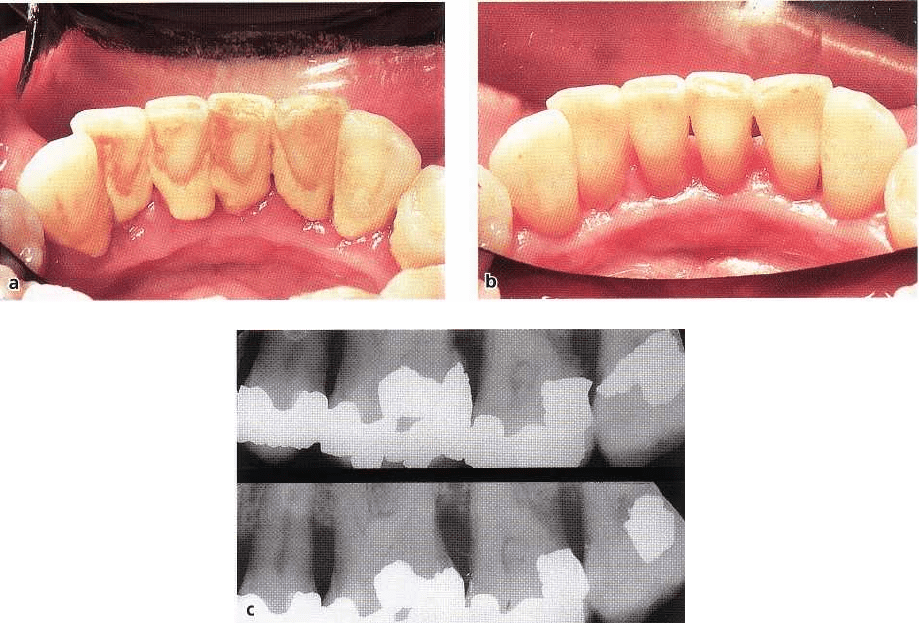
Fig. 20-6. (a) Photograph illustrating the lingual aspect of the mandibular front tooth region with supragingival
plaque and calculus. (b) The same tooth region 3 weeks after supragingival scaling and institution of proper oral
hygiene. (c) Radiographs of left maxilla before and after removal of calculus and amalgam overhangs.
tion is dependent on the material and geometry of the
edge, the sharpness of the edge and the forces used
during instrumentation (Lindhe 1964). Even if sub-
gingival scaling and root planing often are regarded as
two separate procedures with different objectives (see
definition), in clinical work they cannot always be
separated from each other.
Subgingival instrumentation aims at resolving the
inflammation in the gingiva and arresting the progres
-
sive destruction of the attachment apparatus by re-
moving the biofilm present in the gingival pocket.
Coupled with an effective supragingival plaque con-
trol program, therefore, subgingival debridement is
the most important measure in the treatment of peri-
odontitis. As a matter of fact, in many patients who
adopt proper self-performed plaque control habits,
subgingival instrumentation results in gingival health
(
Fig. 20-7).
Prior to the start of subgingival instrumentation,
the presence and extent of gingivitis and breakdown
of the supporting apparatus in all parts of the denti-
tion must have been properly assessed (see Chapter
18). Depending on the severity of the case, and the skill
of the operator, the number of teeth that may be in-
cluded in each treatment session of subgingival scal-
ing and root planing may vary. As a general rule, in a
patient with moderate/severe periodontitis, each ses-
sion should not involve treatment of more than one
quadrant.
The subgingival instrumentation should prefer-
ably be performed under local anesthesia. The root
surface of the diseased site is first explored with a
probe to identify (1) the probing depth, (2) the anat-
omy of the root surface (irregularities, root furrows,
open furcation, etc.), and (3) the location of the calci-
fied deposits.
When all root surfaces selected for treatment in a
given session have been examined, the order in which
the various sites are to be instrumented is determined.
The curette is inserted into the first pocket. The instru
ment is held in a so-called
modified pen grasp
and with
finger rest -
fourth finger rest or third finger rest - with
the face of the blade parallel to and in light contact
with the root surface (Fig. 20-8). It is important that all
root surface instrumentation is performed with a
proper finger rest. This implies that one finger - the
third or the fourth - must act as a fulcrum for the
movement of the blade of the instrument. A proper
finger rest should (1) provide a stable fulcrum, (2)
permit optimal angulation of the blade, and (3) enable
the use of wrist-forearm motion. In addition, to opti-
mize instrumentation and to avoid undue tissue dam
-
age, the finger rest must be secured as close as possible
to the particular root surface selected for treatment.
After the base of the periodontal pocket has been
identified with the distal edge of the blade, the instru-
ment is turned into a proper "cutting" position (Fig.
20-9). The grasp of the instrument is tightened, the
436 • CHAPTER 20
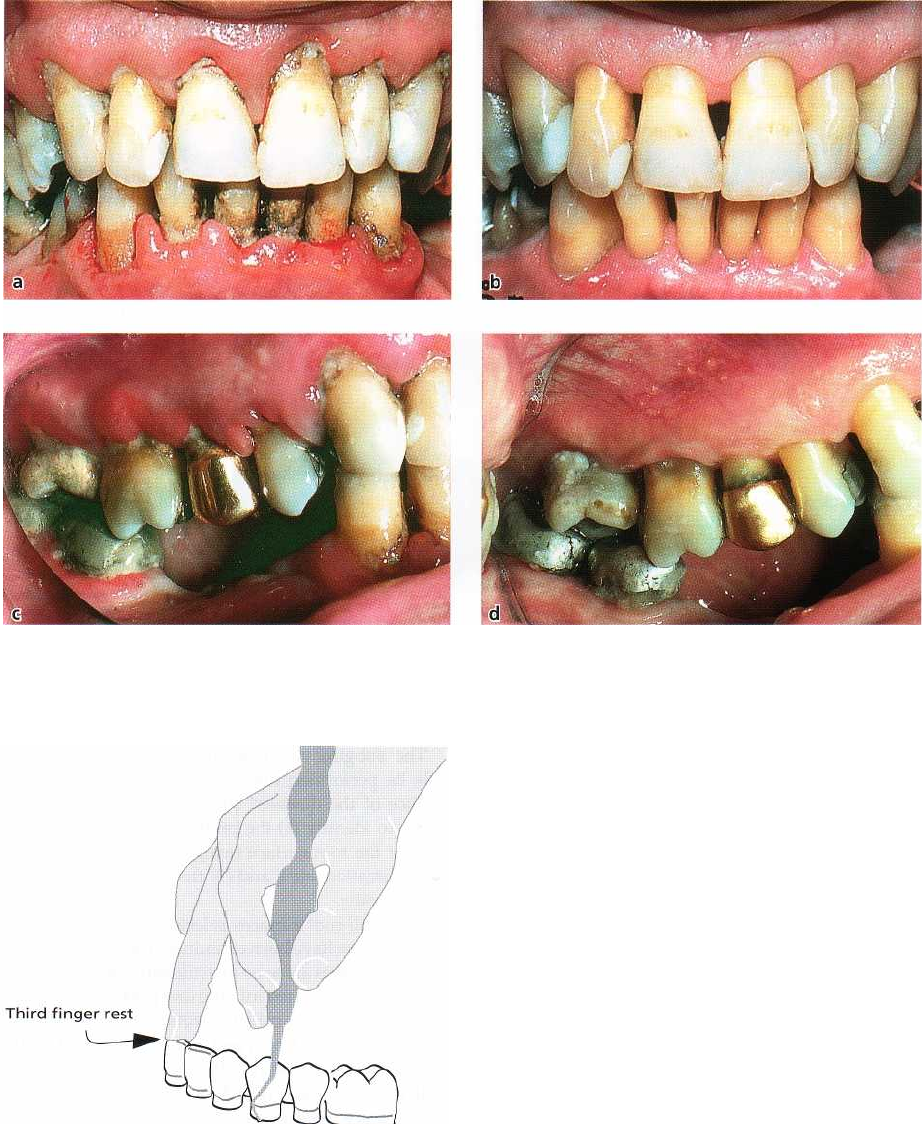
Fig. 20-7a-d. Photographs illustrating the effect of scaling, root planing and proper self-performed plaque control
on the gingival tissues. Note gingival recession and the stippled outer surface of the gingiva after treatment (b,d)
compared to the status before treatment (a,c).
force between the cutting edge and the root surface is
increased, and the blade is moved in a firm stroke
(
working stroke) in coronal direction. Due to the struc-
tural and chemical composition of root cementum and
dentine, the cutting operation
should always be initiated
at the bottom of the pocket and be guided in coronal direc-
tion.
In this movement the edge is moved into the root
surface, and root substance with attached calculus is
removed. The working stroke is followed by a finish
ing
stroke which will produce a smooth root surface.
Working and finishing strokes must be made in differ-
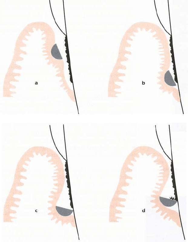
Fig. 20-8. Schematic illustration demonstrating the
proper "third finger rest" using a modified pen grasp
in the molar region in the left mandible.
ent directions to cover all aspects of the root surface
(
crosswise, back and forth) but, as stated above, the
strokes should always start from an apical position
and be guided in coronal direction. After the
working
and
finishing strokes
have been made, the probe is
inserted in the pocket again and the surface of the root
surface assessed anew. The root surface is considered
properly treated when the operator, using a periodon-
tal probe, finds the surface "smooth" and "hard".
The importance of removing "diseased" cementum
during root planing was questioned by several
CAUSE-RELATED PERIODONTAL THERAPY .
437
Fig. 20-9. (a) The curette is in-
serted into the periodontal pocket.
Note the close-to-zero degree an-
gulation of the face of the curette
against the root surface to facili-
tate the accessibility of the pocket.
(b)
The bottom of the periodontal
pocket is identified with the distal
edge of the blade of the curette.
(c)
The curette is turned to a
proper cutting position for scal-
ing. (d) The blade is moved along
the root surface in a scaling stroke
to remove calculus.
authors. Nyman et al. (1986, 1988) monitored the out
-
come of surgical periodontal therapy at sites where
the teeth were exposed to either extensive root instru-
mentation to remove all of the cementum and some
dentin, or gentle removal of plaque to leave most of
the root cementum. The authors found that in patients
with proper self-performed plaque control, both pro-
cedures allowed for excellent soft tissue healing. This
observation was confirmed by Oberholzer et al. (1996)
who concluded – from a clinical study – that the
establishment of a smooth and hard root surface was
not a critical factor in periodontal therapy. This means
that "overinstrumentation" also during non-surgical
therapy may cause more harm than good.
In this context it must also be realized that the
complete removal of subgingival plaque and calculus
in a "non-surgical" procedure is difficult if not impos-
sible. Waerhaug (1978) first assessed the pocket depth
of periodontally involved teeth that were sub-
sequently exposed to comprehensive scaling and root
planing. Following the completion of this treatment,
which was carried out by a skilled periodontist, the
teeth were extracted and the root surfaces examined
under the microscope. It was concluded that at sites
with a pocket depth of > 5 mm, subgingival scaling
and root planing in most cases (about 90%) left depos
-
its of plaque and calculus behind. A similar conclusion
was reached by, for example, Rabbani et al. (1981) and
Magnusson et al. (1984), and also by Sherman et al. (
1990) who in a clinical study demonstrated that
"
closed" subgingival instrumentation was a proce
dure
which consistently failed to eliminate all deposits
of
calculus. In their study, periodontally involved
teeth
were first exposed to subgingival scaling and
root
planing. Following the completion of this treat
ment,
soft tissue flaps were elevated to expose the root
surfaces. The authors observed that small remnants of
calculus had been left behind in several locations.
The studies quoted above imply that residual
plaque and calculus may remain even after careful
and repeated subgingival instrumentation. It is there-
fore the responsibility of the clinician to monitor treat
-
ment outcome. If a site fails to heal properly, i.e.bleed
-
ing on probing persists, and if the clinical attachment
level in deep pocket sites is not improved, additional
therapy such as access flap surgery must be consid-
ered. The reason for this is that findings by, for exam
-
ple, Eaton et al. (1985) and Caffesse et al. (1986) have
shown that scaling during access therapy may im-
prove the efficacy of root instrumentation.
43
8
' CHAPTER 20
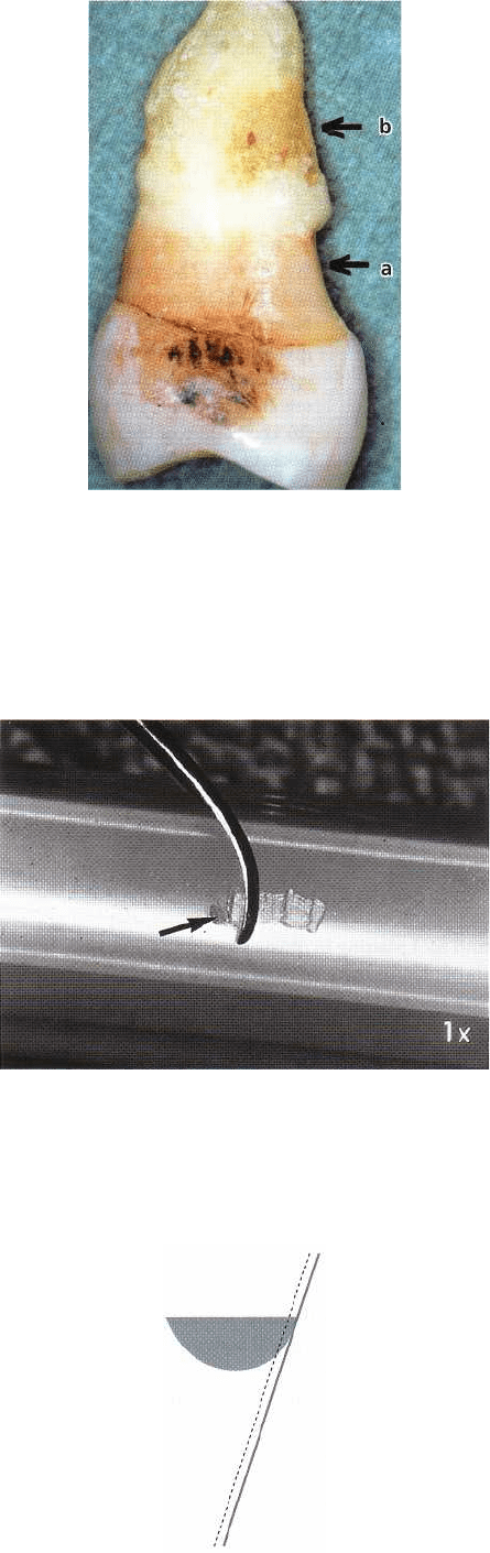
Fig.
20-10. A
tooth has been subjected to subgingival
scaling and root planing and extensive tooth substance
has been removed due to repeated intense instrumenta
tion (arrow a). However, the periodontal pocket was
not
properly diagnosed and calculus has been left (ar
row
b).
Fig.
20-11.
The sharpness of a curette is tested on a plas
-
tic stick.
A
chip (arrow) is easily produced by a sharp
instrument.
Fig. 20-10 shows a tooth that has been subjected to
repeated scaling and root planing. The clinician did not,
however, reach the bottom of the periodontal
pocket,
plaque and calculus were left behind, the periodontitis
lesion progressed and the tooth eventually
had to be
extracted.
Factors important for the outcome of subgingival
instrumentation
Root anatomy:
The surface of a single-rooted tooth is
often easier to reach by subgingival instrumentation
than the furcation complex of multirooted teeth. How-
ever, concavities and tooth furrows exist in booth
single and multirooted teeth. Such root irregularities
may contain small deposits of plaque and calculus
which are difficult to reach. In such sites ultrasonic
instruments with specially designed tips may facili
tate
local therapy (Kocher & Plagmarm 1997). The
technical problems inherent in subgingival instru-
mentation increase with increasing probing depth.
Specially designed instruments with a long shank
(Fig.
20-3) may be used in deep pockets.
Skill of the operator:
The outcome of the subgingival
instrumentation is "operator sensitive". Thus, the
technical skill of the dentist/dental hygienist influ-
ences the result of this procedure. Brayer et al. (1989)
demonstrated that experienced dentists were more
efficient in subgingival debridment than more inexpe-
rienced operators. The difference between the two
categories of therapists was most pronounced in the
treatment of deeper (> 6 mm) pocket sites. Also, in
surgical therapy the technical skill of the operator
remains important for the outcome of the root de-
bridement part of the treatment.
Time allowed:
The time allowed for the instrumentation
will also influence the treatment result. In a study by
Badersten et al. (1981) it was demonstrated that as
much as 6-8 minutes were required for a comprehen-
sive subgingival treatment of one single tooth when
hand instruments were used. When ultrasonic instru-
ments were used 4-6 minutes were required.
Instrument sharpening
The hand instruments must have proper cutting edges
Fig. 20-12. Schematic drawing illustrating the sharpen
ing of a curette. The original geometry of the cutting
edge must be maintained during the sharpening proce
-
dure.
CAUSE-RELATED PERIODONTAL THERAPY • 439
in order to make subgingival instrumentation a pre-
cise and efficient procedure. A curette with a blunt
cutting edge
must be pressed
against the root surface
with a larger force than is required when a sharp
instrument is used. Scaling with instruments with
blunt cutting edges often results in an incomplete
removal of calculus but in the establishment of a
"
smoothened" root surface. Remaining calculus on
such a "smoothened" root surface is difficult to detect
with a periodontal probe. The cutting edge of the hand
instrument, therefore, must be controlled repeatedly
during scaling. This can be done by planing a plastic
stick (Fig. 20-11).
The sharpening of hand instruments can be per-
formed by the use of either "rotating" (cylindrical or
cone-shaped) or "plain" stones (India or Arkansas
stones). Curettes and sickles are sharpened by grind-
ing the lateral surfaces and/or the face of the blade. It
is important that the original geometry of the instru-
ment is not changed by the sharpening procedure (Fig.
20-12).
Ultrasonic and sonic instruments
For many years ultrasonic scalers (e.g. Cavitro
n
®
, Am
-
dent
®
, Odontosonl have been used for the removal
of
plaque, calculus and stain. Scaling with ultrasonic
instruments often results in the establishment of an
uneven root surface. It has been suggested, therefore,
that ultrasonic scaling should be supplemented with
hand instrumentation to establish a smooth root sur-
face (Bjorn & Lindhe 1962). Clinical studies have
evaluated the effect of scaling using ultrasonic or hand
instruments (Torafson et al. 1979, Badersten et al.
1981). The authors found that debridement of 4-7 mm
pockets with ultrasonic instrumentation was as suc-
cessful for healing diseased periodontal sites as was
scaling with hand instruments (curettes). It has also
been questioned whether indeed
a
smooth
root surface
after treatment is important for successful healing
(
Rosenberg & Ash 1974). Waerhaug (1956) found that
a
junctional epithelium readapted and a normal "epi-
thelial cuff" also formed to uneven root surfaces. Prop
-
erly used, therefore, ultrasonic instrumentation must
be regarded as a valuable substitute for conventional
scaling with hand instruments and may even be the
best instrument for scaling at furcation areas (Leon &
Vogel 1987).
Recently a new type of instrument for tooth de-
bridement was introduced —
the sonic scaler (e.g.
Titan
-
s
®
,
Micro-Mega Air Scaler
®
). This instrument is air-
driven and produces vibrations in the sonic range
(
2300-6300 cycles per second). In an
in vitro
study (Lie
& Leknes 1985) and in clinical studies (Loos et al. 1987,
Baehni et al. 1992) it was shown that the sonic scaler
was as effective for calculus removal as the ultrasonic
instrument and, besides, the sonic scaler caused less
root surface roughness than the ultrasonic device.
The removal of plaque and calculus by ultrasonic
and sonic instrumentation is accomplished by (1) the
vibration of the tip of the instrument, and (2) the
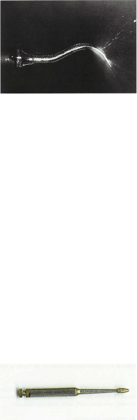
Fig. 20-13. The tip of the ultrasonic instrument. Note
the spraying and cavitation of the fluid used for cool
ing.
spraying and cavitation effect of the fluid coolant. The
vibrations (amplitude ranging from 0.006-0.1 mm) in
the ultrasonic instruments are produced by a metal
core which can change its dimension in an electromag
netic field with an operating frequency between 25
000
and 42 000 cycles per second. In the sonic
instruments,
the vibrations are generated
mechanically. During the
generation of the ultrasonic
vibrations heat is pro
duced, which is why the tip
during instrumentation
always has to be irrigated
with a coolant.
Before use, the ultrasonic instrument must be ad-
justed regarding the power (tuning) and cooling ac-
cording to the manual (Fig. 20-13). The tip should be
applied to the tooth surface with very light pressure
and be moved back and forth over the surface in
sweeping movements and in such a way that its pat-
tern of vibration is orientated parallel to the tooth
surface. This will prevent damage to the root. A peri-
odontal probe should always be used to check the root
surface characteristics after instrumentation.
A novel, diamond-coated,
sonic scaler insert was
introduced by Kocher & Plagmann (1997) The authors
claimed that this type of insert facilitated scaling and
root planing in furcation areas.
Rotating instruments
Root furrows, furcation areas and root surfaces in
deep, narrow, infrabony pockets are difficult to prop-
erly debride with the use of hand instruments. At such
sites, therefore, rotating instruments such as fine-
grained diamonds (or sonic scalers with diamond-
Fig. 20-14. A fine-grained diamond for mechanical de
bridement. Magnification x 1.5.
440 • CHAPTER 20
Fig. 20-15. (a) A diamond tip inserted in a handpiece (The Profin
®
Directional System). Arrows indicate the direc-
tions of the movement. (b) A set of tips (Lamineer
®
) of different design.
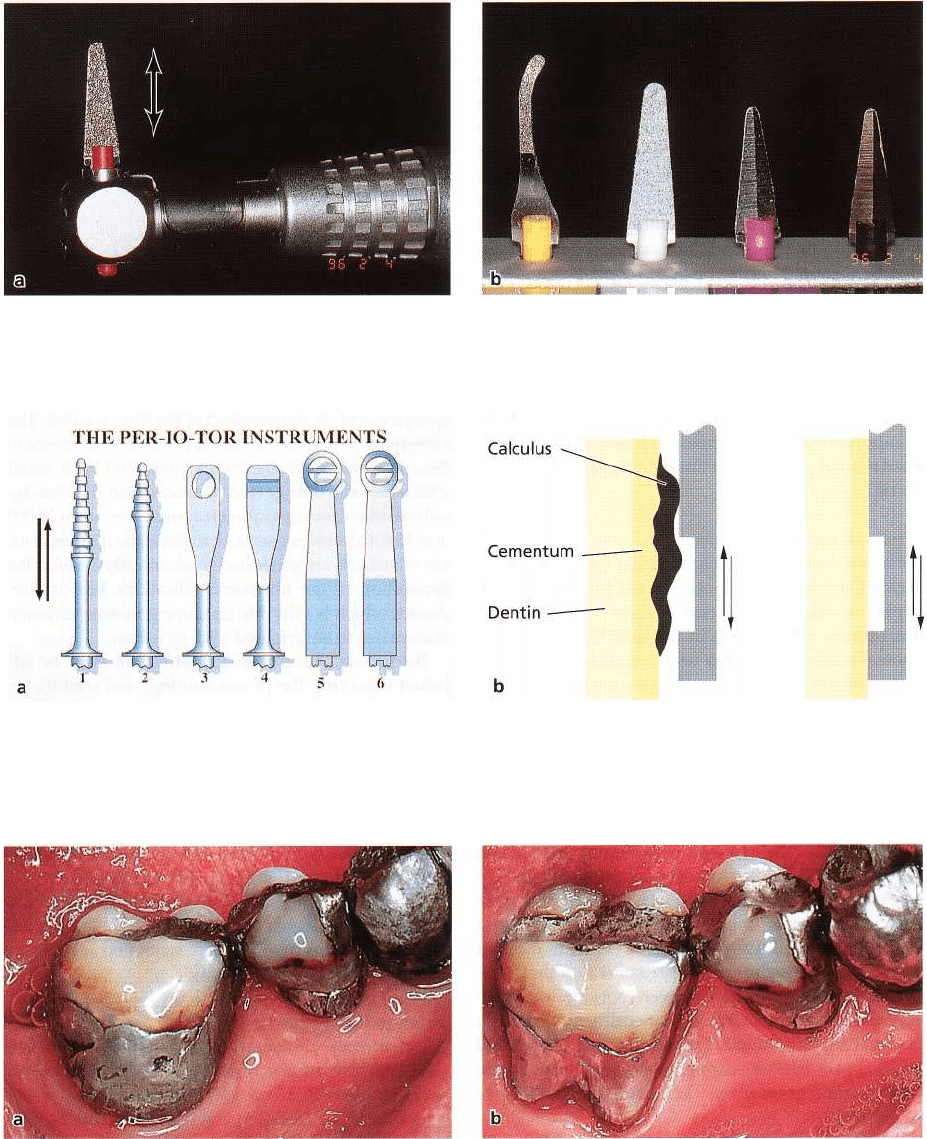
Fig. 20-16. (a) A collection of the PER-IO-TOR
®
instrument tips for scaling and root planing. (b) A schematic illustra-
tion of the effect of the PER-IO-TOR
®
instrument on the root surface with deposits (left). When the root cementum is
clean and planed, no more cementum can be removed because of the design of the instrument (right).
Fig. 20-17. Amalgam restorations before (a) and after (b) the adjustment of the overhangs. The buccal restoration in
tooth 46 is prepared in order to reduce the degree of furcation involvement.
coated inserts, see above) can be used (Fig. 20-14). Care
should be taken, however, to avoid excessive removal
of
tooth substance with such cutting procedures.
Reciprocating instruments
The Profin
®
Directional System offers a specially de-
signed handpiece (a second generation of the so called
Eva
®
system which was introduced in 1969) with a 1.2
mm reciprocating motion of the working tips set in
self-
steering or fixed mode (Fig. 20-15). A recom
mended
engine speed of 10 000-15 000 rpm will give
20 000-30
000 tip strokes per minute. Specially de-
signed working
tips have been developed for the
Profin Directional
System (Axelsson 1993). The PER
IO-TOR
®
instruments
will optimize cleaning and
planing of rough root
cementum surfaces and prevent
CAUSE-RELATED PERIODONTAL THERAPY • 441
further removal of root cementum once the surface is
clean and smooth (Fig. 20-16). Mengel et al. (1994)
evaluated the PER-IO-TO
R
®
instruments in an
in vitro
study. They stated that the PER-10-TOR" instruments
have similar planing properties as manual hand in-
struments, but cause minimal removal of tooth struc
-
tures.
Laser instruments
Laser instruments have been used in dentistry for
cavity preparation since 1964 and for root debride-
ment in combination with periodontal surgery. In a
recent clinical study by Schwarz et al. (2001), the
Er:
YAG (erbium-doped:yttrium, aluminium and gar-net
laser) was used for "closed" subgingival scaling
and
root planing. The laser treatment was compared
to
conventional instrumentation with curettes. The
authors concluded that the laser method gave better
results in terms of pocket reduction at sites with deep
pockets. Further studies are needed, however, to
evaluate the laser instrument and the long-term out-
come of this kind of treatment.
Removal of plaque-retention factors
In an epidemiological study Bjorn et al. (1969, 1970)
observed that ill-fitting artificial crowns and fillings
were associated with a reduced height of the peri-
odontal bone level. Jeffcoat & Howell (1980) reported
that marginal bone loss was more pronounced around
teeth with overhanging amalgam restorations than
around teeth without restorations. Rodriguez-Ferrer
et al. (1980) concluded that "the presence of a sub-
gingival overhanging defective margin may be the
only
important clinically significant feature of an
amalgam
restoration related to the pathogenesis of
chronic
inflammatory periodontal disease". It is not
the
overhang of the restoration
per se,
however, which
causes or maintains periodontal disease. Waerhaug
(
1960) pointed out that the more advanced gingival
inflammation observed in sites with ill-fitting restora-
tions was the result of extensive plaque accumulation
and not of mechanical or chemical irritation caused by
the filling material. Thus, overhangs of restorations
must be removed to (1) facilitate the removal of plaque
and calculus, and (2) to establish an anatomy of the
tooth surface which facilitates self-performed tooth
cleaning.
Overhanging margins of dental restorations can be
removed using a flame-shaped diamond stone
mounted on a handpiece for rotator movements, or a
flat diamond stone mounted on a handpiece for hori-
zontal reciprocal movements (Eva-system'
s
, Profin
e
Directional System) (Fig. 20-15). The overhang is re-
moved and the restoration is given a proper form and
a
smooth surface. The triangular or V-shaped tips
(
Lamineer
,
) are appropriately tailored for use in nar-
row interproximal spaces and can reach 2-3 mm sub-
gingivally for the reshaping and polishing of restora-
tions (Fig. 20-15b).
The adjustment of an improperly designed filling
or
artificial crown is often a difficult and time-consuming
procedure. It is sometimes more convenient to
remove the improper filling (crown) and insert a new
one with a proper marginal fit. Fig. 20-17 illustrates
the effect on the periodontal tissues of the adjustment
of an amalgam overhang.
HEALING AFTER
INITIAL,
CAUSE-RELATED THERAPY
Clinical measurements
The potential of subgingival instrumentation to elimi-
nate inflammation and arrest progression of peri-
odontal disease was studied in a number of early
clinical trials, e.g. Lovdal et al. (1961), Suomi et al.
(
1971), Axelsson & Lindhe (1978, 1981), Hirschfeld &
Wasserman (1978), Morrison et al. (1980), Badersten et
al. (1984).
Lovdal et al. (1961) examined around 1500 indi-
viduals in Norway regarding their oral hygiene status,
gingival conditions, alveolar bone loss and tooth loss.
The patients were subsequently, on an individual ba-
sis, given careful oral hygiene instruction, including
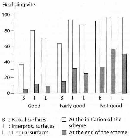
Fig. 20-18. The patients were divided into three groups
after the initial examination according to their oral hy
giene, "good", "fairly good", "not good". During a 5-
year period the patients were subjected to scaling and
root planing measures two to four times per year. In all
three categories of patients there was a remarkable im
provement of gingival conditions after 5 years of re-
peated plaque control measures. Data from Lovdal et
al. 1961.
442 • CHAPTER 20
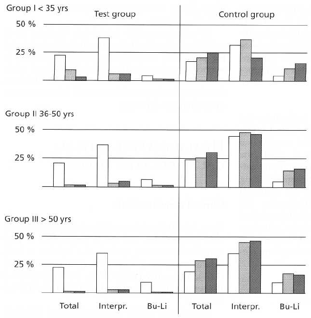
Fig. 20-19. Histogram showing the
frequency distribution of inflamed
gingival units in the test and con
-
trol groups at the baseline (open
bars) and re-examinations after 3
and 6 years (hatched and black
bars respectively). The individual
mean (total) figures describe unal
-
tered gingival conditions in the
control groups but marked im-
provements in the test groups dur
ing the trial. Data from Axelsson
& Lindhe 1981.
detailed information in the proper use of toothbrush,
toothpick, dental floss, etc. In addition, their teeth
were subjected to meticulous supra and subgingival
scaling and root planing. The oral hygiene instruction
as well as the scaling and root planing measures were
repeated two to four times per year over a 5-year
period, at the end of which the patients were re-exam
-
ined. The authors reported (Fig. 20-18) that "the com-
bined effect of subgingival scaling and controlled oral
hygiene definitely reduced the incidence of gingivitis
and tooth loss". The improvement was remarkable
also in patients whose home-care habits did not im-
prove during the 5 years of trial. Similar findings were
reported by Suomi et al. (1971) from a study compris-
ing about 350 subjects in California. A test group of
patients received, during a 3-year period, oral hygiene
instruction and scaling three to four times per year,
while a control group during the same period received
restorative oriented dental care. At the end of the 3
years the test group patients had significantly less
gingivitis and less attachment loss than the controls.
Axelsson & Lindhe (1978, 1981) compared the effect
of a prophylactic program including (1) oral hygiene
control and (2) scaling and root planing repeated once
every 2-3 months over a 6-year period with the effect
of traditional dental therapy, i.e. a therapy which was
directed towards the symptoms of dental disorders
rather than towards the elimination of the etiological
factors. It was observed that in the patients of the
control groups there was only a minor improvement
in oral hygiene, while in the test groups at the re-ex-
aminations after 3 and 6 years, the mean plaque scores
were consistently low (< 20%). In the control groups
there was no improvement of the gingival conditions (
Fig. 20-19), while in the test groups the gingivitis
scores had approached 0 values (meaning gingival
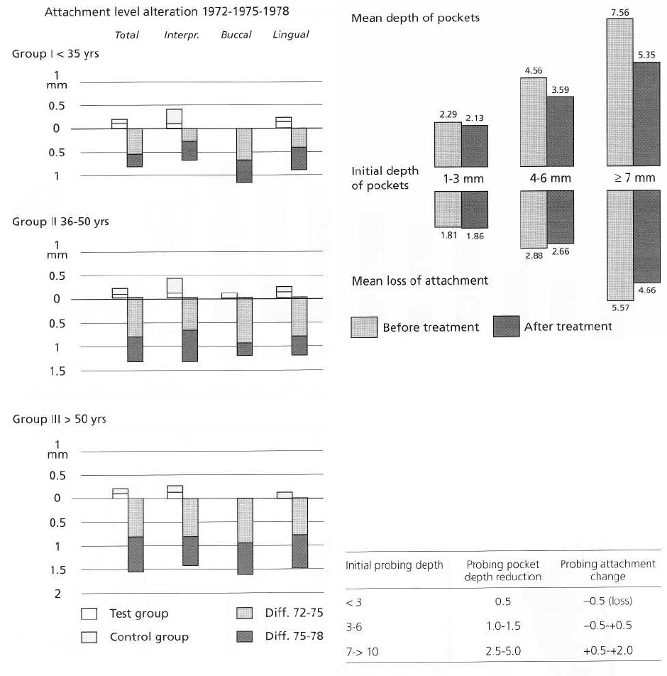
health) at the re-examinations. Fig. 20-20 gives the
attachment level alterations that occurred between
1972 and 1978. It can be seen that, while in the patients
of the control groups there was a substantial further
loss of attachment, in the test patients there was no
further breakdown of the attachment apparatus.
Hirschfeld & Wasserman (1978) presented a long-
term survey of tooth loss in 600 patients treated for
periodontal disease. The patients were re-examined at
an average of 23 years after the active treatment, which
included primarily subgingival scaling, and root plan-
ing followed by careful maintenance therapy. The
authors observed that "during the post-treatment pe-
riod 300 patients had lost no teeth from periodontal
disease", and "199 had lost 1-3 teeth". This means that
periodontal therapy directed towards the elimination
of the subgingival infection using a non-surgical ap-
proach can be effective in preventing tooth loss in
most patients and also in those with advanced peri-
odontal disease.
Morrison et al. (1980) studied the short-term effect
of non-surgical treatment in 90 subjects with moder-
ately advanced periodontal disease. Following an in-
itial examination the patients were given oral hygiene
instruction and the pockets were treated by subgingi-
val scaling and root planing. Re-examination per-
formed 4 weeks after active therapy revealed that (1)
the patients had improved their oral hygiene, (2) the
gingivitis scores had decreased, and (3) the initial
depth of the periodontal pockets was substantially
CAUSE-RELATED PERIODONTAL THERAPY • 443
Fig.
20-20.
Histogram illustrating the alterations in the
clinical attachment levels between the baseline
(1972)
and re-examinations
(1975, 1978)
in various test and
control groups. The attachment level remained unal-
tered in the test groups but was reduced in all the con
-
trol groups (below the 0-line). The average attachment
loss per year in control groups was: I: 0.13 mm, II:
0.
23mm, III: 0.26 mm. Data from Axelsson & Lindhe
1981.
reduced (Fig. 20-21). The authors concluded that al-
ready, 1 month following cause-related periodontal
therapy, the clinical severity of periodontal disease
was
markedly reduced.
Badersten et al. (1984) studied the effect of sub-
gingival scaling and root planing in 16 patients with
moderate-advanced periodontitis. The patients re-
ceived oral hygiene instruction and all single-rooted
teeth were exposed to meticulous subgingival de-
bridement using either handscalers or an ultrasonic
device. Re-examination carried out after 2 years
Fig. 20-21. Histogram describing the reduction of prob
ing depth 4 weeks after active therapy comprising scal
-
ing and root planing. Data from Morrison et al.
1980.
Table 20-1. The mean probing reduction and prob-
ing attachment gain/loss (mm) that can be expected
following scaling and root planing in non-molar
sites
showed that this treatment had resulted in the establ-
ishment of healthy gingival conditions (only about
10%, of the gingival sites bled on probing) and in a
marked reduction in the overall probing depth (Fig.
20-
22; Badersten 1984). The authors concluded that
careful subgingival scaling and root planing was an
effective means to eliminate gingivitis and reduce the
probing depth even at sites with initially very deep
(> 9
mm) periodontal pockets. Similar observations
have
also been reported in later clinical studies (for
review
see Cobb 1996).
It may be concluded that in patients who adopt
proper oral hygiene measures:
• Healing after non-surgical therapy seems to be com-
plete after about 3-6 months
•
The number of sites that bleed on probing will be
markedly reduced
•
More gingival recession and gain of probing attach-
444 • CHAPTER 20
Dimensional
changes for sites with different initial
probing
depths
* Interpretation: For sites initially 8-8.5 mm deep, gingival recession amounted to about 2mm, gain of probing attachment to 1 mm
and residual probing depth to 5 mm.
Fig. 20-22. Histogram illustrating clinical results after non-surgical periodontal therapy. Data from Badersten et al
1981, 1984.
Fig. 20-23. A schematic illustration of a gingival unit (a) before and (b) after periodontal therapy. Probing depth
measurements (blue lines) before (a) and after (b) therapy. The dotted line indicates the "histologic" attachment
level. Recession = green line. Location of the gingival margin before therapy (A) and after therapy (B). ICT = infil-
trated connective tissue, NCT = non-infiltrated connective tissue.
ment will be obtained in initially deeper than more • In sites with initial pockets of < 3 mm, a probing
shallow
pocket sites. See Fig. 20-22 and Table 20-1.
attachment loss of 0.5 mm will occur.
• In sites with initial probing depths of 6-9 mm, the
residual probing depth will be 4-5 mm, and the
amount of gingival recession about 2 mm.