Lowenthal G., Airey P. Practical Applications of Radioactivity and Nuclear Radiations
Подождите немного. Документ загружается.

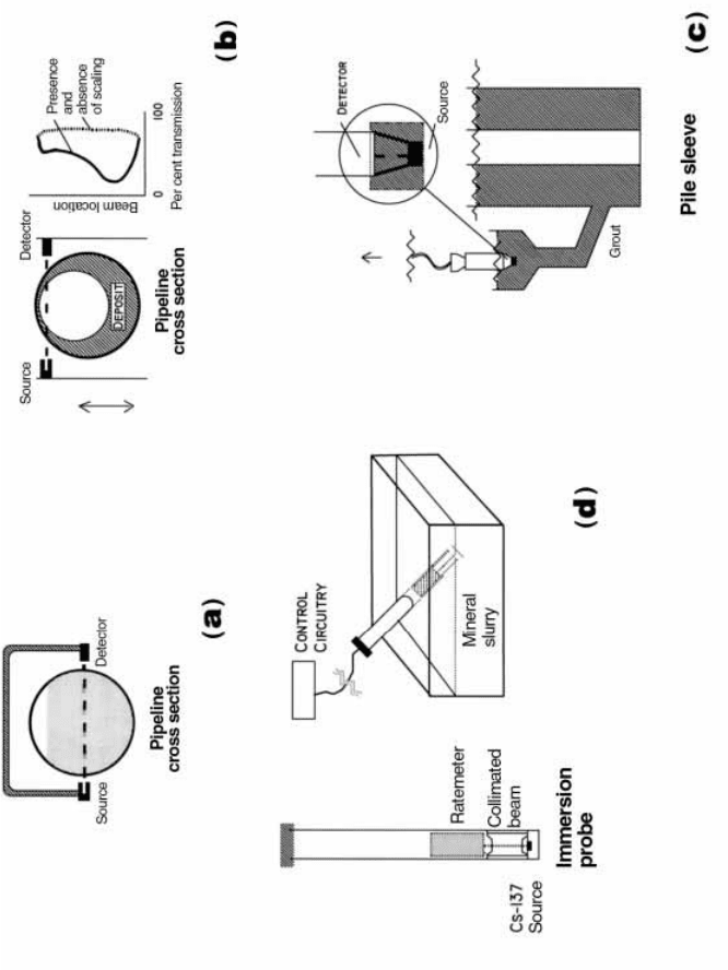
Figure 7.3. Four applications of gamma ray attenuation. (a) The
monitoring of slurry in a pipeline. (b) Investigation of scale
deposits in pipelines. (c) Monitoring of density of grout during the
construction of, for example, offshore platforms (ICI Synetix
Tracerco, product information). (d) The
in situ measurement of the density of mineral slurries (Cutmore
et al., 1993).
the density of concrete grout used in the construction of piles supporting
offshore oil platforms is within speci®cation. Grout is pumped under pressure
into the pile and continuously ®lls the space between the radioactive source
and the detector. The density of the grout is directly related to the response of
the detector which is monitored by personnel on the surface. Such a system is
shown schematically in Figure 7.3(c).
The second application is known as the Gammascan
TN
Flooded Member
Detector System. The principle is simple and robust. Flooding of a sub-sea
member of a platform with water will reduce the transmission of the gamma
beam between the source and the detector. The system is normally mounted
on the arms of an ROV and is capable of rapidly monitoring both vertical
and horizontal members.
Other applications of gamma ray transmission are discussed in the
following paragraphs.
Mineral processing
Density gauges are widely used in the minerals processing industry. The real
time analyses of the levels of valuable minerals in process streams generally
employ radioisotope X ray ¯uorescence or g ray preferential absorption
techniques. As the responses of these detectors depend not only on the
composition of the mineral component, but also on the bulk density of the
aqueous slurry, nucleonic density gauges are an essential element of radio-
isotope on-stream analysis systems. The gauge illustrated here (Figure 7.3(d))
is designed for immersion in the mineral slurry.
Coastal engineering
Nucleonic gauges have been adapted for the measurement of the levels of
sediment in rivers and estuaries. Quantitative information on the mobilisation
of sand and sediment under various conditions is important in the investiga-
tion phase of many coastal engineering projects. Either absorption or back-
scatter gauges may be used for this purpose. They may be linked with other
gauges measuring, for instance, depth, salinity and temperature. This infor-
mation, together with position ®xing data from satellite navigation systems,
is integrated into computerised monitoring systems. Further comments are
made in Section 9.3.5.
Radiography
Gamma radiography has long been one of the most important industrial
applications of radioisotopes. The technology evolved out of the widespread
use of X rays in medical imaging and has been adapted to monitoring the
Industrial applications of radioisotopes and radiation192
internal structure of manufactured components and to checking the integrity
of welds.
The scienti®c principles of g radiography are illustrated in Figure 7.4.
Photographic X ray ®lm housed in a suitable cassette holder serves to detect
the transmitted radiation from the radioisotope source. If an inclusion in the
sample under investigation has a higher electron density than the surrounding
material, fewer g rays are transmitted and a shadow image is produced on the
®lm.
A wide range of radiography sources is commercially available. A selection
is listed in Table 7.4. Factors determining the choice of the source are similar
to those for density gauges as discussed above. Particular attention must be
paid to the gamma ray energy. If the g ray transmission is too high, the image
contrast is inadequate. If the transmission is too low, there could be problems
due to a low detector countrate. About 50% transmission is close to
optimum. In practice,
60
Co,
137
Cs or
192
Ir sources are commonly used.
Computerised tomography (CT)
Computerised tomography (CT) is a sophisticated extension of radiography
in which the detailed internal structure of a component can be obtained in
two or three dimensions by an analysis of the attenuation of the X or g ray
beam in a large number of projections. Cormack and Houns®eld indepen-
dently demonstrated the technique during the early 1960s (Bull, 1981) and
were jointly awarded the Nobel Prize for Medicine and Physiology in 1979.
However, it was not until the coming of suf®ciently powerful computers
during the early 1980s that tomography could be used routinely, ®rst in
medical imaging (Romans, 1995) and later in industry (Section 3.7.2).
When designing a CT instrument, a number of parameters need to be
optimised. They include the energy of the incident beam, the data acquisition
time and the resolution of the image. The criterion for energy selection is
identical to that for density gauges. Data acquisition times should generally
be as short as practicable to permit maximum utilisation of expensive
equipment. Image resolution should be high, but there are intrinsic limits
depending on the wavelengths (energies) of the radiation. Comparison of
computed tomography for medical diagnosis and industrial inspection is
discussed by Martz et al. (1990).
As with nucleonic density gauges and radiography, the principles of
tomography are ultimately based on Eq. (3.7). This equation was derived to
calculate the attenuation of g rays in material made up of components
differing in their linear attenuation coef®cients m
ti
and thicknesses Dt
i
. The
sum
P
m
ti
Dt
i
can be expressed as the line integral
R
m dl. This is known as the
1937.2 Applications of gamma rays
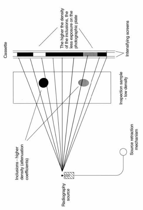
Figure 7.4. The principles of gamma radiography (after Charlton, 1986,
Ch. 13).

Table 7.4.
Radioisotopes used as X or
g ray sources. (Based on Charlton, 1986, Table 14.1. For more details, see
Table 3.2 of this book.)
Isotope
Half life Energy (keV) Applications and comments
55
Fe
2.72 y 5.9 KX ray
Low energy X ray analysis. KX rays usefully excited Al to Cr
238
Pu
87.8 y 12±17 LX ray XRF applications. KX
rays excited; Mn to Y
109
Cd
463 d 22 KX ray
Detector calibration and XRF. KX rays usefully excited Fe to Mo
210
Pb
22.2 y 47 KX ray
Used as an energy standard and for XRF
241
Am
432 y 59.5
g ray
Used for continuous gauging applications e.g. ash in coal measure-
ments (Section 7.2.1);
Used with target elements (e.g. copper or silver) to generate
sources of (Section 7.2.3) pure ¯uorescent X rays
XRF applications. KX rays usefully excited I to Lu
170
Tm
127 d 84 KX ray
Low-energy radiography
57
Co
272 d 122 and 136
g rays Mo
È
ssbauer spectroscopy
T(
3
H)/Zirconium 12.3 y 5 to 9
Bremsstrahlung sources. The X rays are generated as a conse-
quence
147
Pm/Aluminium 2.6 y
12 to 45
of the deceleration of the negatrons (
b
particles) emitted by the
90
Sr/Aluminium 28 y
60 to 150
source (Section 3.8.1)
Radon integral after the mathematician who, in 1917, laid the foundation for
later work on the mathematical reconstruction of tomographic images.
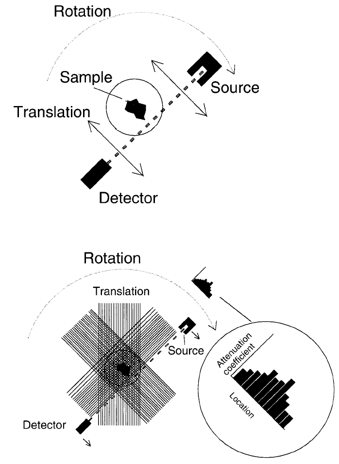
Tomographic systems have gone through several stages or generations of
development. The principle is here illustrated using a ®rst generation system
shown in Figure 7.5 (Zatz et al., 1981). Repeated measurements of the
attenuation of a highly collimated beam of g or X rays are made as the
source±detector system traverses the sample. Similar data are then obtained at
a number of projections by rotating the sample over a range of at least 1808.
This information is accumulated by the computer which uses it to reconstruct
a two or three dimensional image of the internal features of the sample.
These ®rst generation systems produced excellent images free of artefacts.
However, the systems were slow with data acquisition times sometimes
upwards of several hours. More advanced and faster systems were designed
to reduce imaging time by using an incident fan beam arrangement of X or g
rays and multiple detectors. Current instruments are based on those designed
for hospital use. In a fully developed industrial system, more than a million
measurements over 180
o
are used to develop a single CT image. The
measurements can be made and the image reconstructed and displayed on a
computer screen in about a minute. CT imaging has been applied to the study
of machine components and to the condition of castings. It has been
demonstrated that the CT-based measurements of the dimensions of complex
castings are as accurate as those obtained by conventional means. However,
for very complex or accurate work, imaging times ranging from a few
minutes to a few hours may still be needed.
The size of the component that can be inspected depends on the energy of
the incident beam. At one extreme, X ray sources from linear accelerators up
to 15 MV in energy are being used commercially for a range of applications,
including the examination of rocket motors.
At the other extreme, high-resolution micro-tomographic systems can
provide information complementary to microscopy. Detailed information
can be obtained on the internal structure of materials (such as wood and
polymers) which would be destroyed during normal sample preparation.
Using a low-energy X ray source, images with resolution of 5 mm have been
obtained using small samples of wood (2 mm across). With this resolution,
the cellular structure of the sample can be readily seen (Wells et al., 1992).
High-speed systems are being developed in the steel industry for the real
time gauging and control of products that are manufactured in a continuous
process. This requires very fast computing capability able to generate output
each 1 to 10 ms. Such systems have been demonstrated in the production of
pipes and a range of structural materials with complex cross sections.
Industrial applications of radioisotopes and radiation196
7.2 Applications of gamma rays 197
Figure 7.5. The scanning, rotation con®guration for the ®rst generation CT
scanner (modi®ed from Zatz, 1981).
Finally, on-line applications of tomography to the milling of timber are
also being developed. It is now possible to tomograph an uncut log and
mathematically reconstruct the grain patterns on veneers sawn at any angle.
When fully developed, it will be possible to ensure that each log is milled in a
way that optimises the use of the valuable resource.
Column scanning
The scanning of large distillation columns in the oil re®ning industry is a long
established example of the application of g ray density pro®ling. The columns
are typically tens of metres high and two to three metres in diameter and are
designed to separate the lighter petroleum fractions from the crude feed
stock. They comprise an interconnected series of trays spaced at intervals of
about one metre. Columns operate continuously and should require a
minimum of maintenance.
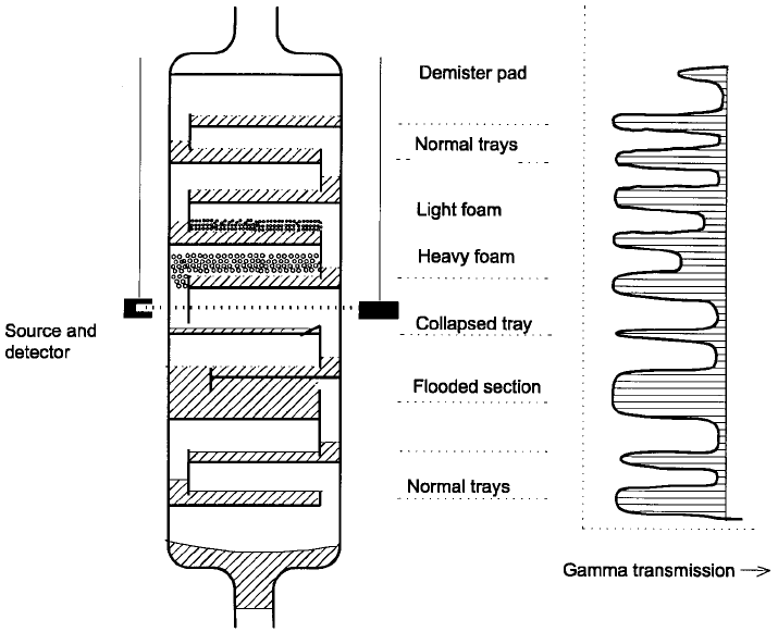
Failure to meet the design speci®cation of a column arises from a number
of causes, notably the collapse of individual trays, the development of
blockages leading to the ¯ooding of trays and the formation of foams in the
vapour phases. A number of these features are illustrated schematically in
Figure 7.6. Many of these problems can be diagnosed using g ray transmis-
sion techniques with no interference to plant operation.
The radioactive source and the detector are lowered in parallel on opposite
sides of the column to develop a scan such as illustrated in the ®gure.
However, a full understanding of the results of such scans in terms of column
malfunction requires extensive experience. Many thousands of scans are
performed annually, the vast majority by international service companies.
Other applications of radiotracers in the oil re®ning industry will be discussed
in Section 8.4.2.
On-line measurement of ash in coal
(1) Dual isotopes applications: The applications described in the previous
sections involved monitoring the density and the distribution of material
between the radiation source and the detector. However, there is often a need
for additional information. For instance, for the optimum performance of
coal ®red boilers in the electricity generating industry, it is desirable that the
feedstock be blended to a constant calori®c value. Although the total ash and
moisture contents must be known, the detailed elemental composition of the
ash may be less important.
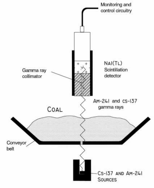
A widely used gauge employs two g ray emitting radionuclides,
137
Cs (662
keV) and
241
Am (59.5 keV) as shown schematically in Figure 7.7. Attenuation
of the 662 keV g rays from
137
Cs is due mainly to Compton scatter. As shown
Industrial applications of radioisotopes and radiation198
earlier (Section 3.4.6), Compton attenuation at that energy is almost indepen-
dent of the composition of the material, so that the 662 keV g rays can be
used to monitor the mass of coal on the conveyor belt as it passes through the
radiation beam.
By contrast, the attenuation of the 59.5 keV g rays from
241
Am is due far
more to photoelectric than to Compton interactions (Figure 3.7(a)) and
hence increases rapidly with increasing concentrations of elements of higher
atomic number such as iron and silicon (Eq. (3.3)). Coal heaped loosely on to
conveyor belts is of fairly low density, permitting the transmission of some of
the 59.5 keV g rays through relatively thick layers. However, it may require
241
Am activities of order 10 to 20 GBq to ensure that the detected signal is
strong enough for suf®ciently precise measurements.
The percentage of ash in coal is calculated from a computer analysis of the
transmission data of both the
137
Cs and the
241
Am g rays. The overall
uncertainty in the determination is normally in the range of 0.7 to 1.5 weight
per cent (Cutmore et al., 1993). Because the information is obtained in real
7.2 Applications of gamma rays 199
Figure 7.6. Gamma transmission scanning of a distillation column illustrating
identi®able features (after Charlton, 1986, Ch. 13).
time, the outputs from a series of gauges installed on different conveyor belts
can be used to optimise the blend of coal fed into the power station furnaces.
(2) Moisture in coke and coal, hybrid technology: The on-line monitoring of
the calori®c value of coke and coal requires knowledge of the moisture
content. Gamma transmission or backscatter gauges cannot be used to
distinguish between the hydrogen in moisture and that associated with the
organic component of the coal. One approach to overcome this dif®culty has
been to develop a gauge based on a combination of microwave phase shift
and g ray transmission techniques (Cutmore et al., 1991).
(3) Multi element analysis of coal: A still higher level of sophistication
makes use of an elemental on-line analysis of the coal. Gauges have been
developed commercially and are available from nuclear equipment manufac-
turers (Section 4.3.3) which can determine ash, moisture, density as well as
the levels of key elements such as sulphur. The operating principles combine
Industrial applications of radioisotopes and radiation200
Figure 7.7. The `on-belt' analysis of ash in coal using the dual-energy gamma
transmission technique (after Cutmore et al., 1993).