Marcus P. Corrosion mechanisms in theory and practice
Подождите немного. Документ загружается.

28. Y. Ishikawa, T. Shibamoto, and I. Nakamichi, Jpn. J. Appl. Phys 31:L750 (1992).
29. A. M. Goodman and J. M. Breece, J. Electrochem. Soc. 117:982 (1970).
30. F. P. Fehlner, J. Electrochem. Soc. 119:1723 (1972).
31. R. J. Archer and G. W. Gobeli, J. Phys.Chem. Solids 26:343 (1965).
32. W. A. Weyl, Coloured Glasses, Dawson’s of Pall Mall, London, 1959, pp. 108ff.
33. M. J. Graham, S. I. Ali, and M. Cohen J. Electrochem. Soc. 117:513 (1970).
34. M. J. Graham, Int. Corrosion Conf. Ser. NACE-6, p. 139 (1981).
35. M. J. Graham and M. Cohen, J. Electrochem. Soc. 119:879 (1972).
36. C. L. McBee and J. Kruger, Electrochem. Acta 17:1337 (1972).
37. D. J. Young and M. Cohen, J. Electrochem. Soc. 124:769, 775 (1977).
38. J. S. Arlow, D. F. Mitchell, and M. J. Graham. J. Vac. Sci. Technol. A5:572 (1987).
39. B. Onay, J. Electrochem. Soc. 136:1578 (1989).
40. D. A. Vermilyea, Acta Metall 6:166 (1958).
41. P. B. Sewell, D. F. Mitchell, and M. Cohen, Surf. Sci. 29:173 (1972).
42. G. F. Cerofolini, G. La Bruna, and L. Meda, Appl. Surf. Sci. 89:361 (1995).
43. H. C. Lu, T. Gustafsson, E. P. Gusev, and E. Garfunkel, Appl. Phys. Lett. 67:1742 (1995).
44. L. Verdi and A. Miotello, Phys. Rev. B. 51:5469 (1995).
45. P. C. J. Graat and M. A. J. Somers, Appl. Surf. Sci. 100/101:36 (1996).
46. T.-C. Lin, G. Seshadri, and J. A. Kelber, Appl. Surf. Sci. 119:83 (1997).
47. S. J. Roosendaal, B. van Asselen, J. W. Elsenaar, A. M. Vredenberg, and F. H. P. M.
Habraken, Surf. Sci. 442:329 (1999).
48. J. A. Slezak, B. D. Zion, and S. J. Sibener, Surf. Sci. 442:L983 (1999).
49. A. Stierle, P. Bödeker, and H. Zabel, Surf. Sci. 327:9 (1995).
50. A. Stierle and H. Zabel, Surf. Sci. 385:167 (1997).
51. A. Stierle and H. Zabel, Surf. Sci. 385:310 (1997).
52. M. Müller and H. Oechsner, Surf. Sci. 387:269 (1997).
53. J. C. Yang, B. Kolasa, J. M. Gibson, and M. Yeadon, Appl. Phys. Lett. 73:2841 (1998).
54. J. C. Yang, M. Yeadon, B. Kolasa, and J. M. Gibson, Appl. Phys. Lett. 70:3522 (1997).
55. J. C. Yang, M. Yeadon, B. Kolasa, and J. M. Gibson, Scr. Mater. 38:1237 (1998).
56. J. C. Yang, M. Yeadon, B. Kolasa, and J. M. Gibson, Proceedings of Spring ECS
Meeting, Seattle, Washington, May 1999.
Thin Oxide Film Formation on Metals 187
Copyright © 2002 Marcel Dekker, Inc.
6
Growth and Stability of Passive Films
Barry MacDougall and Michael J. Graham
National Research Council of Canada, Ottawa, Ontario, Canada
INTRODUCTION
Interest in passivity started with the studies of Faraday [1] and Schönbein [2] over
150 years ago. The lack of metallic corrosion in the case of iron immersed in certain
solutions was attributed to either the presence of an oxide film or an electronic
change in the metal. This basic argument has persisted in various forms to this day,
although the majority of scientific evidence suggests protection by a three-
dimensional oxide film. Much has been published on passivity and its breakdown
over the last 50 years. This chapter does not attempt to cover all the literature but
concentrates on work over the past 10–15 years, emphasizing the passivity of iron,
nickel, iron-chromium, and iron-nickel alloys in aqueous environments. Examples
are given from the authors’ and other selected laboratories.
Iron becomes passive in the absence of an applied current in a variety of
solutions ranging all the way from concentrated nitric or sulfuric acids to basic
solutions containing oxygen. In more recent years iron has been protected from
corrosion in acid solutions by the addition of inhibitors usually containing nitrogen,
sulfur, or hydroxyl groups. This complex subject is dealt with in Chapter 14. Here
we concentrate on anodic passivity, dealing with the nature of passive films—their
composition, thickness, growth, and stability—and their role and influence on pit
initiation. Much of the information is obtained from surface analytical techniques
like Auger electron spectroscopy (AES), X-ray photoelectron spectroscopy (XPS),
and secondary ion mass spectrometry (SIMS).
ANODIC PASSIVITY
Although there is general agreement today that anodic passivity of metals such as
iron and nickel is associated with the formation of a three-dimensional oxide film
on the surface and that breakdown of passivity is due to the disappearance of this
protective film either locally or generally, there is still considerable controversy
concerning the nature, composition, and structure of the passive film. Here the
most prominent models for passivity will be presented and the nature of the
passive oxide film on common metals such as iron and nickel will be discussed.
189
Copyright © 2002 Marcel Dekker, Inc.
The use of Pourbaix diagrams in predicting the stability of metals in various
environments will not be detailed in this chapter. The subject has been discussed
on many occasions over the past 40 years (see, e.g., Refs. 3–5). It should, however,
be pointed out that these equilibrium potential-pH diagrams can be extremely useful
in establishing the regions of metal immunity as well as (possible) corrosion and
passivation. Since the diagrams do not provide any direct kinetic information, the
real rate of corrosion and extent of passivation are not evident from a simple
examination of the diagrams. Some oxides (e.g., NiO) dissolve only very, very
slowly in certain solutions (e.g., neutral borate buffer) for kinetic as opposed to
thermodynamic reasons. It should also be pointed out that the oxide stoichiometries
and thermodynamic information given in the Pourbaix diagrams are for thick, bulk
oxides, which may be quite different from the very thin (often ≤ 1 nm) surface
oxide films found on passivated metal surfaces. For this reason, it should not be
surprising that the actual composition of passive oxide films is sometimes not
identical to that found in the Pourbaix diagrams [e.g., the diagram for nickel does
not show the presence of NiO but rather Ni(OH)
2
; see below].
NATURE OF THE PASSIVE FILM ON IRON
Iron and nickel are examples of metals that display an active-passive transition
when anodically polarized in many aqueous solutions. Passivity is generally
ascribed to the presence of a thin oxide film 1–4 nm thick that isolates the metal
surface from the corrosive aqueous environment. The resistance of this anodic
oxide film to dissolution is related to its physical and chemical nature, which
determines the corrosion resistance of the metal. The other major factor influencing
the rate of metallic corrosion is the aggressiveness of the aqueous environment,
i.e., the pH, temperature, and anion content of the solution.
Iron can be passivated in aqueous solutions by the application of either a
constant potential or a constant current. In both cases a certain minimum potential or
current is required before passivation occurs and a finite time is required to attain
the passive (i.e., very slow dissolution) state. Early work dealing with the
electrochemistry of iron was done in acid solution, but over the last 30 years studies
have been carried out mainly in neutral buffered solution. The major impetus for
work on neutral solutions came from the research of Nagayama and Cohen [6] using
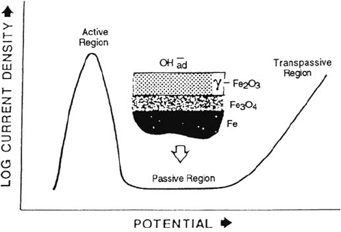
a pH 8.4 sodium borate–boric acid buffer solution. In Figure 1, a schematic of the
passivation curve for iron in this solution is shown. Starting with an oxide-free
surface, the current first increases as the potential is increased, reaches a maximum,
and then decreases again. There is a region (of over a volt) in which iron does not
dissolve, or is passive, and then the current rises again due to oxygen evolution and/or
transpassive metal dissolution. In the passive region iron is covered by a thin film of
cubic oxide of the γ-Fe
2
O
3
/Fe
3
O
4
type, which is probably formed by reactions such as
3Fe + 4H
2
O → Fe
3
O
4
+ 8H
+
+ 8e
2Fe + 3H
2
O → Fe
2
O
3
+ 6H
+
+ 6e
The film thickness increases with anodic potential to a limiting value approaching
5 nm. This is the same type of film that is formed by the reaction of clean iron with
oxygen or dry air. The dissolution of iron by corrosion processes usually takes place
190 MacDougall and Graham
Copyright © 2002 Marcel Dekker, Inc.
because of film breakdown and the rate of corrosion depends on the factors leading
to film breakdown and repair.
The γ-Fe
2
O
3
/Fe
3
O
4
structure for the passive film presupposes that the film is
formed by a heterogeneous reaction between iron and the solution, probably, as in
the case of inhibitors, involving adsorption (of H
2
O or OH
–
ions), reaction, and
thickening steps. Other compositions and structures have been proposed for the
passive film, some involving the inclusion of hydrogen [7,8] or the presence of
water [9]. In fact, the composition of the passive film on iron depends on the type of
electrochemical treatment for forming the film and the nature of the solution in
which it is formed. If iron is anodized in either a neutral solution without good buffer
capacity or an acid solution, e.g., Na
2
SO
4
or H
2
SO
4
, respectively, the passivation
process is very inefficient and most probably occurs with the participation of an
FeSO
4
-type salt film, which eventually precipitates on the iron surface. This surface
oxide film cannot be described as a “good” passive film and may contain
incorporated species from the passivating solution. Indeed, there are suggestions that
true passivity (i.e., anodic currents in the range of 1 μAcm
–2
) cannot be achieved in
solutions such as sulfate unless a prior oxide film exists on the surface and is not
removed before anodic polarization is applied [10,11]. There may even be problems
with passivation of iron in pH 8.4 borate buffer if the anodic treatment allows the
dissolution of substantial amounts of Fe
2+
. In such a situation, the ferrous ion in
solution may anodically deposit on the surface to give an outer γ-FeOOH layer. Sato
and co-workers [12] suggest that such a film tends to arise in neutral solutions, and
Seo et al. [13], using Auger spectroscopy, found incorporated boron in the oxide
films formed in borate buffer solutions containing Fe
2+
. If the passivation of iron is
carried out in pH 8.4 borate buffer using a potential step to anodic values well into
the passive region, the surface oxide film forms with a current efficiency of
essentially 100% [14] and can be described as a “true” passive film.
One of the most important developments over the past 20 years in the study
of passivity has been the use and application of surface analytical techniques.
Growth and Stability of Passive Films 191
Figure 1 Anodic polarization curve (determine potentiostatically) for iron exhibiting
passivity.
Copyright © 2002 Marcel Dekker, Inc.
Techniques such as AES, XPS, and SIMS have been used with great advantage to
obtain detailed information regarding the composition of thin passive oxide films on
metals and alloys. However, these are ex situ techniques which involve removal of
the sample from solution and installation into an ultrahigh-vacuum system. It has
been suggested that the vacuum environment may cause dehydration of the passive
film and remove bound water, which could play a vital role in conferring passivity.
Because of this concern, many studies have been carried out using devices for
transfer from solution to the vacuum system or have involved in situ measurements.
Using a transfer device for Auger analysis, Bockris and co-workers [15] concluded
that the passive film on iron is Fe(OH)
2
in a polymeric layered structure. In one of
the earliest in situ structural investigations, O’Grady [16] used Mössbauer
spectroscopy to examine both in situ and “dried” passive films at room and liquid
helium temperatures. The in situ film was described as amorphous and polymeric,
consisting of chains of iron atoms bonded together by dioxy and dihydroxy briding
bonds further linked by water to form a continuous film. However, the film was
reported to change character on removal from the passivating medium and long-term
drying to more closely resemble γ-Fe
2
O
3
. Eldridge and co-workers [17] performed
experiments similar to those of O’Grady but were unable to reproduce his
parameters. They confirmed that the film was primarily Fe
3+
but could not rule out the
possibility that it was microcrystalline. Eldridge and Hoffman [18] also reported that
with the exception of those formed at very low passivating potentials, passive films do
not seem to undergo significant local structural changes upon drying in the air. Graham
and co-workers [19] used the more surface-sensitive electron back-scattering
Mössbauer spectroscopy to examine ex situ passive films. They found Mössbauer
parameters somewhat different from those of O’Grady’s in situ film but close to those
of his dried film and within the error limits of data obtained by Eldridge et al. for in situ
films. Although low-temperature Mössbauer data resembled those for amorphous
iron oxides or hydroxides, interpretation in terms of a small particle size crystalline
oxide, probably similar to γ-Fe
2
O
3
, appears more plausible. Complementary XPS
data for films formed in Fe
2+
-free solutions supported the model obtained from the
Mössbauer measurements that the films resemble γ-Fe
2
O
3
. The lack of hydroxyl ions
within passive films has been confirmed by Mitchell and Graham [20] using SIMS.
Figure 2 shows experimental SIMS data for a passive film together with “dry” Fe
2
O
3
and “wet” FeOOH standards. As seen in the figure, the profile for the passive film
is very similar to that for the Fe
2
O
3
standard until the oxide-metal interface is
reached after ~7 min of sputtering (i.e., removal of the film by ion bombardment).
The hydroxyl content within the film, calculated from the SIMS data, is zero
(±0.1%); a fraction of a monolayer of OH
–
is adsorbed on the oxide surface. From
these Mössbauer, XPS, and SIMS data, and also from reflection high-energy electron
diffraction (RHEED) measurements, it can be concluded that the passive film on iron
is a small particle size γ-Fe
2
O
3
/Fe
3
O
4
-type film without any incorporated OH
–
. These
data from modern surface-analytical techniques therefore confirm the structure pro-
posed by earlier workers. Hydrogen may be incorporated in the outer layer of
the cation-deficient oxide. In films as thin as these there is probably no phase
boundary between Fe
3
O
4
and γ-Fe
2
O
3
but a constant oxygen lattice with a varying
ion concentration from the metal-oxide interface to the solution-oxide interface.
Although there may indeed be no sharp boundary between Fe
3
O
4
and γ-Fe
2
O
3
,
the sandwich model does underscore the fact that there are significant differences
192 MacDougall and Graham
Copyright © 2002 Marcel Dekker, Inc.

between the inner and outer layers. One of the important experimental results
supporting the bilayer view is the two-stage cathodic reduction of the passive oxide
film [6]. It has also been suggested [21] that the oxide film is a single-phase oxide
and that the two cathodic reduction arrests discussed in the following are simply
due to a two-stage reduction process, much like that seen with AgO or CuO.
However, the experimental evidence in favor of the two-layer model is considerable,
and the prevalent view at the present time favors the γ-Fe
2
O
3
/Fe
3
O
4
structure.
SIMS can be used to study in detail the cathodic reduction (i.e., removal) of
passive films formed on iron in borate solutions enriched with
18
O [14]. Figure 3a
shows the typical cathodic reduction profile with two arrests, the first likely due to
the reduction of γ-Fe
2
O
3
and the second representing reduction of Fe
3
O
4
[6]. Figure
3b shows the oxide thickness as determined by SIMS as a function of cathodic
reduction. Experimentally, samples were anodized in 10%
18
O-enriched solution
and, after rinsing, were reduced to various extents in nonenriched solution. The
break point in Figure 3b corresponds to the sharp drop in potential in the reduction
curve and provides direct evidence for two different reduction mechanisms
corresponding to the two arrests in the cathodic reduction profile. Comparison of the
slopes in Figure 3b with calculated charge efficiencies for various reactions indicates
Growth and Stability of Passive Films 193
Figure 2 Passivity of iron. SIMS profiles from (a) a passive oxide film formed in pH 8.4
borate buffer solution at 0.14 V (vs. Hg/Hg
2
SO
4
) for 1 h; (b) a “dry” sputter-deposited
Fe
2
O
3
film; (c) a “wet” FeOOH film. Sputtering was by 1-keV xenon ions. (From Ref. 20.)
Copyright © 2002 Marcel Dekker, Inc.
that the first reduction represents γ-Fe
2
O
3
→Fe
2+
in solution (conversion rate, 1.56
nm/mC/cm
2
). The observed efficiency is not in agreement with reductions of
γ-FeOOH, supporting the SIMS data discussed earlier indicating that the film
contains no bound OH
–
. The second reduction is most probably associated with
Fe
3
O
4
→Fe (metal), with a current efficiency ~ 60%.
Additional support for the bilayer model comes from ring-disk experiments
which indicate that reduction of the outer γ-Fe
2
O
3
layer generates Fe
2+
in solution
[22] and the in situ ellipsometric work of Ord and DeSmet [23], which identifies an
outer, electrically limiting layer of γ-Fe
2
O
3
and an inner conducting layer of Fe
3
O
4
.
An alternative model for the passive film is the chemiconductor model of Cahan and
Chen [24], which suggests that the oxide is not a classical semiconductor but a
highly doped film with Fe
2+
and Fe
4+
as defects; the stoichiometry of this film can
vary over a wide, continuous range in response to changes in the electrode potential.
It is evident that even with this model, the oxide film near the iron electrode
contains Fe
2+
and is therefore akin to Fe
3
O
4
. It is also interesting to note that the Fe
4+
species discussed by Cahan and Chen is essentially equivalent to the Fe
6+
defect
species suggested by Nagayama and Cohen [6], each being a surface species which
could influence film behavior such as breakdown. Schultze and co-workers [25,26]
194 MacDougall and Graham
Figure 3 Passivity of iron. (a) Cathodic reduction profile (10 μA cm
–2
) of a passive film
formed in pH 8.4 borate buffer solution of 0.4 V (vs. Hg/Hg
2
SO
4
) for 1 h. (b) Oxide
thickness as determined from
18
O/SIMS as a function of cathodic reduction; the slopes of
the two lines are indicated. (From Ref. 14.)
Copyright © 2002 Marcel Dekker, Inc.
have shown that the semiconductor model of the passive film can be used as a
good approximation for the passive film in the steady state.
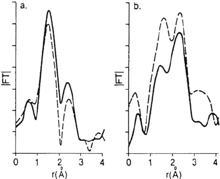
In a series of experiments designed to investigate directly the structure of
oxide films on iron, Kruger et al. [27–31] employed the sophisticated technique
known as EXAFS (extended X-ray absorption fine structure). The technique
permitted spectra to be obtained for films in in situ conditions and for comparisons
to be made with ex situ measurements (i.e., out of the electrolyte solution in
the “dry” condition). It should be mentioned here that the majority of the films
examined were not anodically formed but rather chemically prepared by exposure
of iron to such passivating solutions as nitrite or chromate. Nevertheless, the
results indicated very significant differences between the in situ and ex situ films,
suggesting that “drying” of the films for ex situ examination does lead to major
structural changes. Comparisons of ex situ and in situ films formed in nitrite and
chromate are shown in Figure 4. The implication is that great care has to be taken
when employing ex situ techniques, such as the ultrahigh-vacuum (UHV) spectro-
scopies or RHEED, because some films being studied may be structurally different
from those present when the electrode is immersed in the electrolyte. In more
recent research employing the spin-polarized neutron reflectivity technique,
Krebs, Kruger, and co-workers [32,33] have studied in situ the passive film
formed anodically on iron in the pH 8.4 borate buffer. The passive film was
observed to be different from the air-formed film. If the passive film proves to be
ferromagnetic, it would support an Fe
3
O
4
or Fe
3
O
4
/γ-Fe
2
O
3
bilayer model. However,
as mentioned previously [19], there is some concern that what may appear as
ferromagnetism in a bulk oxide would instead become superparamagnetism in a
thin film. As pointed to by Krebs et al. [33], if this is the case for the passive film
on iron, then it will be extremely difficult to apply a polarizing magnetic field
strong enough to observe any changes in magnetic scattering density.
Growth and Stability of Passive Films 195
Figure 4 Passivity of iron. Magnitude of the Fourier transforms of EXAFS spectra for
(a) ex situ and (b) in situ films. Nitrite-formed film results are given by the dashed lines
and chromate formed film results by the solid lines. (From Ref. 30.)
Copyright © 2002 Marcel Dekker, Inc.
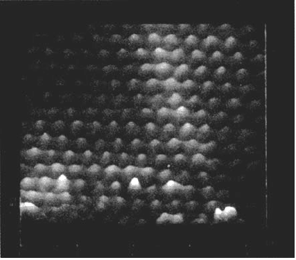
More recent studies have used in situ and ex situ scanning tunneling
microscopy (STM) [119] X-ray scattering [120,121] to study the passive film formed
on iron in borate buffer solution. Ex situ STM examination in air (Fig. 5) showed
long-range crystalline order on the film surface with the same triangular lattice of
spacing 0.30 ± 0.01 nm appearing on all the crystallites studied. In situ STM at inter-
mediate potentials after passivation at high potentials showed the same lattice on a
large number of areas. The structure is consistent with a γ-Fe
2
O
3
/Fe
3
O
4
structure of
the passive film and the results refute the suggestion of a highly disordered or amor-
phous passive film. X-ray scattering data [120,121] of the passive film are consistent
with a spinel oxide (γ-Fe
2
O
3
, Fe
3
O
4
, or related structure) and inconsistent with other
crystalline bulk oxides, hydroxides, or oxyhydroxides. However, the data demon-
strate that neither Fe
3
O
4
, γ-Fe
2
O
3
, nor any combination of these phases can adequately
describe the experimental data. The model proposed [120,121] assumes a uniform
distribution of vacancies and interstitials and the best-fit structure has a stoichiometry
of Fe
1.9±0.2
O
3
, which indicates that most of the Fe cations are in the Fe
3+
state. In the
proposed structure, the octahedral vacancies are randomly distributed but occupy
specific sites in γ-Fe
2
O
3
and are absent in Fe
3
O
4
; in γ-Fe
2
O
3
and Fe
3
O
4
the tetrahedral
sites are fully occupied and there are no octahedral interstitials.
NATURE OF THE PASSIVE FILM ON NICKEL
The situation with nickel is quite different from that for iron, passivity being readily
achieved in a wide variety of solutions over a large range of pH [34]. In the case of
nickel, there is not the necessity to have a highly buffered neutral solution in order to
achieve good passivity, and this may be due to the fact that the potential for anodic
deposition of an NiOOH film is fairly high [35]. For example, nickel passivates much
more readily than iron in unbuffered pH 3.0 Na
2
SO
4
[34]. Iron is much more
196 MacDougall and Graham
Figure 5 Atomically resolved STM image recorded in air of the passive film on iron in
pH 8.4 borate buffer at 1040 mV (NHE) for 15 min. (From Ref. 119.)
Copyright © 2002 Marcel Dekker, Inc.
sensitive to the conditions of passivation than nickel, and good passivity can be
achieved with iron only under rather specific conditions. The fundamental reason
for this difference is most probably associated with differences in the nature of the
passive oxide film present on each metal.
As with iron, a considerable amount of works has been performed on nickel
to investigate the nature of the passive oxide film. The exact composition of this
film is still under discussion, but there is general agreement that its thickness is
between 0.9 and 1.2 nm [35,36] and that anodic potential does not have a strong
influence on thickness. In contrast, with iron and oxide film thickness varies
from 1.5 to 4.5 nm depending on the electrode potential. The two most prevalent
views of the passive film on nickel are that it is either entirely NiO with a small
amount of nonstoichiometry giving rise to Ni
3+
and cation vacancies [37] or that
it consists of an inner layer of NiO and an outer hydrous layer of Ni(OH)
2
[38].
In the latter view, the film structure is similar to that observed when value metals
(e.g., aluminum and tantalum) are anodized in acid solution. (A review of the
subject is given in Ref. 39.) Evidence in support of the two-layer model comes
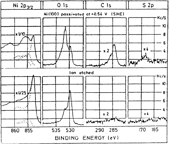
mainly from XPS investigations [38,40,41]. Figure 6 shows XPS data of Marcus
et al. [38]. The O 1s region for the passive film shows two fairly well-resolved
peaks at 529.8 eV and 531.6 eV with a shoulder at higher binding energy. These
two peaks correspond to the positions of oxygen in NiO and Ni(OH)
2
. (The
shoulder is assigned to sulfate.) There may, however, be other interpretations of
these XPS data as suggested by Roberts and co-workers [42], who point out that
Ni
3+
defects can give rise to spectral features very similar to those of Ni(OH)
2
.
Another view is that the NiO film has some adsorbed hydroxyl ions on the
Growth and Stability of Passive Films 197
Figure 6 Passivity of nickel. XPS spectra of a nickel(100) electrode after passivation in
0.1 N H
2
SO
4
at 0.54 V (vs. SHE) for 30 min. (row 1); row 2 shows spectra obtained after
ion etching (0.5 keV, 1 min, P
Ar
= 4 × 10
–5
torr). (From Ref. 38.)
Copyright © 2002 Marcel Dekker, Inc.