Mark James E. (ed.). Physical Properties of Polymers Handbook
Подождите немного. Документ загружается.


CHAPTER 23
Small Angle Neutron and X-Ray Scattering
George D. Wignall
Neutron Scattering Sciences Division, Oak Ridge National Laboratory, Oak Ridge,
Tennessee TN 37831-6393
23.1 Introduction . ............................................................. 407
23.2 Contrast ................................................................. 411
23.3 Examples of the Application of SANS and SAXS to Polymers ................. 411
Acknowledgments ........................................................ 420
References . . ............................................................. 420
23.1 INTRODUCTION
Scattering in the context of this article means the deflec-
tion of a beam of radiation (neutrons/x-rays/light, etc.) from
its original direction by interaction with the nuclei or elec-
trons of polymer/solvent molecules in a sample. The angular
distribution of the intensity reflects the structure of the
sample and such techniques have been employed since
the beginnings of polymer science to provide information
on the spatial arrangements of macromolecules [1]. The first
measurements were made in the 1920s using x-rays to
determine crystal structures via the Bragg law
nl ¼ 2D sin u , (23:1)
where D is the distance between crystallographic planes, l is
the wavelength of the radiation used, 2u is the angle of
scatter, and n is the (integer) order of reflection. The inten-
sity is conventionally measured as a function of the momen-
tum transfer, Q, which is related to 2u via
Q ¼ 4l
1
sin u (23:2)
although several different symbols have been used to denote
this parameter in the literature for the different types of
radiation used (e.g., Q, K, h, k, s, q, m etc.). Combining
Eqs. (23.1) and (23.2) gives
D ¼ 2=Q (23:3)
which indicates the distance scale probed by a measurement
at a given value of Q. Experiments in the range
0:6 < Q < 15 A
˚
1
contain most of the information relevant
for the determination of unit cell dimensions and are con-
ventionally referred to as wide-angle scattering, which
probe a distance scale 0:4 < D < 10 A
˚
. Wide-angle
x-ray scattering (WAXS), with a wavelength 1A
˚
, has
been the principal technique for the determination of poly-
mer crystal structures [2]. Unit cell dimensions [3], along
with details of the WAXS technique [4] are given in stand-
ard reference works [2–4]. Subsequently, neutron diffraction
or wide-angle neutron scattering (WANS) has supplemented
these measurements of crystal structures [5,6].
In the amorphous state, the intermolecular correlations
are more diffuse, and the information available from wide-
angle scattering is less precise. A Fourier transform of the
data gives a radial distribution function (RDF) which is a
weighted sum of interatomic pair correlation functions
g
ij
(r), which express the probability of finding atomic spe-
cies i and j separated by a distance r. In the crystalline
regions of polymers the g
ij
(r) reduce to a series of delta-
functions defining the interatomic distances in the unit cell.
For amorphous materials, the RDFs are generally featureless
for r > 10 A
˚
, indicating the absence of long range order
between neighboring chains [7].
Although Bragg’s law does not apply to amorphous ma-
terials, the Fourier or inverse relationship between the struc-
ture in real-space (r) and the scattering in Q-space, means
that Eq. (23.3) may be applied to first order for all types of
scattering. Thus, data at lower Q-values probe longer length
scales, and x-ray methods have been widely used to deter-
mine chain dimensions in dilute solution, lamellar spacings
in crystalline polymers, etc. These measurements are con-
ventionally referred to as small-angle x-ray scattering
(SAXS), though it is the Q-range (typically 10
3
< Q <
10
1
A
˚
1
) which determines the size of objects studied and
radiation with other wavelengths (e.g., light, neutrons) can
407
provide similar information in different angular ranges. For
example, light scattering (LS), with l 2---6,000 A
˚
, probes a
much smaller Q-range ( 2 10
6
< Q < 2 10
3
A
˚
1
)
than SAXS, even though the angular range can be quite
large (up to 2u 160
). Hence, the measurements probe
distance scales, via Eq. (23.3), up to 10 mm and the tech-
nique has been used extensively since the 1940s, to deter-
mine the molecular weight and global dimensions of polymer
molecules, for example in dilute solution.
For over two decades, small-angle neutron scattering
(SANS), with a wavelength l 5---20 A
˚
, has proven to be
extremely useful for the evaluation of polymer chain con-
formation. Due to a combination of high bulk penetrating
power, the ability to manipulate scattering amplitudes
through isotopic labeling (e.g., deuteration), or an appropri-
ate choice of solvent (contrast variation), SANS has devel-
oped into a powerful tool for the study of polymers,
particularly in systems inaccessible to SAXS or LS (e.g.,
bulk polymers, concentrated solutions etc.).
For most applications in polymer science, neutron, x-ray,
and light scattering are examples of predominantly elastic
scattering, where the incident and scattered radiation have
the same energy or wavelength. Such experiments give
information on the time-averaged structure and conform-
ation of polymer molecules and form the bulk of the work
undertaken on polymers. There has been less work involv-
ing inelastic processes, where there is a change of energy on
scattering, and the incident and scattered radiation have
different wavelengths. This technique gives valuable infor-
mation on polymer dynamics, though this methodology is
beyond the scope of this article [8–10]. Also, due to space
limitations, is not possible to survey all contributions to the
understanding of polymer structure by all types of radiation
(neutrons, x-rays, light, electrons etc.) in different Q-ranges
(small-angle, wide-angle, etc.). Similarly, it is not possible
to derive the scattering theory, which will be quoted from
existing reviews of neutron [8,11–13], x-ray [14,15], and
light scattering techniques [12,16]. Most of the work on
polymers has been undertaken at small Q-values to probe
the longer length scales associated with these materials. The
article will illustrate the type of information provided by
SANS and SAXS, along with analogies and differences
between neutron and photon scattering.
The treatment will emphasize the importance of placing
data on an absolute scale, typically in the form of a differ-
ential scattering cross section dS=dV(Q), per unit sample
volume (in units of cm
1
) for SAXS and SANS. The equiva-
lent quantity for LS is the Rayleigh ratio, which is directly
analogous to d S=dV [8,17,18]. The use of absolute units is
not essential for the measurement of spatial dimensions
(e.g., the determination of the radius of gyration of polymer
molecules). However, it forms a valuable diagnostic tool for
the detection of artifacts, to which scattering techniques are
particularly vulnerable.
Because the cross-section varies as the sixth power of the
dimensions [14], it is a very sensitive indicator of whether
an appropriate structural model has been chosen. Thus,
absolute SANS measurements of melt-crystallized blends
of normal (hydrogenous) and deuterium-labeled polyethy-
lenes showed that the scattering could exceed the expected
intensity for randomly mixed molecules by three orders of
magnitude. This indicated that some kind of previously
unsuspected aggregation or clustering phenomenon was
taking place [19]. Similarly, scattering studies of colloidal
micellar solutions may be modeled by calculation of the
interparticle correlations between spherical micelles as a
function of a set of parameters describing the particle struc-
ture (inner/outer radius, degree of ionization etc.). On an
arbitrary intensity scale, it is possible to produce excellent
fits of the particle shape, which may be in error by as much
as 3–4 orders of magnitude in intensity [20]. Thus, absolute
calibration allows such artifacts to be recognized, and the
model parameters may be restricted to those which repro-
duce the observed cross section. Because the literature often
contains general formulae, as opposed to practical examples
of how such calculations are actually accomplished, this
article will illustrate such comparisons via a range of ex-
amples on different polymeric systems.
Figure 23.1 illustrates the relationship between the neu-
tron energy and wavelength. The kinetic energy of a neutron
of particle velocity 750 m=sec (wavelength l ¼ 5:3A
˚
)is
3 meV or 4:7 10
15
ergs [8]. Such energies are of the
same order as the vibrational and diffusional energies of
molecular systems and much lower than x-ray photons
( 10 keV). For LS, the scattering patterns are very depen-
dent on the polarization directions, though because of the
much higher energies of x-rays, chemical bonding has little
effect on SAXS and there is negligible influence of the
differences between the directions of radiation polarization
and molecular orientation [21]. Hence polarization effects,
0.3
1
1.2
9
8
7
6
5
4
3.5
3
2.5
2
1.5
1.2
9
8
7
6
5
4
3.5
3
2.5
2
1.5
9
8
7
6
5
4
3
2
1.5
9
8
7
6
5
4
3
2
1.5
9
8
7
6
5
4
3
2
1.5
9
8
7
6
5
4
3
2
1.5
10
−4
10
−3
10
3
10
−2
10
−1
10
4
0.35
0.4
0.5
0.6
0.7
0.8
0.9
1
1.2
1.5
2
2.5
3
3.5
4
5
6
7
8
9
10
15
20
25
1.2
Wavelength (Å)
ORNL-DWG 94M-11823
Energy (eV)
Velocity (msec
−1
)
FIGURE 23.1. Conversion chart for neutron wavelength, energy, and velocity.
408 / CHAPTER 23

which are important for LS, can be neglected in SAXS and
also for SANS experiments on polymers
1
.
SANS, SAXS, and LS all involve interference phenom-
ena between the wavelets scattered by different elements in
the system. When a plane wave, described by a wave func-
tion of unit density [8] interacts with a single nucleus, the
scattered wave is given by
C
1
¼
b
r
exp (ikr): (23:4)
The quantity b has the dimensions of length and is called
the scattering length, which may be regarded as a real
(known) constant for a given nucleus (isotope). The scat-
tered single atom cross section is given [8,22,23] by
¼ 4pb
2
: (23:5)
It can be seen from Eq. (23.5) that has the dimensions of
area. The magnitude of b is typically of the order of
10
12
cm, and this gives rise to the usual unit for a cross
section which is called a barn (10
24
cm
2
).
Neutrons are scattered isotropically from individual nu-
clei, whereas for LS and SAXS, the scattering originates in
the electron cloud, so the atomic form factors are in principle
Q-dependent. However, the variation is very small in practice
(< 1% for Q < 0:1A
˚
1
) for SAXS and LS, and is usually
neglected [4]. The Thompson scattering amplitude of a clas-
sical electron is r
T
¼ 0:282 10
12
cm [24], so the x-ray
scattering length of an atom, f, is proportional to the atomic
number ( f ¼ r
T
Z) and increases with the number of electrons
per atom. For neutrons, there is no general trend throughout
the periodic table in the values of b, which vary from isotope
to isotope. If the nucleus has nonzero spin, it can interact with
the neutron spin, and the total cross section (
coh
) splits
into coherent and incoherent components defined by
coh
¼ 4p < b >
2
, (23:6)
inc
¼
tot
coh
¼ 4p[ < b
2
> < b >
2
], (23:7)
where the brackets <> represent a thermal average over the
spin state population.
If the isotope has no spin, then < b
2
> ¼ < b >
2
as
< b > ¼ b and there is no incoherent scattering for neu-
trons. Only coherent scattering contains information on the
structure of the sample. The incoherent cross section contains
no information on interference effects and forms an isotropic
(flat) background which must be subtracted off in SANS
structural investigations. While most of the atoms encoun-
tered in neutron scattering from polymers are mainly coher-
ent scatterers (e.g., carbon, oxygen, deuterium), there is one
important exception [8,22,23]. In the case of hydrogen (H
1
)
s
coh
¼ 1:76 10
24
cm
2
, (23:8)
s
inc
¼ 79:7 10
24
cm
2
: (23:9)
For photons, there is no strict analog of incoherent scat-
tering of neutrons due to nonzero spin in the scattering
nucleus. Compton scattering which occurs for x-rays is
similar in that it contains no information on interference
effects, i.e., the structure of the sample, and forms a back-
ground to the coherent signal. However, to a good first
approximation this background goes to zero in the limit
Q ! 0 and is usually neglected in SAXS and LS studies.
Table 23.1 gives the cross sections and scattering lengths
for atoms commonly encountered in synthetic and natural
polymers. These cross sections refer to bound protons and
neglect inelastic effects arising from interchange of energy
with the neutron. For coherent scattering which is a collect-
ive effect arising from the interference of scattered waves
over a large correlation volume, this approximation is rea-
sonable [8]. However, for incoherent scattering, inelastic
effects become increasingly important for long wavelength
neutrons with the result that the H
1
incoherent cross section,
and hence the sample transmission, is a function of both the
TABLE 23.1. Bound atom scattering lengths and cross sections for typical elements in synthetic and natural polymers.
Atom Nucleus b
coh
(10
12
cm)
coh
¼ 4pb
2
coh
(10
24
cm
2
)
inc
(10
24
cm
2
)
abs
(10
24
cm
2
) f
x-ray
(10
12
cm)
Hydrogen
1
H 0.374 1.76 79.7 0.33** 0.28
Deuterium
2
H(D) 0.667 5.59 2.01 0 0.28
Carbon
12
C 0.665 5.56 0 0 1.69
Nitrogen
14
N 0.930 11.1 0 1.88** 1.97
Oxygen
16
O 0.580 4.23 0 0 2.25
Fluorine
19
F 0.556 4.03 0 0 2.53
Silicon
28
Si 0.415 2.16 0 0.17** 3.94
Chlorine Cl* 0.958 11.53 5. 9 33.6** 4.74
*Values are for the naturally occuring element and are an average over the mixture of isotopes; f
x-ray
is given for Q ¼ 0, though
the angular dependence is small (<1%) for Q < 0:1A
˚
1
.
**Values of the absorption cross section (
abs
) are a function of wavelength (l) and are given at ¼ 1:8A
˚
.As
abs
, values at
other wavelengths may be estimated by scaling via the ratio =1:8.
1
Except in the hypothetical case of a material containing elements
with unpaired spins (e.g., Fe, Mn, rare earths etc.), where polarization
effects can theoretically occur, due to the interaction with the neutron spin.
In practice, polymers do not contain such elements, so polarization effects
can also be neglected in SANS.
SMALL ANGLE NEUTRON AND X-RAY SCATTERING / 409

incident neutron energy and sample temperature [25]. In
addition, because of inelastic effects due to torsion, rotation,
and vibration, the effective incoherent cross section is a
function of the particular chemical group (methyl, hydroxyl
etc.) in which the proton is situated [26]. This is illustrated in
Table 23.2, which shows the total hydrogen atom cross sec-
tion (s
tot
) in various liquids and polymers. s
tot
is dominated
by the incoherent component (s
inc
), and hence is also a strong
function of l and only approaches 80 barns at l 4:5A
˚
.
Thus, the cross sections given in Table 23.1 cannot be used to
calculate the incoherent background because although
s
inc
¼ 79:7 10
24
cm
2
is widely quoted in the literature,
this value almost never applies to real polymer systems.
It may be seen from Table 23.1 that there is a large
difference in the coherent scattering length between deuter-
ium and hydrogen and that the latter value is actually nega-
tive. This arises from a change of phase of the scattered
wave and results in a marked difference in scattering power
(contrast) between molecules synthesized with deuterated or
protonated monomer units.
The majority of neutron scattering experiments under-
taken on polymers fall into the category of SANS from
a fraction of deuterated chains in a matrix of normal (pro-
tonated) polymer and such experiments are examples of
predominantly coherent elastic scattering, which gives
information on the time-averaged structure (e.g., chain
configuration or orientation in the bulk, polymer compati-
bility, segregation etc.). Similarly, for x-ray scattering, the
energy changes are much less than the incident energy, so
SAXS and WAXS are effectively elastic processes, which
give complementary information (e.g., lamellar spacings,
chain configuration in solution, crystal structures etc.).
As LS probes longer length scales, the most commonly
observed pattern for unoriented samples is due to spherul-
ites, which may be interpreted in terms of the scattering of
anisotropic spheres to investigate the spherulite size as a
function of the crystallization conditions [21]. For liquids, it
is well known that LS is particularly sensitive to contamin-
ants (dirt, dust etc.), and that samples must be carefully
filtered. This has meant that LS methods have been largely
restricted to dilute solutions, though SANS is much less
sensitive to this artifact. Accordingly, SANS has been the
preferred technique to investigate concentrated solutions
[27,28] and bulk polymers [8,13,21,29].
For such experiments, we can define a coherent scattering
length of the repeat monomer unit (segment) by
a
H
¼ S
k
b
k
, (23:10)
where the summation runs over all the atoms in an unlabeled
monomer unit and a similar equation may be written for the
coherent scattering length of a labeled monomer unit a
D
.If
the two polymers, with polymerization index (N) and seg-
ment (monomer) volume (V) are blended together so that the
volume fraction of H- and D-labeled components are w
H
and
w
D
, respectively, the coherent cross section is given [8] by
dS
dV
(Q) ¼ V
1
Nw
H
w
D
(a
H
a
D
)
2
P(Q) (23:11)
after subtracting off the incoherent signal (principally due
to H
1
atoms) and the coherent background due to heterogene-
ities (voids, catalyst residues) or density fluctuations (crystal–
TABLE 23.2. Experimental (total) hydrogen atom cross sections (s
tot
) in various liquids and polymers at room temperature.
Cross section per H
1
atom s
tot
(10
24
cm
2
)
Compound Formula l ¼ 9:0A
˚
l ¼ 4:75 A
˚
Methanol CH
3
OH 137
Ethanol CH
3
CH
2
OH 124
Isopropanol CH
3
CHOHCH
3
123
n-Butanol CH
3
CH
2
CH
2
CH
2
OH 117
n-Propanol CH
3
CH
2
CH
2
OH 113
Ethanediol HOCH
2
CH
2
OH 108
Propanetriol HOCH
2
CHOHCH
2
OH 100
Polyvinylalcohol –(CH
2
CHOH)
n
–97
Polymethylmethacrylate CH
3
j
–(CH CH
2
)
n
– 115 92
a
j
–CO
2
CH
3
–
Polyethylene –(CH
2
CH
2
)
n
– 113 89
b
Water (T ¼ 23 8C) H
2
O 114 89
c
Values of at ¼ 9:0A
˚
are taken
2
from reference [26].
a
Reference [68].
b
Reference [69].
c
Reference [66].
2
By permission of Butterworth-Heinemann Ltd.
ß
410 / CHAPTER 23

amorphous boundaries, thermal vibrations). The coherent
scattering is governed by the single chain form factor, P (Q),
which originates from monomer pairs belonging to
the same chain [P(0) ¼ 1]:dS=dV(Q) is directly analogous
to the Rayleigh ratio, used in light scattering [18] and contains
information on the single chain (intramolecular) scattering
function, P(Q). The mole fraction of each component
modulates the coherent cross section and P(Q) may be
obtained from the measured intensity at labeling levels up to
50%, though this result was not appreciated in the earliest
SANS studies of bulk polymers and concentrated solutions.
These experimentsrelied on analogieswith LS wherethe limit
of zero concentration was required to eliminate interchain
interference. It may be seen that for w
D
51, w
H
1 and the
cross section is proportional to the concentration [14,32], as
assumed in the Guinier [14] and Zimm [30] approximations.
23.2 CONTRAST
The quantity (a
D
a
H
)
2
is related to the difference in
scattering power between labeled and unlabeled chains and
is called the contrast factor. In general, radiation incident on
a medium whose scattering power is independent of position
is scattered only into the forward direction (u ¼ 0), and all
scattering cancels unless the scattering power fluctuates
from point-to-point in the sample. X-rays and light photons
interact with electrons in the sample and hence are scattered
by fluctuations in the electron density (r
e
). Neutrons on the
other hand, have no interaction with electrons, so the con-
trast arises from fluctuations in scattering length density
(r
n
). Because each nucleus has a different scattering ampli-
tude (Table 23.1), the scattering length density (SLD) is
defined as the sum of coherent scattering lengths over all
atoms lying in a given volume DV, divided by DV [31]. For
example, in bulk polymers, the SLD is given by the coherent
neutron scattering length [Eq. (23.10)] divided by the mono-
mer volume. The coherent cross sections of a system of
uniform scattering length density is zero, though fluctu-
ations may be introduced by means of isotopic substitution,
thus giving rise to a finite cross section which is proportional
to (a
H
a
D
)
2
. Table (23.3) shows values of the SLDs of
some H- and D-labeled polymers and solvents.
The parameter used to describe the overall size of a poly-
mer chain is the radius of gyration (R
g
), the root mean square
distance of all scattering elements from the center of gravity
R
2
g
¼
Sf
k
r
2
k
Sf
k
: (23:12)
The summation runs over all scattering elements (k),
which are the electrons in the case of SAXS or LS. For
SANS, the summation runs over all nuclei, and is weighted
by the scattering length of each atom. Thus in principle, the
R
g
may be different when measured via different techniques.
However, in practice each monomer has the same scattering
power for a given incident radiation, so for large polymer-
ization indices the differences between SANS, SAXS, and
LS radii are negligible. R
g
may be derived by expanding
P (Q) [Eq. (23.11)] in a power series for low Q (Q < R
1
g
)
and plotting dS
1
(Q)=dV versus Q
2
(30). Alternatively,
these parameters may be obtained by plotting ln
[dS(Q)=dV] versus Q
2
at low Q [14]. These types of plots
are conventionally referred to as Zimm and Guinier plots,
respectively, and the former is generally used for investigat-
ing polymer configurations as it has been found to be linear
over a wider Q-range. As mentioned earlier, the first meas-
urements in bulk amorphous polymers and concentrated
solutions were generally performed in the limit of low rela-
tive labeling (w
D
51), and extrapolated to zero concentra-
tion. In this range, Eq. (23.11) may be expanded to give
dS
1
dV
(Q) ¼
V
(a
H
a
D
)
2
Nw
D
1 þ
Q
2
R
2
g
3
þ ...
"#
: (23:13)
23.3 EXAMPLES OF THE APPLICATION OF SANS
AND SAXS TO POLYMERS
23.3.1 SANS from Amorphous Polystyrene
Figure (23.2) shows a Zimm plot for 5.0 wt%
(w
D
¼ 0:047) deuterated polystyrene (PSD) in a matrix
of hydrogenous (normal) polymer (PSH). The coherent
TABLE 23.3. Comparison of the scattering length densities
of various polymers and solvents.
Polymer or solvent
Density
3
, r
(at T ’ 23 8C unless
otherwise stated)
(gm cm
3
)
Scattering
length
density, r
n
(10
10
cm
2
)
Carbon disulfide 1.63 1.24
Water 1.0 0.56
D
2
O 6.4
Xylene 0.880 0.79
Xylene-d
10
6.04
Toluene 0.867 0.94
Toluene-d
8
5.66
Benzene 0.8765 1.18
Benzene-d
6
5.4
Polybutadiene 0.89 0.41
Polyethylene 0.78 (T ¼ 145 8C) 0.28
Polyethylene-d
4
6.71
Polymethylmethacrylate 1.2 1.06
PMMA-d
8
7.09
Polystyrene 1.05 1.41
Polystyrene-d
8
6.47
3
The values of SLD are calculated at the indicated densities, which may
vary slightly with temperature, tacticity (e.g., for PMMA), degree of crys-
tallinity (e.g., for polyethylene) etc. For different densities, the SLD is
proportional to r and may be scaled from the values shown. For deuterated
materials, it is assumed that the number of monomers per unit volume is
independent of deuteration.
SMALL ANGLE NEUTRON AND X-RAY SCATTERING / 411
scattering lengths of the labeled (C
8
D
8
) and unlabeled (C
8
H
8
)
are 2:32810
12
cm and 10: 66 10
12
cm, respectively, [via
Eq. (23.10) and Table 23.1]. Given a density, r ’ 1:05 gm
cm
3
, the segment volume is 164:5 10
24
cm
3
and the
extrapolated cross section [dS=dV(0) ¼ 17:4 0:5cm
1
],
leads to a polymerization index of the labeled chains of
N
D
¼ 928 30 or a molecular weight of (96:5 3) 10
3
,
in reasonable agreement with independent determinations via
osmometry [32]. The radius of gyration is close to that meas-
ured in ideal Q-solvents [8,21] and this supports the unper-
turbed Gaussian coil as a good approximation to the
molecular configuration in amorphous polymers [8,13,32].
23.3.2 SANS and SAXS from Melt-Crystallized
Polyethylene
Figure 23.3 shows a Zimm plot of the SANS differential
scattering cross section for 6.0 wt% (w
D
¼ 0:053) of deuter-
ated polyethylene (PED) in a matrix of unlabeled PEH after
rapidly quenching from the melt. The coherent scattering
lengths of C
2
H
4
and C
2
D
4
are 0:166 10
12
and 4:00
10
12
cm
1
, respectively, [via Eq. (23.10) and Table 23.1],
and based on an average density of r ’ 0:94 gm cm
3
, the
segment volume is 49:5 10
24
cm
3
. Thus the extrapolated
cross section [dS=dV(0) ¼ 28:0 2cm
1
] leads to a poly-
merization index (N) of 1,600, which is of the same order as
the value from gel permeation chromatography [33]. How-
ever, when the same sample is slow cooled from the melt [Fig.
23.4], the extrapolated cross section increases by over an
order of magnitude. It is clear that these data do not originate
in the scattering from single molecules, and it has been shown
that the excess intensity is caused by aggregation or clustering
of the labeled molecules [19], though this would not be
apparent if the data were in arbitrary units. This behavior
illustrates the point referred to above that the intensity is
extremely sensitive to the particle or molecular dimensions
and even an approximate (25%) absolute calibration is
sufficient to reveal the presence of such artifacts.
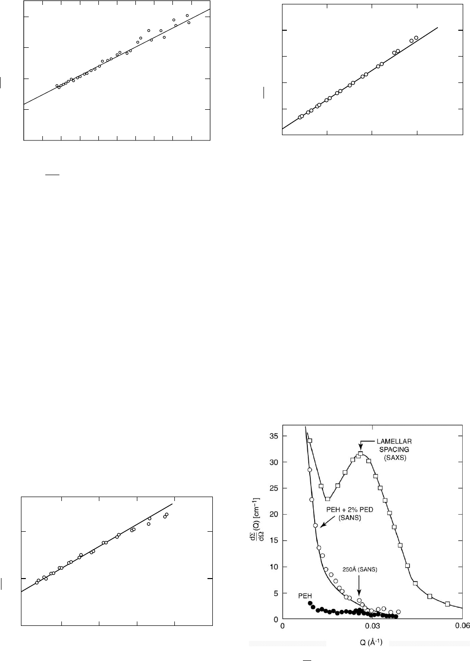
Figure 23.5 shows SAXS data from the polyethylene
sample described above. Because PED and PEH have the
same electron density, there is no contrast between
the different isotopes and PEH, PED and partially labeled
0
0
0.05
0.1
0.15
0.2
10 20 30
Radius of gyration, R
g
= <S
2
>
½
z
= 87 Å
MW(SANS) = 96,500;MW (Osmometry) = 97,200
40 50
Q
2
⫻10
5
[Å
−2
]
60 70 80 90 100
(Q) (cm)
dS
−1
dW
FIGURE 23.2.
d
1
d
(Q)VSQ
2
for sample containing 5 wt %
labeled PSD molecules in polystyrene (PSH).
0
0
5
10
Quenched
MW = 46 ⫻ 10
3
(SANS)
MW = 60 ⫻ 10
3
(GPC)
Rg = 132 Å
123
Q
2
⫻10
−4
(Å
−2
)
45
(Q) ⫻ 10
2
(cm)
dS
−1
dW
FIGURE 23.3. Typical Zimm plot for 6 WT % PED molecules
in PEH matrix quenched from the melt.
0
0
1
2
3
Slow cooled (1°C
−1
)
MW = 693 ⫻ 10
3
(SANS)
MW = 60 ⫻ 10
3
(GPC)
R
g
= 368 Å
4
5
12
Q
2
⫻ 10
−4
(Å
−2
)
34
(Q)⫻ 10
2
(cm)
dS
−1
dW
FIGURE 23.4. Typical Zimm plot for 6 wt% PED molecules in
PEH matrix slow cooled (18C
1
) from the melt.
FIGURE 23.5.
dS
dV
(Q)vsQ for SAXS and SANS data from
melt crystallized polyethylenes.
412 / CHAPTER 23
samples all have the same SAXS profile. The background
due to Compton scattering is virtually zero in this Q-range
[4] and the signal arises from density fluctuations [13]. The
interlamellar peak at Q 0:025 A
˚
1
is proportional to the
square of electron density difference between the amorph-
ous and crystalline regions (lamellae). The upturn as Q ! 0
probably arises from voids and other large scale structures
such as spherulites. Figure 23.5 also shows the SANS data
for PEH (solid circles), where the coherent signal is super-
imposed on a flat (incoherent) background 1cm
1
. The
open circles show the extra (coherent) cross section pro-
duced by adding 2% deuterated molecules (PED), which is
proportional to the contrast difference (a
H
a
D
)
2
between
deuterated and protonated segments.
Departures from the flat incoherent background of the
PEH sample (solid circles) are caused by density fluctu-
ations in the sample and it is just possible to see the peak
at Q 0:025 A
˚
1, due to the periodic stacking of crystal-
line lamellae alternating with amorphous regions. The
SANS coherent signal in PEH is very weak, however, due
to the cancellation between the scattering lengths of carbon
and hydrogen (Table 23.1), which makes the SLD very
small for PEH (Table 23.3).
In the case of PED, there is no cancellation between
the coherent scattering lengths of carbon and deuterium
(Table 23.1), and the incoherent background is very much
smaller than for PEH. Thus, PED should have virtually
identical SAXS and SANS cross sections apart from a
scale factor. Figure 23.6 shows absolute SAXS and SANS
data for the same sample of PED, which should scale as the
ratio of the electron density to the scattering length density. As
the number of segments per unit volume is the same for SAXS
and SANS, this term cancels and the ratio (R)reducesto
R ¼
(0:282 10
12
16)
2
(4:00 10
12
)
2
¼ 1:27, (23:14)
where r
T
¼ 0:282 10
12
cm is the Thompson scattering
factor of one electron, and 4:00 10
12
cm is the neutron
scattering length of a C
2
D
4
monomer, which contains 16
electrons. Thus the measured (1.31 + 0.1) and theoretical
ratios are in good agreement [34].
23.3.3 Application of Contrast Variation Methods
to Core–Shell Latex Structures
Contrast variation methods can sometimes be used to
remove a component of the scattering by matching its scat-
tering power with that of the medium in which it is dis-
persed. This principle can be used in SANS experiments via
isotopic solvent mixtures (e.g. H
2
O=D
2
O) to adjust the
scattering power of the medium, as for example in studies
of polymer latexes. Grancio and Williams [35] postulated a
polymer-rich spherical core surrounded by a monomer-rich
shell which serves as the major locus of polymerization, thus
giving rise to core–shell morphology. Thus, the first formed
polymer constitutes the core and the second formed polymer
makes up the shell, and neutron scattering has been used to
test this hypothesis by isotopically labeling chains generated
at specific points in the polymerization process [36–40].
For a homogeneous particle, suspended in a solvent, the
neutron scattering cross section is given by
dS
dV
(Q) ¼ (r
np
r
ns
)
2
N
p
V
2
p
P(Q), (23:15)
where r
np
and r
ns
are the neutron scattering length densities
of the particle and solvent, respectively, N
p
is the number of
particles per unit volume, V
p
is the particle volume, and P (Q)
is the particle form factor [P(0) ¼ 1]. According to Grancio
and Williams [35], polymerization takes place in a surface
shell and thus if the monomer feed is changed from proto-
nated to deuterated material, this will result in a predomin-
antly D-labeled shell. When examined by SANS in an
H
2
O---D
2
O mixture which matches the scattering length
density of the protonated core, the scattering will arise from
a hollow sphere with a particle form factor [41] given by
P(Q) ¼ 9
[ sin (QR) sin (QR1) QR cos (QR)
þQR1 cos (QR1)]
2
Q
6
R
6
(1 l)
6
, (23:16)
where R and a are the outer and inner radii, respectively,
(l ¼ a=R).
Figure 23.7 shows SANS data from a 4.6 vol% latexes
with a fully deuterated PMMA-D shell, (thickness 30 A
˚
)
polymerized on a PMMA-H core (radius, a ¼ 498 A
˚
), after
desmearing corrections for the finite instrumental resolution
[37,42]. The absolute intensity at zero scattering angle is
given by Eq. (23.15) with P(0) ¼ 1 and Vp equal to the
volume of the D-labeled polymer in the shell with SLD
4
,
r
np
¼ 6:97 10
10
cm
2
. The SLD of the solvent is close to
that of the PMMA-H core (r
ns
¼ 1:06 10
10
cm
2
) and thus
0
10
−1
10
−1
10
−2
10
0
10
1
10
2
10
3
10
4
10
2
10
1
10
0
0.02 0.04
SANS
SAXS
0.06 0.08
Q (Å
−1
)
0.10 0.12
dW
dS
(Q) for SANS data (cm
−1
)
dW
dS
(Q) for SAXS data (cm
−1
)
FIGURE 23.6.
dS
dV
(Q)vsQ for deuterated polyethylene sam-
ple after subtraction of incoherent background.
4
This value is slightly different to that quoted in Table 23.3, based on a
density of 1:2gm cm
3
, which is an average over the atactic, isotactic and
syndiotactic homopolymers. For atactic PMMA, r ’ 1:18 gm cm
3
.
SMALL ANGLE NEUTRON AND X-RAY SCATTERING / 413
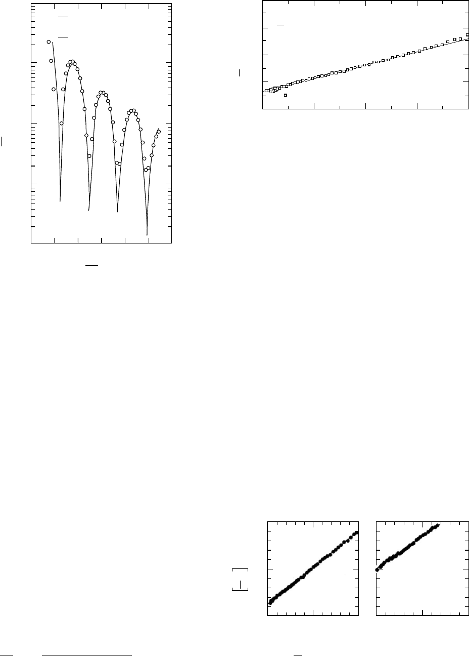
the core contrast is negligible compared to the PMMA-D
shell. The extrapolated cross section, dS=dV(0) ¼ 1:7
10
3
cm
1
, is in good agreement with the calculated value
of (2:3 10
3
cm
1
), in view of the extreme sensitivity of the
calculations to slight variations in shell thickness, mis-
matches in SLD, surface roughness etc. [37,42]. Similarly
the particle dimensions from SANS are in excellent agree-
ment with independent techniques (e.g., LS).
23.3.4 SAXS and SANS from 2-Phase Systems
Blends of High- and Low-Density Polyethylenes
SANS experiments have indicated that blends of high
density (linear) and long-chain branched low density poly-
ethylenes (HDPE/LDPE) are homogeneous in the melt,
though the components may separate on slow cooling due
to the difference in crystallization mechanisms [43]. The
semicrystalline blends form effectively two-phase systems
in the solid state, and it was shown [43, 44] that the Debye–
Bueche (DB) [45,46] model was appropriate to describe the
morphology, with a SANS cross section of the form
dS
dV
(Q) ¼
8pa
3
w
1
w
2
[r
n1
r
n2
]
2
(1 þQ
2
a
2
1
)
2
, (23:17)
where a
1
is a length characterizing the structure, w
1
and w
2
are the volume fractions, and r
n1
and r
n2
are the neutron
scattering length densities of the two phases [43,44]. Figure
23.8 shows a DB plot [d S=d V(Q)
1=2
vsQ
2
] of the data
for a 50/50 blend after cooling from the melt at 0:75
C
min
1
. The extrapolated cross section [d S=d V(0)
¼ 24:510
3
cm
1
] is well over an order of magnitude higher
than in the melt, indicating that the components have phase-
separated on cooling. The plot is reasonably linear and
the (Q ¼ 0) cross section is given by Eq. (23.17) where
the correlation length (a) is derived from the ratio of slope/
intercept of the DB plot [44–47]. Assuming complete
separation of the H- and D-labeled components, the SLDs
in the solid state can be scaled (via the density) from the melt
values shown in Table 23.3, to give a calculated cross section
of 28:2 10
3
cm
1
. In view of the fact that the experiments
are independently calibrated with no arbitrary fitting factors
in the intensity scale, the agreement with the absolute cross
sections calculated from the DB theory is excellent.
Interpenetrating Polymer Networks
Figure 23.9 shows DB plots of SANS data from polystyr-
ene–polybutadiene interpenetrating polymer networks [47].
0.005
10
−1
10
0
10
1
10
2
10
3
dS
dW
(O)=1.7⫻10
3
cm
−1
(Experiment)
(O)=2.3⫻10
3
cm
−1
(Calculated)
0.010 0.015 0.020 0.025
dS
dW
(Q) (cm
−1
)
dS
dW
4π
λ
Q =
sin q [Å
−1
]
FIGURE 23.7. Desmeared SANS data (O) for PMMA Latex
CI (4.6 vol%) with 30 A
˚
D-DMMA shell on surface (core
contrast matched) compared with theoretical hollow shell
scattering. Reprinted from the Journal of Colloid and Interface
Science, 123, L. W. Fisher, S. Melpolder, J. M. O’reilly,
V. Ramakrishnan, and G. D. Wignall, ‘‘Neutron Scattering
from Interfacially polymerized Latexes’’, 29–33, Copyright
(1988) with permission of Elsevier.
0.00
0.00
4.00
a = 67.3 Å
(0) = 24.5 x 10
3
cm
−1
= 28.2 x 10
3
cm
−1
calculated for 2-phase
structure of pure PED and PEH
x10
-2
dW
dS
1.0 2.0
10
4
Q
2
(Å
−2
)
3.0 4.0
(Q) (cm
½
)
dW
dS
−½
FIGURE 23.8. Debye–Bueche plot for phase separated
blend of deuterated HDPE and protonated LDPE slow cooled
from the melt AT 0:75 8C
1
. Reprinted with permission from
R. G. Alamo, J. D. Londono, L. Mandelkern, F. C. Stehling and
G. D. Wignall, Macromolecules, 27, 411 (1994). Copyright
(1994) American Chemical Society.
0
0
3.5
a = 106 Å a = 53 Å
7.0
2.5 5 0
Q
2
x10
4
Å
−2
0
3
6
2.5 5
PB(0.2% DICUP)/PS
semi-I IPN
Fully deuterated PS
PB(0.1 % DICUP)/PS
semi-I iPN
Fully deuterated PS
(Q)
(cm
½
)
dS
dW
−½
FIGURE 23.9.
dS
dV
(Q)
1=2
vs: Q
2
for two PB/PS semi-I-IPN
systems with different amounts of cross-linker (DICUP).
414 / CHAPTER 23
Assuming complete segregation of the components,
d S=d V(0) may be calculated from Eq. (23.17), via the
measured correlation lengths (Fig. 23.9) and the SLDs
given in Table 23.3. For the data from the two samples
shown in Fig. 23.9, this leads to calculated values of
17:2 10
3
cm
1
and 2:7 10
3
cm
1
, compared to experi-
mental determinations of 21:6 10
3
cm
1
and 2:0
10
3
cm
1
. The discrepancies are not unreasonable in view
of the strong dependence of the cross section on the domain
dimensions, which is a general feature of absolute intensity
comparisons. However, this illustrates the point made earl-
ier that even an approximate ( + 25%) absolute calibration
is sufficient to test the assumption of complete phase separ-
ation of the blend components.
In the limit Qa41, Eq. (23.17) reduces to d S=dV
PQ
4
P ¼ 2P(r
n1
r
n2
)
2
S=V, (23:18)
where P is the Porod constant, which contains information
on the specific surface of the material, i.e., the total inter-
phase surface (S) area per unit volume (V). By comparison
of Eqs. (23.17) and (23.18)
S=V ¼ 4w
1
w
2
=a: (23:19)
For the data shown in Fig. 23.9, this leads to specific surface
values in the range (58---150) 10
4
cm
1
or (58---150)m
2
gm
1
(r ’ 1:0gm cm
3
).
Void Content of Hompolymers via SAXS Invariant
Analysis
Figure 23.10 shows a Kratky plot [(Q
2
d S=d V(Q)vsQ]
for a polyimide sample made from the condensation of
pyromellitic-dianhydride and oxydianiline (PMDA-ODA).
The integrated area under this curve is the invariant which
for a 2-phase system is given by
Q
0
¼
Z
1
0
Q
2
d S=dV(Q)dQ
¼ 2p
2
w
1
w
2
r
2
T
[r
e1
r
e2
]
2
, (23:20)
where w
1
, w
2
, and r
e1
, r
e2
are the volume fractions and
electron densities of the two phases, respectively. PMDA-
ODA may be regarded as a 2-phase system consisting of
polymer and voids [48], with w
1
51 and (1 w
1
) ’ 1. The
polymer has a density of 1:4gm cm
3
and the repeat unit
(mass 382) contains 196 electrons, so r
e2
¼ 0:43 10
24
electrons cm
3
and r
e1
¼ 0. From Fig. 23.10 the invariant,
Q
0
¼ 0:25 10
4
cm
1
A
˚
3
,or0:25 10
20
cm
4
, giving a
void fraction, w
1
’ 8:7 10
5
, which is typical for such
materials [48].
An alternative estimate for w may be obtained via Guinier
analysis if the voids are reasonably monodisperse, as indi-
cated in Fig. 23.11. Assuming that the voids are spherical,
the radius (R) may be obtained from the measured R
g
via
R ¼ (5=3)
0:5
R
g
’ 348 A
˚
. The extrapolated cross section
dS=dV(0) ’ 142 cm
1
is given by
dS
dV
(0) ¼ N
P
V
2
r
2
T
w
1
w
2
[r
e1
r
e2
]
2
, (23:21)
where V ¼ 4=3pR
3
¼ 176 10
6
A
˚
3
(or 176 10
18
cm
3
)is
the particle (void) volume and N
p
is the number of particles
per unit volume. For w
1
51, N
p
V ’ w
1
and Eq. (23.21)
gives w
1
’ 5:6 10
5
. The two estimates from the invari-
ant and Guinier analysis are of the same order, and the
difference probably results from the Guinier assumption of
a relatively monodisperse void distribution. Departures
from nonlinearity in the Guinier plot observed at the
higher Q-values in Fig. 23.11 may reflect polydispersity
effects, and thus the estimate via invariant analysis
(which is independent of such assumptions) is probably
more accurate.
0
0
0.005
0.0015
0.0020
0.0035
0.0040
0.010 0.015
Q (Å
−1
)
0.020 0.025
Q
2
(Q) (cm)
−1
Å
−2
)
dS
dW
FIGURE 23.10. Integrated scattering curve for PMDA–ODA
imidized at 350 8C.
0
−2
−1
0
1
2
3
4
5
6
R
g
= 270 Å
7
0.4 0.8 1.2
Q
2
x 10
4
(Å
−2
)
1.6 2.0
In (Q)
dS
dW
FIGURE 23.11. SAXS Guinier plot for PMDA–ODA Imidized
at 350 8C.
SMALL ANGLE NEUTRON AND X-RAY SCATTERING / 415

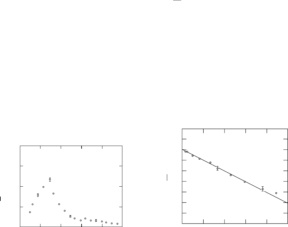
23.3.5 Characterization of Multiphase Systems by SANS
and SAXS
Blends of High-Density and Linear Low-Density
Polyethylenes
Equations (23.17)–(23.20) are strictly valid only for
two-phase morphologies, and for multiphase systems, an
extension of the SAXS invariant analysis may be employed
by generalizing Eq. (23.20) and summing over the number
of phases involved
Q
0
¼ 2p
2
X
i6¼j
r
2
T
w
i
w
j
[r
e1
r
e2
]
2
: (23:22)
Such an analysis has been applied to semicrystalline
blends of polycaprolactone and polycarbonate via SAXS
[49] and complementary SANS experiments [50] have
employed the DB analysis described above [see Sections
‘‘Blends of High-and Low-Density Polyethylene’’ and
‘‘Interpenetrating Polymer Networks’’]. For semicrystalline
blends of high density and short-chain branched linear low
density polyethylenes (LLDPE), complementary DSC and
TEM techniques indicate that the compositions of the vari-
ous crystals and surrounding amorphous regions are such
that the system cannot be described by a two-phase model
[51]. The (Q ¼ 0) cross section [dS=dV(0)] cannot be cal-
culated for multiphase systems, though it may be estimated
via a ‘‘pseudo two-phase’’ model to a good approximation.
For example, with deuterated HDPE-D and protonated
LLDPE-H (to provide SANS contrast), the SLD of the
HDPE-D crystal is 8:57 10
10
cm
2
, whereas the SLDs of
the mixed (HDPE-D/LLDPE-H) crystals and amorphous are
0.44 and 0.46 (10
10
cm
2
). Thus, the SANS cross section
[dS=dV(0)] can be calculated to a good approximation by
grouping the mixed phases into an average background
(r
av
¼ 0:45 10
10
cm
2
) surrounding pure HDPE-D crystal
in a pseudo two-phase model. The SANS invariant may be
calculated for a multiphase morphology [Eq. (23.22)] by
substituting the neutron scattering length densities (r
n
) for
the x-ray scattering length density (r
T
r
e1
) and summing over
the various phases [51]. The (Q ¼ 0) cross section may be
estimated via Eq. (23.17) for the pseudo two phase model.
For series of HDPE-D/LDPE-H samples isothermally crys-
tallized from the met at 117 8C, the experimental data are
compared with calculations for two possible morphologies
suggested by DSC and TEM analysis:
A. A fraction of the HDPE-D component segregates dur-
ing isothermal crystallization and the remainder co-
crystallizes with LLDPE-H on cooling.
B. HDPE-D partially segregates from the LLDPE-H dur-
ing isothermal crystallization and the remainder also
segregates on cooling. A compositionally mixed homo-
geneous amorphous phase was assumed to surround the
crystals in both cases.
The experimental and calculated Q
0
values are listed in
Table 23.4, and for the 18/82 (vol%) blend the calculated Q
0
and the experimental data are identical for morphology type
A. Similarly, the value of d S=dV(0) calculated from the
pseudo two-phase model and Eq. (23.1) is 41:4 10
3
cm
1
for morphology type A, which agrees closely with the ex-
perimental value of 39:3 10
3
cm
1
. When morphology
type B is assumed, the calculated values do not agree with
the experimental data for this blend. Thus, SANS supports
the idea that predominantly LLDPE-rich blends crystallize
isothermally with morphology A, where a fraction of
the HDPE-D component segregates during isothermal
crystallization and the remainder co-crystallizes with the
LLDPE-H on cooling.
For the linear-rich, 78/22 blend the agreement between
the experimental and calculated Q
0
and d S=dV (0) values is
closer for morphology type B. SANS indicates that for this
blend the intensity and invariant conforms a more segre-
gated morphology of the linear and branched components
than for the LLDPE-rich blend. For 50/50 blends, the meas-
ured and calculated values of Q
0
and d S=dV(0) indicate
an intermediate between the A and B types, where part of
the HDPE component that crystallizes on cooling is co-
crystallized with the branched LLDPE and part crystallizes
as pure HDPE [51].
In view of the fact that the experiments are independently
calibrated with no arbitrary fitting factors in the intensity
scale and that the crystal/amorphous compositions are
obtained from DSC, the general agreement with the SANS
data is excellent. Thus, the two-phase approximation is able
to reproduce not only the SANS invariant, but also the
(Q ¼ 0) cross section with good accuracy.
TABLE 23.4. Measured and calculated cross sections and invariants for HDPE-D/LDPE-H blends isothermally crystallized at
117 8C.
Composition (% volume)
HDPE-D/LLDPE-H
dS=dV(Q ¼ 0)
10
3
(cm
1
) expt.
Q
0
cm
1
A
˚
3
expt.
Proposed
morphology
dS=dV(Q ¼ 0)
10
3
(cm
1
) calc.
Q
0
cm
1
A
˚
3
calc.
18/82 41.4 0.009 A 39.2 0.009
B 58.1 0.013
78/22 36.1 0.0158 A 18.4 0.007
B 33.1 0.013
416 / CHAPTER 23