Мир М.Афзал Атлас клинического диагноза
Подождите немного. Документ загружается.


1
ATLAS
OF
CLINICAL
DIAGNOSIS
42
These scaling papular lesions
of
tinea corporis occur
in
an
annular formation, spreading peripherally
and
clearing
centrally
(1.213).
The
outer
border
is red and
irregular
with
fine
scaling.
The
lesions occur
on the
face,
limbs
and
trunk.
Herpes zoster
is a
common
and
easily recognizable con-
dition caused
by the
varicella virus
and
characterized
by a
localized
vesicular
or
bullous eruption, usually limited
to a
dermatome
(often
the
ophthalmic branch
of the
trigemi-
nal
nerve; 1.214), which
is
innervated
by the
corresponding
sensory ganglion. Along
the
distribution
of a
cutaneous
nerve there
are
usually clusters
of
vesicles with erythema,
oedema
and
scabs
(1.215).
Intense
pain
can be a
very
distressing
and
chronic feature
of
this condition.
Blistering
diseases
Of
the
common blistering disorders, erythema multiforme
and
herpes zoster have been mentioned already,
and
herpes simplex
will
be
discussed
in the
next chapter. Der-
matitis herpetiformis
and
porphyria
are
referred
to
later
in
this chapter (see Systemic Disorders). Since pemphigus
often,
and
pemphigoid
sometimes,
can
affect
the
face,
these
will
be
presented here
briefly.
Pemphigus
vulgaris usually starts
in the
oral mucosa
and
then many months later involves
the
skin
of the
face
(1.216). Large, thin bullae occur usually
on
normal skin
and
on
rupture leave non-healing,
red
erosions
on the
face
(1.217)
and
scalp (1.218).
The
epidermis
can be
made
to
1.213
Tinea
corporis:
advancing
erythema
with
scaling
1.214
Herpes zoster:
ruptured
blisters
with
crusting
1.215
Clusters
of
vesicles
with
scabs
1.216
Pemphigus: ruptured
bullae
and
mucosal
lesions
THE
FACE
1
43
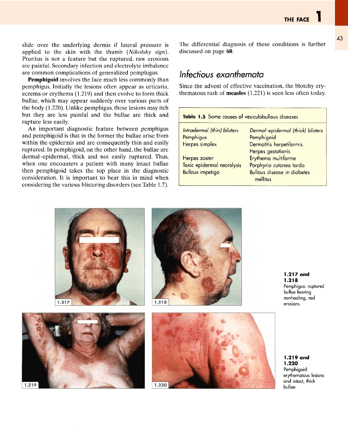
slide over
the
underlying
dermis
if
lateral pressure
is
applied
to the
skin with
the
thumb (Nikolsky sign).
Pruritus
is not a
feature
but the
ruptured,
raw
erosions
are
painful.
Secondary infection
and
electrolyte imbalance
are
common
complications
of
generalized pemphigus.
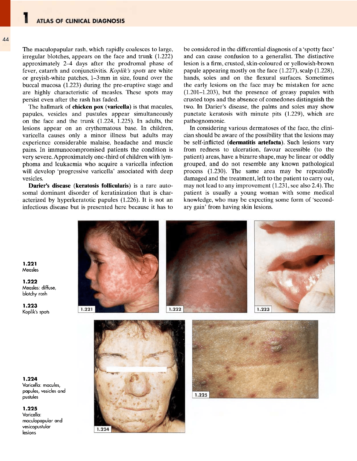
Pemphigoid involves
the
face much less commonly than
pemphigus. Initially
the
lesions
often
appear
as
urticaria,
eczema
or
erythema
(1.219)
and
then
evolve
to
form thick
bullae,
which
may
appear suddenly over various parts
of
the
body (1.220). Unlike pemphigus, these lesions
may
itch
but
they
are
less painful
and the
bullae
are
thick
and
rupture less easily.
An
important diagnostic feature between pemphigus
and
pemphigoid
is
that
in the
former
the
bullae
arise
from
within
the
epidermis
and are
consequently thin
and
easily
ruptured.
In
pemphigoid,
on the
other hand,
the
bullae
are
dermal-epidermal,
thick
and not
easily ruptured. Thus,
when
one
encounters
a
patient with many intact bullae
then pemphigoid takes
the top
place
in the
diagnostic
consideration.
It is
important
to
bear
this
in
mind when
considering
the
various blistering disorders (see Table 1.7).
The
differential
diagnosis
of
these conditions
is
further
discussed
on
page
60.
Infectious
exanthemata
Since
the
advent
of
effective
vaccination,
the
blotchy ery-
thematous rash
of
measles
(1.221)
is
seen
less often today.
Table
1.5
Some
causes
of
vesiculobullous
diseases
Intradermal
(thin)
blisters
Dermal-epidermal
(thick) blisters
Pemphigus
Herpes
simplex
Herpes
zoster
Pemphigoid
Dermatitis
herpetiformis
Herpes
gestationis
Erythema
multiforme
Toxic
epidermal
necrolysis
Porphyria
cutanea
tarda
Bullous
impetigo
Bullous
disease
in
diabetes
mellitus
1.217
and
1.218
Pemphigus:
ruptured
bullae leaving
nonhealing,
red
1.219
and
1.220
Pemphigoid:
erythematous
lesions
and
intact, thick
bullae
ATLAS
OF
CLINICAL
DIAGNOSIS
44
The
maculopapular
rash, which rapidly coalesces
to
large,
irregular
blotches, appears
on the
face
and
trunk (1.222)
approximately
2-4
days
after
the
prodromal phase
of
fever,
catarrh
and
conjunctivitis.
Koplik's
spots
are
white
or
greyish-white patches,
1-3
mm in
size,
found
over
the
buccal mucosa (1.223) during
the
pre-eruptive stage
and
are
highly characteristic
of
measles.
These
spots
may
persist even
after
the
rash
has
faded.
The
hallmark
of
chicken
pox
(varicella)
is
that macules,
papules,
vesicles
and
.pustules
appear simultaneously
on
the
face
and the
trunk (1.224, 1.225).
In
adults,
the
lesions
appear
on an
erythematous base.
In
children,
varicella
causes only
a
minor illness
but
adults
may
experience considerable malaise, headache
and
muscle
pains.
In
immunocompromised patients
the
condition
is
very
severe. Approximately one-third
of
children with
lym-
phoma
and
leukaemia
who
acquire
a
varicella infection
will
develop
'progressive
varicella'
associated with
deep
vesicles.
Darier's disease
(keratosis
follicularis)
is a
rare
auto-
somal
dominant disorder
of
keratinization that
is
char-
acterized
by
hyperkeratotic papules (1.226).
It is not an
infectious
disease
but
is
presented
here
because
it has to
be
considered
in the
differential
diagnosis
of a
'spotty
face'
and
can
cause confusion
to a
generalist.
The
distinctive
lesion
is a firm,
crusted, skin-coloured
or
yellowish-brown
papule appearing mostly
on the
face
(1.227), scalp (1.228),
hands,
soles
and on the flexural
surfaces. Sometimes
the
early lesions
on the
face
may be
mistaken
for
acne
(1.201-1.203),
but the
presence
of
greasy papules with
crusted
tops
and the
absence
of
comedones distinguish
the
two.
In
Darier's
disease,
the
palms
and
soles
may
show
punctate keratosis with minute pits (1.229), which
are
pathognomonic.
In
considering various dermatoses
of the
face,
the
clini-
cian
should
be
aware
of the
possibility that
the
lesions
may
be
self-inflicted
(dermatitis
artefacta).
Such lesions vary
from
redness
to
ulceration,
favour
accessible
(to the
patient) areas, have
a
bizarre shape,
may be
linear
or
oddly
grouped,
and do not
resemble
any
known pathological
process (1.230).
The
same area
may be
repeatedly
damaged
and the
treatment,
left
to the
patient
to
carry
out,
may
not
lead
to any
improvement
(1.231,
see
also 2.4).
The
patient
is
usually
a
young woman with some medical
knowledge,
who may be
expecting some
form
of
'second-
ary
gain'
from
having skin lesions.
1.221
Measles
1.222
Measles:
diffuse,
blotchy
rash
1.223
Koplik's
spots
1.224
Varicella:
macules,
papules,
vesicles
and
pustules
1.225
Varicella:
maculopapular
and
vesicopustular
lesions
THE
FACE
1
45
1.226
Darier's disease:
papules
with
hyperkeratotic
crusts
1.227
Hyperkeratotic
papules
1.228
Yellowish-brown
papules
and
scattered
pits
1.229
Punctate
keratosis
with
pits
1.230
Dermatitis
artefacta
1.231
Clean-cut,
self-
inflicted
ulcers
1
ATLAS
OF
CLINICAL
DIAGNOSIS
46
Cutaneous
manifestations
of
systemic
disorders
The
skin
as a
system,
or an
organ, shares
the
manifesta-
tions
of
some diseases with other systems
but it
does
so in
its own
special, pictorial way.
It
thereby provides
a
window
through
which
one can
see, suspect and,
at
times,
identify
the
offending
underlying disorder.
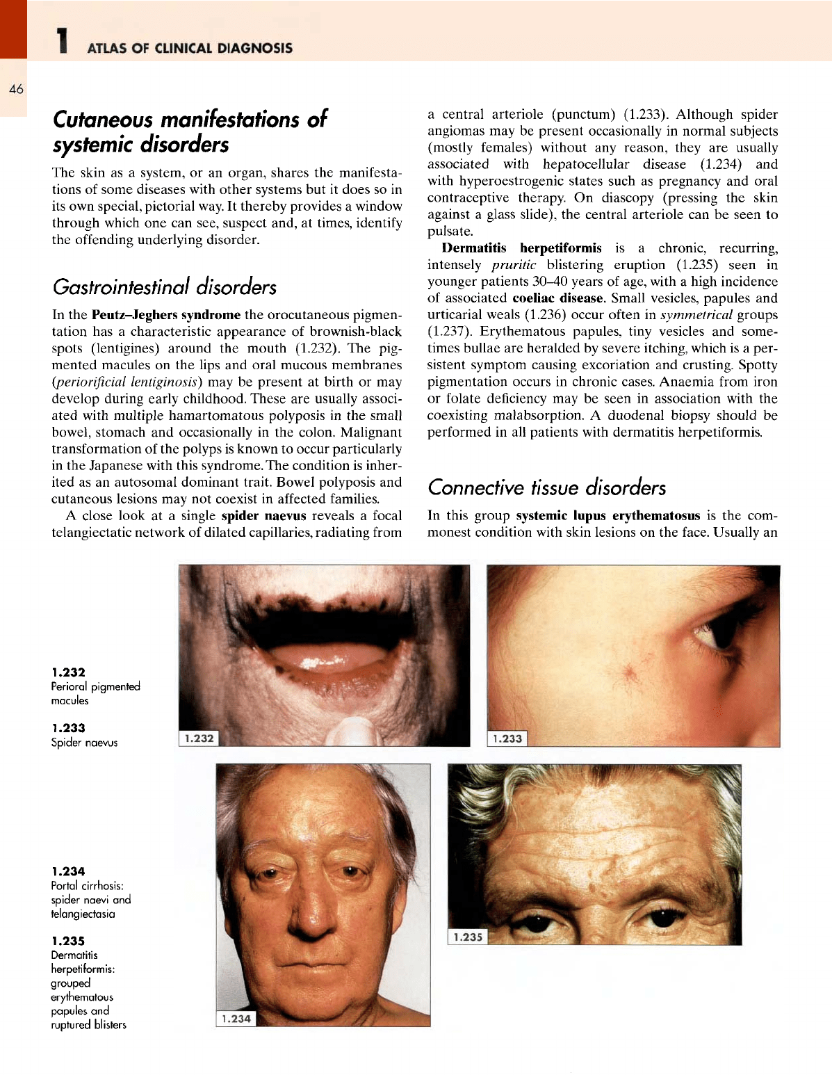
Gastrointestinal disorders
In
the
Peutz-Jeghers
syndrome
the
orocutaneous pigmen-
tation
has a
characteristic appearance
of
brownish-black
spots (lentigines) around
the
mouth
(1.232).
The
pig-
mented macules
on the
lips
and
oral mucous membranes
(periorificial
lentiginosis}
may be
present
at
birth
or may
develop during early childhood.
These
are
usually associ-
ated
with multiple
hamartomatous
polyposis
in the
small
bowel,
stomach
and
occasionally
in the
colon. Malignant
transformation
of the
polyps
is
known
to
occur particularly
in
the
Japanese with this syndrome.
The
condition
is
inher-
ited
as an
autosomal dominant trait. Bowel polyposis
and
cutaneous lesions
may not
coexist
in
affected
families.
A
close look
at a
single
spider
naevus reveals
a
focal
telangiectatic
network
of
dilated capillaries, radiating
from
a
central arteriole (punctum)
(1.233).
Although spider
angiomas
may be
present occasionally
in
normal subjects
(mostly
females) without
any
reason, they
are
usually
associated
with
hepatocellular
disease
(1.234)
and
with
hyperoestrogenic states such
as
pregnancy
and
oral
contraceptive therapy.
On
diascopy (pressing
the
skin
against
a
glass slide),
the
central
arteriole
can be
seen
to
pulsate.
Dermatitis
herpetiformis
is a
chronic, recurring,
intensely pruritic blistering eruption (1.235)
seen
in
younger patients
30-40
years
of
age, with
a
high incidence
of
associated coeliac disease. Small vesicles, papules
and
urticarial
weals (1.236) occur often
in
symmetrical groups
(1.237). Erythematous papules, tiny vesicles
and
some-
times bullae
are
heralded
by
severe itching, which
is a
per-
sistent
symptom causing excoriation
and
crusting. Spotty
pigmentation occurs
in
chronic cases. Anaemia
from
iron
or
folate
deficiency
may be
seen
in
association with
the
coexisting
malabsorption.
A
duodenal
biopsy
should
be
performed
in all
patients with dermatitis herpetiformis.
Connective
tissue
disorders
In
this group systemic lupus
erythematosus
is the
com-
monest condition with skin lesions
on the
face.
Usually
an
1.232
Perioral
pigmented
macules
1.233
Spid
er
naevus
1.234
Portal cirrhosis:
spider
naevi
and
telangiectasia
1.235
Dermatitis
herpetiformis:
grouped
erythematous
papules
and
ruptured
blisters
THE
FACE
1
47
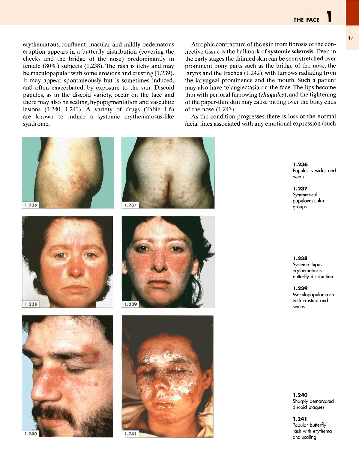
erythematous,
confluent, macular
and
mildly oedematous
eruption appears
in a
butterfly
distribution (covering
the
cheeks
and the
bridge
of the
nose) predominantly
in
female
(80%) subjects (1.238).
The
rash
is
itchy
and may
be
maculopapular with some erosions
and
crusting (1.239).
It may
appear spontaneously
but is
sometimes induced,
and
often exacerbated,
by
exposure
to the
sun. Discoid
papules,
as in the
discoid variety, occur
on the
face
and
there
may
also
be
scaling, hypopigmentation
and
vasculitic
lesions (1.240,
1.241).
A
variety
of
drugs (Table 1.6)
are
known
to
induce
a
systemic
erythematosus-like
syndrome.
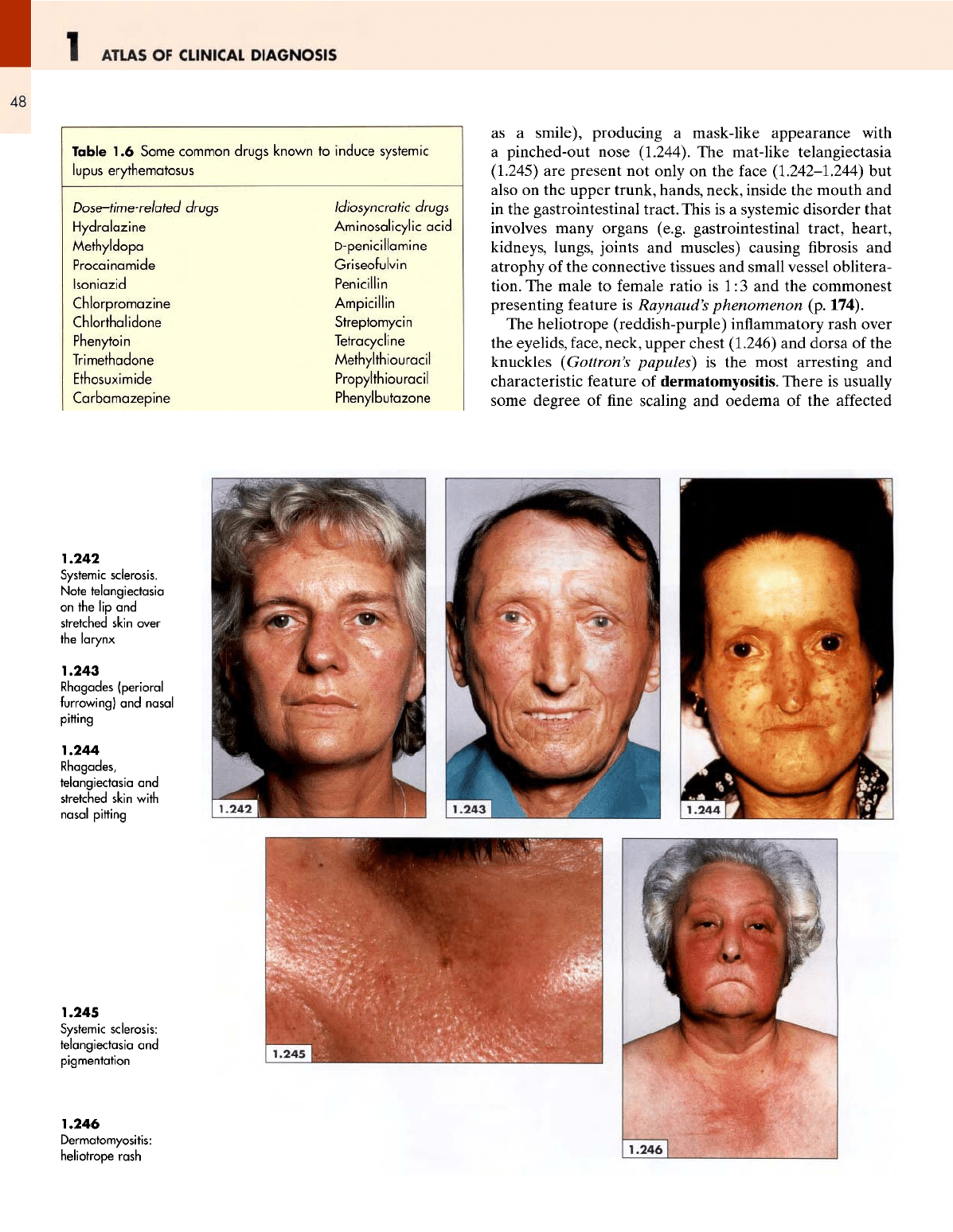
Atrophic contracture
of the
skin
from
fibrosis of the
con-
nective tissue
is the
hallmark
of
systemic sclerosis. Even
in
the
early stages
the
thinned skin
can be
seen stretched over
prominent bony parts such
as the
bridge
of the
nose,
the
larynx
and the
trachea (1.242), with
furrows
radiating
from
the
laryngeal prominence
and the
mouth. Such
a
patient
may
also have telangiectasia
on the
face.
The
lips become
thin
with perioral
furrowing
(rhagades),
and the
tightening
of
the
paper-thin skin
may
cause pitting over
the
bony ends
of
the
nose (1.243).
As the
condition progresses there
is
loss
of the
normal
facial
lines associated with
any
emotional expression (such
1.236
Papules, vesicles
and
weals
1.237
Symmetrical
papulovesicular
groups
1.238
Systemic
lupus
erythematosus:
butterfly
distribution
1.239
Maculopapular
rash
with
crusting
and
scales
1.240
Sharply
demarcated
discoid
plaques
1.241
Papular butterfly
rash
with
erythema
and
scaling
ATLAS
OF
CLINICAL
DIAGNOSIS
48
Table
1.6
Some common drugs known
to
induce
systemic
lupus
erythematosus
Dose-time-related
drugs
Hydralazine
Methyldopa
Procainamide
Isoniazid
Chlorpromazine
Chlorthalidone
Phenytoin
Trimethadone
Ethosuximide
Carbamazepine
Idiosyncratic
drugs
Aminosalicylic
acid
D-penicillamine
Griseofulvin
Penicillin
Ampicillin
Streptomycin
Tetracycline
Methylthiouracil
Propylthiouracil
Phenylbutazone
as
a
smile), producing
a
mask-like appearance with
a
pinched-out nose (1.244).
The
mat-like telangiectasia
(1.245)
are
present
not
only
on the
face
(1.242-1.244)
but
also
on the
upper trunk, hands, neck, inside
the
mouth
and
in
the
gastrointestinal tract. This
is a
systemic disorder that
involves
many organs (e.g. gastrointestinal tract, heart,
kidneys,
lungs, joints
and
muscles) causing
fibrosis and
atrophy
of the
connective tissues
and
small vessel oblitera-
tion.
The
male
to
female ratio
is
1:3
and the
commonest
presenting feature
is
Raynaud's
phenomenon
(p.
174).
The
heliotrope (reddish-purple) inflammatory rash over
the
eyelids,
face,
neck, upper chest (1.246)
and
dorsa
of the
knuckles
(Gottron's
papules}
is the
most arresting
and
characteristic feature
of
dermatomyositis.
There
is
usually
some
degree
of fine
scaling
and
oedema
of the
affected
1.242
Systemic
sclerosis.
Note
telangiectasia
on
the lip and
stretched skin
over
the
larynx
1.243
Rhagades
(perioral
furrowing)
and
nasal
pitting
1.244
Rhagades,
telangiectasia
and
stretched
skin
with
nasal
pitting
1.245
Systemic
sclerosis:
telangiectasia
and
pigmentation
1.246
Dermatomyositis:
heliotrope
rash
THE
FACE
1
49
areas.
The
oedema
is
particularly noticeable periorbitally
when
viewed
from
the
side (1.247).
The
upper eyelids
are
almost invariably involved
and
there
may be
telangiecta-
sia
scattered
on the
erythematous
rash (1.248).
Raynaud's
phenomenon
occurs
in
approximately
20% of
patients.
Fatiguability,
muscle
pains
and
proximal
muscular weak-
ness
occur
in all
patients.
There
is no
agreement
on the
inci-
dence
of an
associated malignancy
(from
6 to
50%)
but a
search should
be
made, especially
in
patients over
40
years
of
age and in
those
who
respond poorly
to
conventional
treatment.
Genetic
and
metabolic disorders
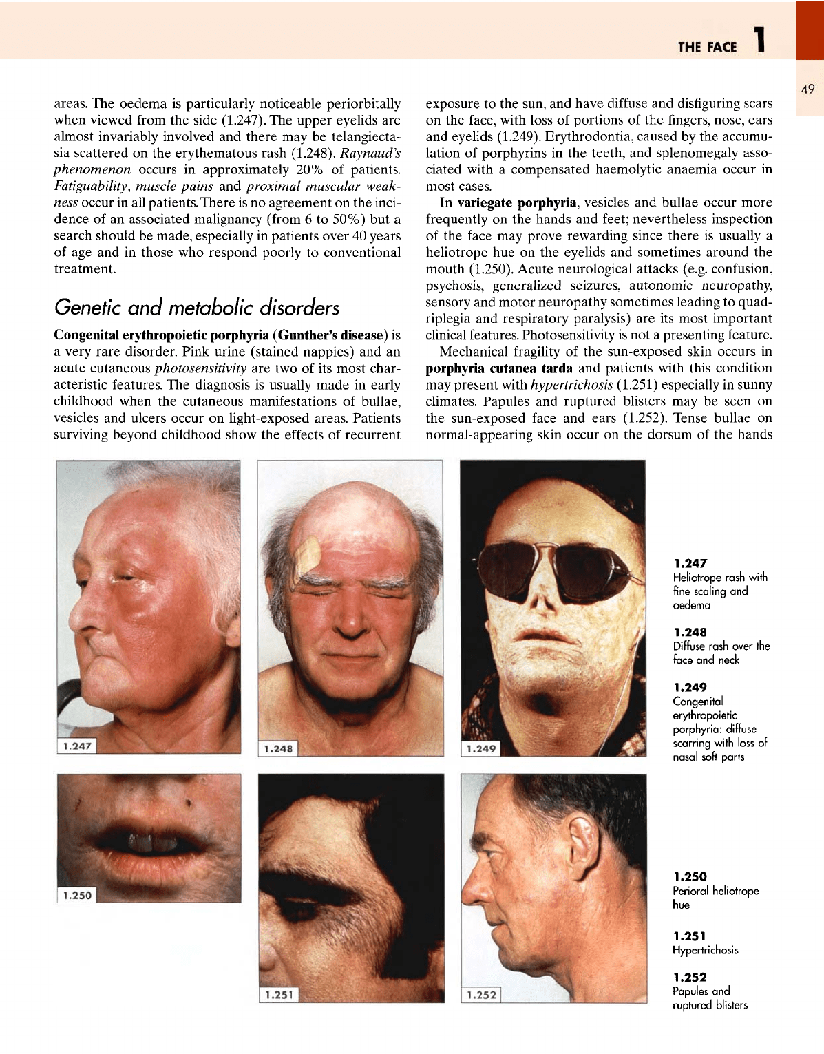
Congenital erythropoietic porphyria (Gunther's disease)
is
a
very rare disorder. Pink urine (stained nappies)
and an
acute cutaneous photosensitivity
are two of its
most char-
acteristic features.
The
diagnosis
is
usually made
in
early
childhood when
the
cutaneous manifestations
of
bullae,
vesicles
and
ulcers occur
on
light-exposed areas. Patients
surviving
beyond childhood show
the
effects
of
recurrent
exposure
to the
sun,
and
have
diffuse
and
disfiguring
scars
on the
face,
with loss
of
portions
of the
fingers,
nose, ears
and
eyelids (1.249). Erythrodontia, caused
by the
accumu-
lation
of
porphyrins
in the
teeth,
and
splenomegaly asso-
ciated
with
a
compensated haemolytic anaemia occur
in
most cases.
In
variegate
porphyria,
vesicles
and
bullae occur more
frequently
on the
hands
and
feet;
nevertheless inspection
of
the
face
may
prove rewarding since there
is
usually
a
heliotrope
hue on the
eyelids
and
sometimes around
the
mouth (1.250). Acute neurological attacks (e.g. confusion,
psychosis,
generalized
seizures,
autonomic
neuropathy,
sensory
and
motor neuropathy sometimes leading
to
quad-
riplegia
and
respiratory paralysis)
are its
most important
clinical
features. Photosensitivity
is not a
presenting
feature.
Mechanical
fragility
of the
sun-exposed skin occurs
in
porphyria
cutanea
tarda
and
patients with this condition
may
present with hypertrichosis
(1.251)
especially
in
sunny
climates. Papules
and
ruptured blisters
may be
seen
on
the
sun-exposed
face
and
ears (1.252). Tense bullae
on
normal-appearing skin occur
on the
dorsum
of the
hands
1.247
Heliotrope
rash
with
fine scaling
and
oedema
1.248
Diffuse
rash over
the
face
and
neck
1.249
Congenital
erythropoietic
porphyria:
diffuse
scarring
with
loss
of
nasal
soft
parts
1.250
Perioral
heliotrope
hue
1.251
Hypertrichosis
1.252
Papules
and
ruptured
blisters
1
ATLAS
OF
CLINICAL
DIAGNOSIS
50
(Chapter
9). As in the
variegate variety,
a
purple-red
hue
may
be
seen
on the
eyelids
and
around
the
mouth. Acute
life-threatening
attacks
do not
occur
in
this condition
but
it
needs
to be
distinguished
from
variegate
porphyria
with
which
it
shares cutaneous lesions. During acute attacks
both conditions have increased levels
of
uroporphyrin
in
the
urine,
but the
diagnostic hallmark
in
variegate
porphyria
is its
markedly elevated
faecal
protoporphyrin
level.
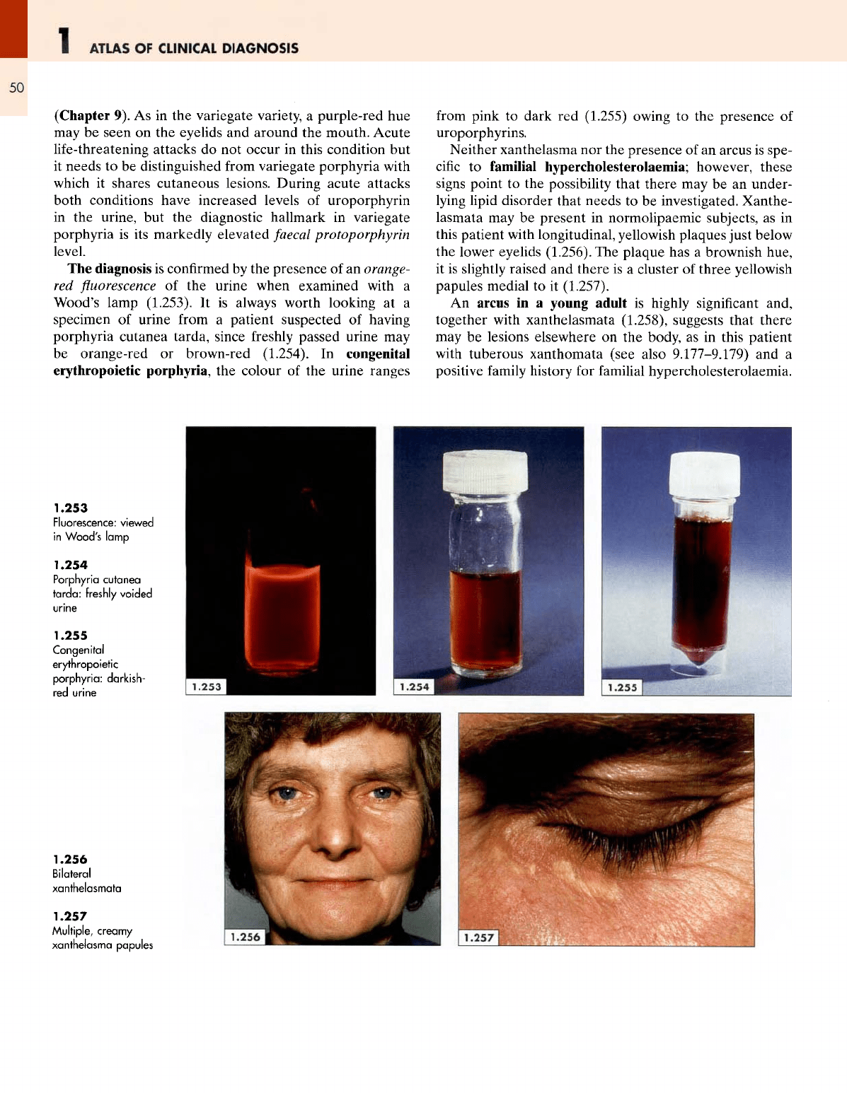
The
diagnosis
is
confirmed
by the
presence
of an
orange-
red
fluorescence of the
urine when examined with
a
Wood's lamp (1.253).
It is
always worth looking
at a
specimen
of
urine
from
a
patient suspected
of
having
porphyria
cutanea tarda, since
freshly
passed urine
may
be
orange-red
or
brown-red (1.254).
In
congenital
erythropoietic
porphyria,
the
colour
of the
urine ranges
from
pink
to
dark
red
(1.255) owing
to the
presence
of
uroporphyrins.
Neither
xanthelasma
nor the
presence
of an
arcus
is
spe-
cific
to
familial
hypercholesterolaemia; however, these
signs
point
to the
possibility that there
may be an
under-
lying
lipid
disorder that needs
to be
investigated. Xanthe-
lasmata
may be
present
in
normolipaemic subjects,
as in
this
patient
with
longitudinal, yellowish plaques
just
below
the
lower eyelids (1.256).
The
plaque
has a
brownish
hue,
it
is
slightly raised
and
there
is a
cluster
of
three yellowish
papules medial
to it
(1.257).
An
arcus
in a
young adult
is
highly
significant
and,
together with xanthelasmata (1.258), suggests that
there
may
be
lesions elsewhere
on the
body,
as in
this patient
with
tuberous xanthomata
(see
also
9.177-9.179)
and a
positive
family
history
for
familial
hypercholesterolaemia.
1.253
Fluorescence:
viewed
in
Wood's
lamp
1.254
Porphyria
cutanea
tarda:
freshly
voided
1.255
Congenital
erythropoietic
porphyria:
darkish-
red
urine
1.256
Bilateral
xanthelasmata
1.257
Multiple,
creamy
xanthelasma papules
THE
FACE
51
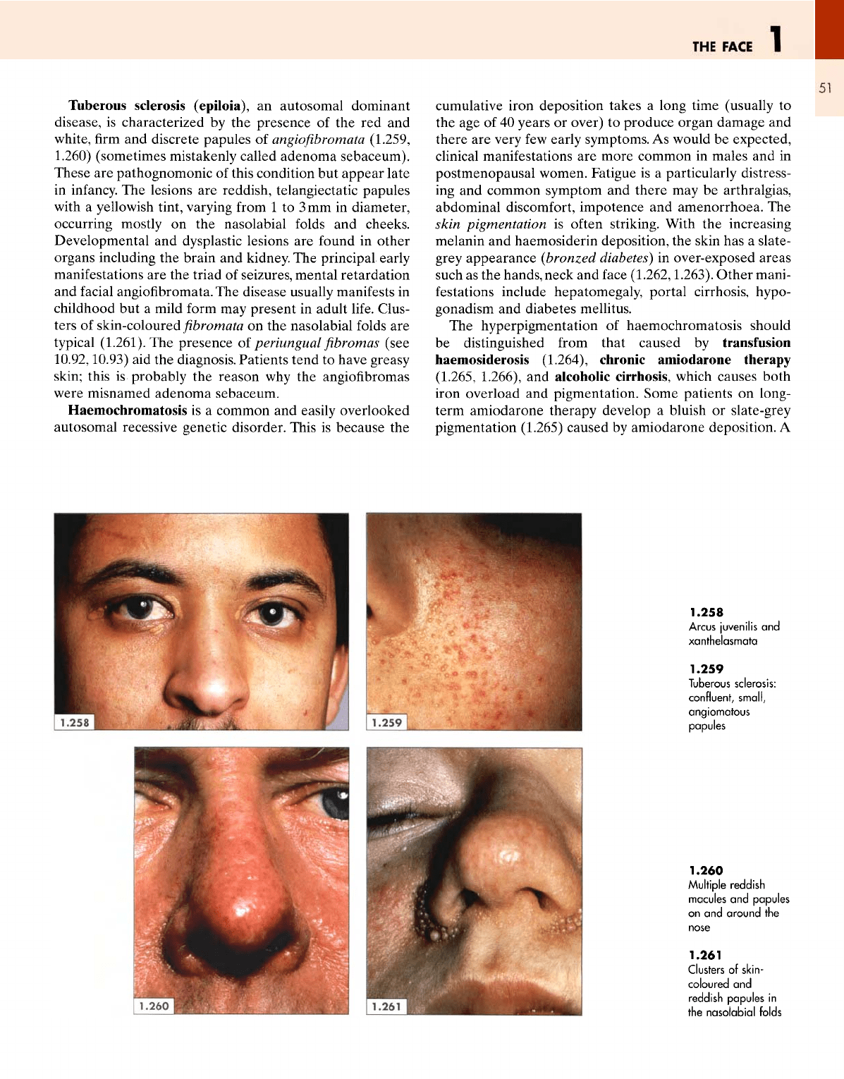
Tuberous sclerosis (epiloia),
an
autosomal dominant
disease,
is
characterized
by the
presence
of the red and
white,
firm and
discrete papules
of
angiofibromata
(1.259,
1.260)
(sometimes mistakenly called adenoma
sebaceum).
These
are
pathognomonic
of
this condition
but
appear late
in
infancy.
The
lesions
are
reddish, telangiectatic papules
with
a
yellowish tint, varying
from
1 to 3 mm in
diameter,
occurring mostly
on the
nasolabial
folds
and
cheeks.
Developmental
and
dysplastic lesions
are
found
in
other
organs
including the brain and kidney. The
principal
early
manifestations
are the
triad
of
seizures, mental retardation
and
facial
angiofibromata.
The
disease usually manifests
in
childhood
but a
mild form
may
present
in
adult
life.
Clus-
ters
of
skin-coloured
fibromata on the
nasolabial
folds
are
typical
(1.261).
The
presence
of
periungual
fibromas
(see
10.92,10.93)
aid the
diagnosis. Patients tend
to
have greasy
skin;
this
is
probably
the
reason
why the
angiofibromas
were
misnamed adenoma sebaceum.
Haemochromatosis
is a
common
and
easily overlooked
autosomal recessive genetic disorder. This
is
because
the
cumulative
iron deposition takes
a
long time (usually
to
the age of 40
years
or
over)
to
produce organ damage
and
there
are
very
few
early symptoms.
As
would
be
expected,
clinical
manifestations
are
more common
in
males
and in
postmenopausal women. Fatigue
is a
particularly distress-
ing
and
common symptom
and
there
may be
arthralgias,
abdominal discomfort, impotence
and
amenorrhoea.
The
skin
pigmentation
is
often striking. With
the
increasing
melanin
and
haemosiderin deposition,
the
skin
has a
slate-
grey
appearance
(bronzed diabetes)
in
over-exposed areas
such
as the
hands, neck
and
face
(1.262,1.263).
Other
mani-
festations
include hepatomegaly, portal cirrhosis, hypo-
gonadism
and
diabetes mellitus.
The
hyperpigmentation
of
haemochromatosis should
be
distinguished
from
that caused
by
transfusion
haemosiderosis
(1.264), chronic amiodarone therapy
(1.265, 1.266),
and
alcoholic cirrhosis, which causes both
iron overload
and
pigmentation. Some patients
on
long-
term amiodarone therapy develop
a
bluish
or
slate-grey
pigmentation (1.265) caused
by
amiodarone deposition.
A
1.258
Arcus
juvenilis
and
xanthelasmata
1.259
Tuberous
sclerosis:
confluent, small,
angiomatous
papules
1.260
Multiple
reddish
macules
and
papules
on
and
around
the
1.261
Clusters
of
skin-
coloured
and
reddish
papules
in
the
nasolabial
folds