Мир М.Афзал Атлас клинического диагноза
Подождите немного. Документ загружается.

1
ATLAS
OF
CLINICAL
DIAGNOSIS
52
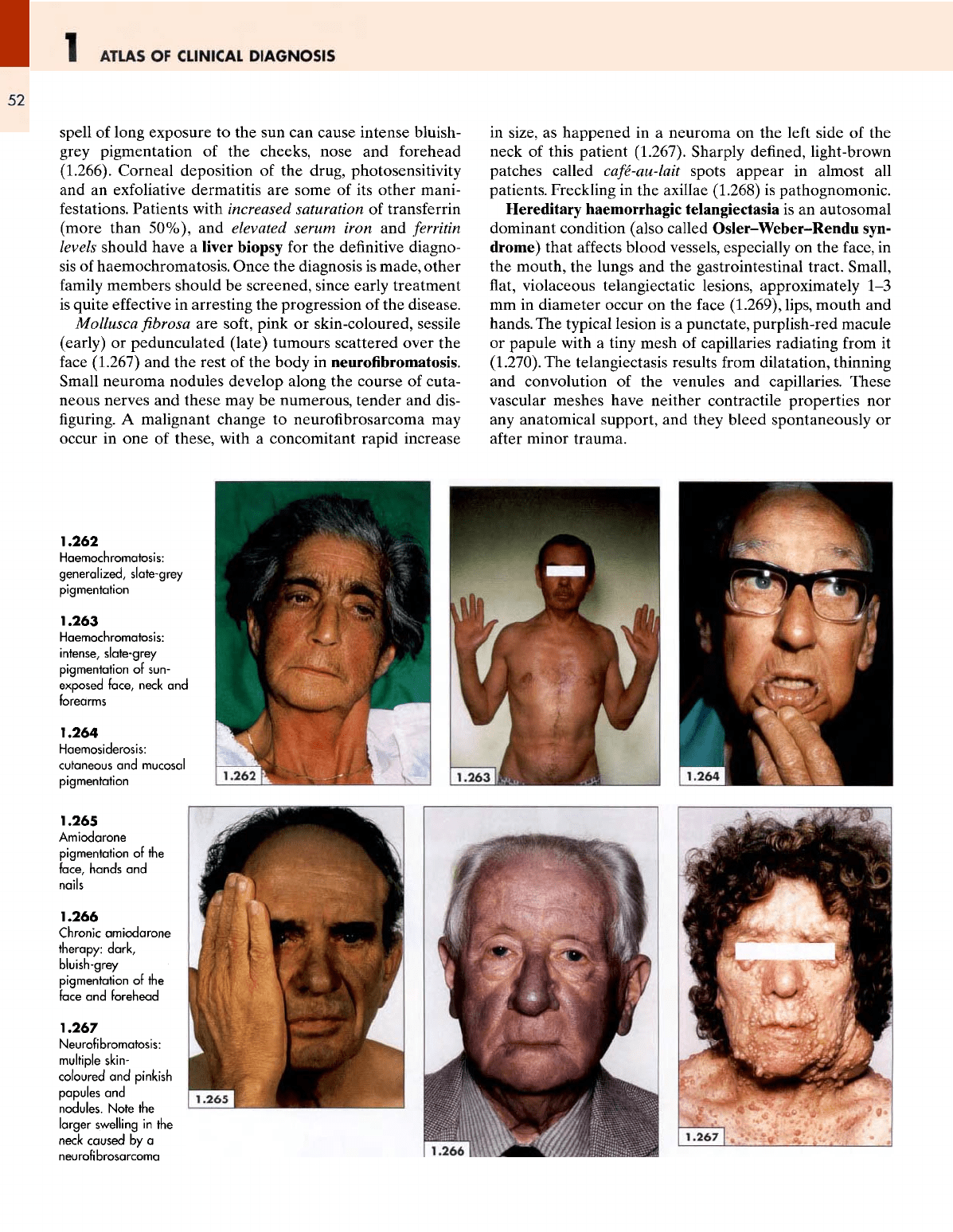
spell
of
long exposure
to the sun can
cause intense bluish-
grey
pigmentation
of the
cheeks, nose
and
forehead
(1.266). Corneal deposition
of the
drug,
photosensitivity
and an
exfoliative dermatitis
are
some
of its
other mani-
festations.
Patients with increased saturation
of
transferrin
(more than 50%),
and
elevated serum iron
and
ferritin
levels
should have
a
liver
biopsy
for the
definitive
diagno-
sis
of
haemochromatosis.
Once
the
diagnosis
is
made, other
family
members should
be
screened, since early treatment
is
quite
effective
in
arresting
the
progression
of the
disease.
Mollusca
fibrosa are
soft,
pink
or
skin-coloured, sessile
(early)
or
pedunculated (late) tumours scattered over
the
face
(1.267)
and the
rest
of the
body
in
neurofibromatosis.
Small
neuroma nodules develop along
the
course
of
cuta-
neous nerves
and
these
may be
numerous, tender
and
dis-
figuring.
A
malignant change
to
neurofibrosarcoma
may
occur
in one of
these, with
a
concomitant rapid increase
in
size,
as
happened
in a
neuroma
on the
left
side
of the
neck
of
this patient (1.267). Sharply
defined,
light-brown
patches called
cafe-au-lait
spots appear
in
almost
all
patients. Freckling
in the
axillae
(1.268)
is
pathognomonic.
Hereditary
haemorrhagic telangiectasia
is an
autosomal
dominant condition (also called
Osier-Weber-Rendu
syn-
drome) that
affects
blood vessels, especially
on the
face,
in
the
mouth,
the
lungs
and the
gastrointestinal tract. Small,
flat,
violaceous telangiectatic lesions, approximately
1-3
mm
in diameter occur on the face (1.269), lips, mouth and
hands.
The
typical lesion
is a
punctate, purplish-red macule
or
papule with
a
tiny
mesh
of
capillaries radiating
from
it
(1.270).
The
telangiectasis results
from
dilatation, thinning
and
convolution
of the
venules
and
capillaries.
These
vascular
meshes have neither contractile properties
nor
any
anatomical support,
and
they bleed spontaneously
or
after
minor trauma.
1.262
Haemochromatosis:
generalized,
slate-grey
pigmentation
1.263
Haemochromatosis:
intense, slate-grey
pigmentation
of
sun-
exposed
face,
neck
and
forearms
1.264
Haemosiderosis:
cutaneous
and
mucosal
pigmentation
1.265
Amiodarone
pigmentation
of the
face,
hands
and
nails
1.266
Chronic
amiodarone
therapy:
dark,
bluish-grey
pigmentation
of the
face
and
forehead
1.267
Neurofibromatosis:
multiple
skin-
coloured
and
pinkish
papules
and
nodules.
Note
the
larger
swelling
in the
neck caused
by a
neurofibrosarcoma

THE
FACE
1
53
Vascular
malformations
in the
lung result
in
pulmonary
arteriovenous
fistulas
in
approximately
one-fifth
of
cases.
These
malformations increase
in
size
and
frequency
with
age
and may
cause recurrent haemoptysis, infections,
hypoxaemia, clubbing
and
polycythaemia.
The
condition
should
be
sought
in
patients with unexplained haemopty-
sis
and
anaemia.
Endocrine
disorders
The
endocrine disorders that cause major changes
to the
facial
appearance have already been discussed
(p. 3).
Non-scarring
alopecia
and
vitiligo, possibly associated
with
some endocrine
and
autoimmune disorders,
and a
hormone-secreting tumour with cutaneous manifestations
(glucagonoma) will need
to be
considered here.
Alopecia areata
is a
localized, well-circumscribed, oval
or
circular loss
of
hair mostly
affecting
the
scalp (1.271)
but
sometimes occurring
on the
beard, eyebrows
or
eye-
lashes.
Erythema
may be
present
in the
early stages
but in
well-developed cases there
are no
inflammatory changes.
Characteristically,
the
peripheral areas
of
hair loss
are
studded
with
the
diagnostic broken-off hairs called
'excla-
mation mark
hairs'.
Occasionally
the
hair
loss
may
spread
to the
entire
scalp
(alopecia
totalis)
(1.272).
This
may be
difficult
to
recognize
in
a
patient wearing
a
wig, although
the
profuseness
of the
hair
on the
wig,
and the
difference
between
the
colour
of
the
native hairs
in
front
of the
ears
and
those
of the wig
(1.273)
may be
obvious
to the
discerning clinician.
Alo-
1.268
Neurofibromatosis:
axillary
freckling
1.269
Osler-Weber-Rendu
syndrome:
multiple,
discrete,
red
macular
and
papular
telangiectases
1.270
Telangiectasis:
a
mesh
of
capillaries
radiates
from
the
central punctum
(arteriole)
1.271
Alopecia
areata
1.272
Alopecia
totalis
1.273
Camouflaged
alopecia
totalis
1
ATLAS
OF
CLINICAL
DIAGNOSIS
54
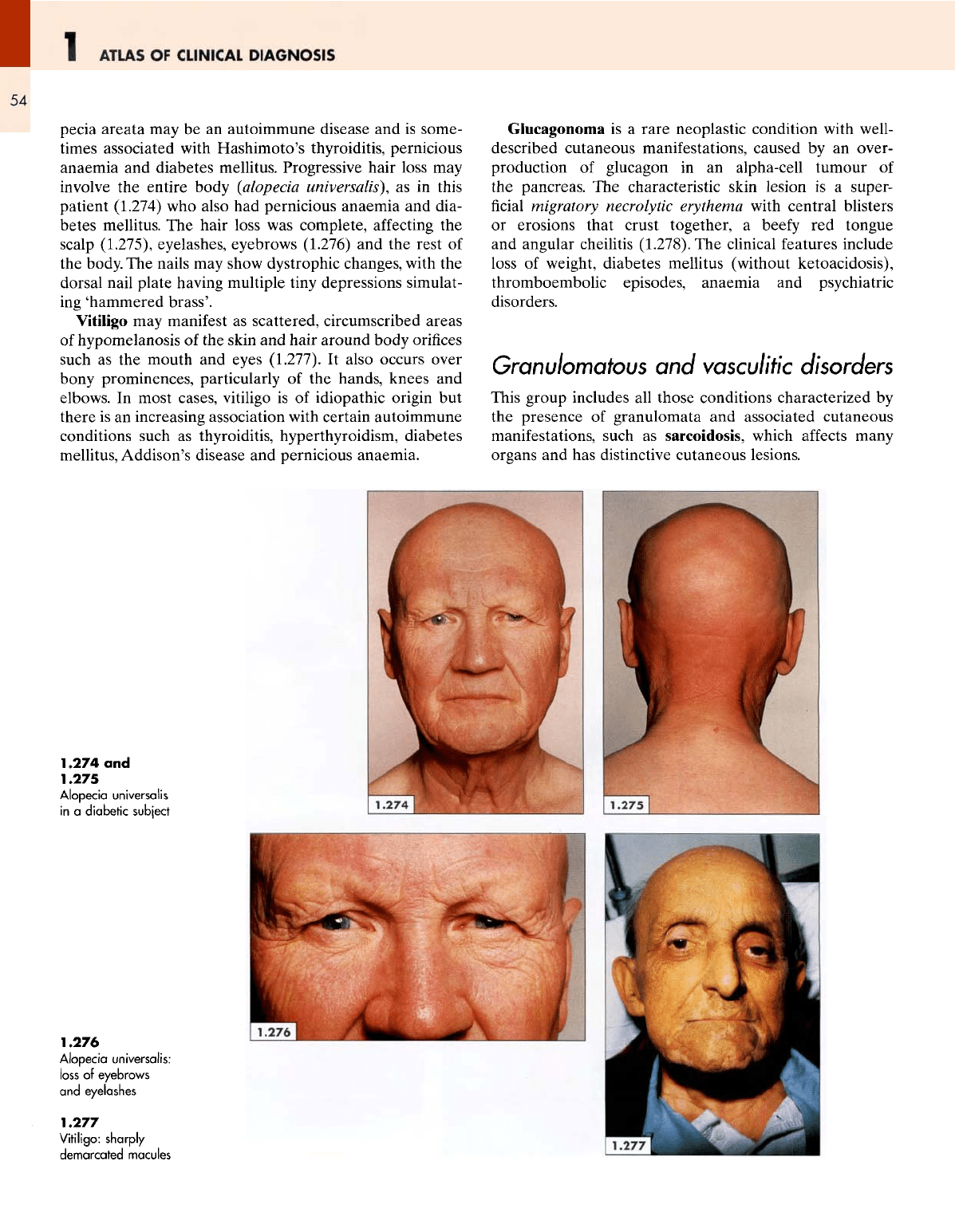
pecia areata
may be an
autoimmune disease
and is
some-
times associated with Hashimoto's
thyroiditis,
pernicious
anaemia
and
diabetes mellitus. Progressive hair loss
may
involve
the
entire
body
(alopecia
universails},
as in
this
patient (1.274)
who
also
had
pernicious anaemia
and
dia-
betes mellitus.
The
hair loss
was
complete,
affecting
the
scalp (1.275), eyelashes, eyebrows (1.276)
and the
rest
of
the
body.
The
nails
may
show dystrophic changes, with
the
dorsal nail plate having multiple tiny depressions simulat-
ing
'hammered
brass'.
Vitiligo
may
manifest
as
scattered, circumscribed areas
of
hypomelanosis
of the
skin
and
hair
around
body orifices
such
as the
mouth
and
eyes (1.277).
It
also occurs over
bony
prominences, particularly
of the
hands, knees
and
elbows.
In
most cases, vitiligo
is of
idiopathic origin
but
there
is an
increasing association with certain autoimmune
conditions such
as
thyroiditis,
hyperthyroidism,
diabetes
mellitus,
Addison's disease
and
pernicious anaemia.
Glucagonoma
is a
rare neoplastic condition with well-
described cutaneous manifestations, caused
by an
over-
production
of
glucagon
in an
alpha-cell tumour
of
the
pancreas.
The
characteristic
skin
lesion
is a
super-
ficial
migratory necrolytlc erythema
with
central blisters
or
erosions that crust together,
a
beefy
red
tongue
and
angular cheilitis
(1.278).
The
clinical features include
loss
of
weight, diabetes mellitus (without ketoacidosis),
thromboembolic episodes, anaemia
and
psychiatric
disorders.
Cranulomatous
and
vasculitic
disorders
This group includes
all
those conditions characterized
by
the
presence
of
granulomata
and
associated cutaneous
manifestations,
such
as
sarcoidosis, which
affects
many
organs
and has
distinctive cutaneous lesions.
1.274
and
1.275
Alopecia
universalis
in a
diabetic
subject
1.276
Alopecia
universalis:
loss
of
eyebrows
and
eyelashes
1.277
Vitiligo:
sharply
demarcated
macules

THE
FACE
1
55
Sarcoidosis
is a
chronic
granulomatous
disease
of
unknown aetiology that
affects
young adults
and
presents
with
skin lesions, pyrexia, ocular involvement, bilateral
hilar
adenopathy
and
pulmonary infiltrations. Erythema
nodosum
is the
commonest cutaneous manifestation
but
multiple
maculopapular lesions (1.279), lupus pernio
and
sarcoid
infiltration
of
scars also occur.
The
maculopapular
and
nodular lesions spread peripherally
in an
annular
fashion
and
some
of the
lesions coalesce into brownish-
purple plaques
on the
face
(1.280).
On
closer examination,
papular invasion
of the
epidermis with
a
resulting purplish
hue
can be
seen clearly
(1.281).
On
diascopy, these lesions
may
look somewhat like
the
'apple-jelly'
(yellowish-
brown)
of
lupus vulgaris (see 1.207, 1.210
and
1.211)
but
there
is no
scarring
in
sarcoidosis. Lupus pernio
(or
chilblain)
is a
more homogeneous, dense,
firm or
soft,
vio-
laceous
infiltration
of the
exposed
parts
such
as the
cheek,
nose (1.282)
and
earlobes (1.283).
Wegener's
granulomatosis
is a
distinct clinicopathologi-
cal
entity
and
consists
of a
triad
of: (i) a
necrotizing granu-
lomatous vasculitis
of the
upper
and
lower respiratory
tracts;
(ii)
a
focal necrotizing glomerulitis;
and
(iii)
a
sys-
temic small vessel vasculitis involving numerous organs.
Cutaneous manifestations occur
in
approximately
half
the
cases
but in
only approximately
10% of
patients
at
initial
presentation.
In
almost
all
patients
a
history
of
symptoms
referable
to
upper
respiratory
catarrh
or
infection
is
obtain-
able. Common symptoms
and
signs include
a
persistent
nasal
discharge,
fever,
cough, haemoptysis
and
nasal
granulomata.
1.278
Migratory
necrolytic
erythema:
inflammatory
papules,
angular
cheilitis
and
beefy
tongue
1.279
Sarcoidosis:
maculopapular
reddish
pepules
1.280
Sarcoidosis:
brownish-purple
granulomatous
plaque
1.281
Yellowish-brown
granulomatous
infiltration
of the
cheek
and
nose
1.282
Lupus
pernio
of the
cheeks
1.283
Lupus
pernio:
swelling
and
congestion
of the
cold-exposed
ear

1
ATLAS
OF
CLINICAL
DIAGNOSIS
56
Granulomatous involvement
of the
nasal mucosa
may
be
betrayed
by a
reddish
hue of the
skin overlying
the
granulomata
(1.284).
The
granulomata
can be
readily seen
by
looking
up the
patient's nostrils (1.285).
Renal
disease
is
seen
in
approximately
80% of
patients
and
virtually
dictates
the
outcome.
The
diagnosis should
be
suspected strongly when
an
upper respiratory
com-
plaint
is
accompanied with haematuria and/or vasculitic
lesions.
Vasculitides
constitute
a
heterogeneous group
of
disor-
ders that includes Wegener's granulomatosis
and can be
loosely
classified
under various subheadings:
•
Hypersensitivity
angiitis
(e.g.
Henoch-Schonlein
purpura, cryoglobulinaemia,
hypocomplementaemic
vasculitis);
•
Allergic
vasculitis
from
drug reactions;
•
Underlying infections
(e.g.
infective endocarditis,
meningococcaemia);
•
Neoplastic diseases
(e.g.
carcinoma, lymphoma,
leukaemia);
•
Connective tissue diseases
(e.g.
systemic lupus
erythematosus, rheumatoid arthritis, polyarteritis
nodosa);
•
Granulomatous vasculitis
(e.g.
Wegener's,
Churg-Straus
disease);
•
Giant
cell
arteritis
(e.g.
temporal arteritis,
poly
myalgia
rheumatica, Takayasu's disease).
The
hallmark
of the
group
is the
predominance
of
cuta-
neous involvement, which
may
appear
as the
classical
palpable purpura
on the
face
(1.286)
and
elsewhere
on
the
body.
In
addition, there
may be
macules, papules,
nodules, bullae, ulcers
and
recurrent urticaria.
The
crusted
lesions represent cutaneous infarction associated with
necrotizing vasculitis.
The
cutaneous manifestations,
together with
the
involvement
of one or
more systems,
suggest
the
possible presence
of one of the
vasculitides.
For
example,
in
meningococcaemia
the
tell-tale cutaneous
vasculitic
lesions
may be red
papules
or
petechiae (1.287),
in
association with
fever,
malaise, headache
and
meningism.
1.284
Wegener's
granulomatosis:
inflammatory
granulomatous
infiltration
of the
1.285
Wegener's
granulomata
1.286
Vasculitis:
multiple
purpuric
papules
with
same crusted
(cutaneous
infarction) lesions
1.287
Meningococcal
.
meningitis: reddish
papules
THE
FACE
1
57
Vascular
and
haemovascular disorders
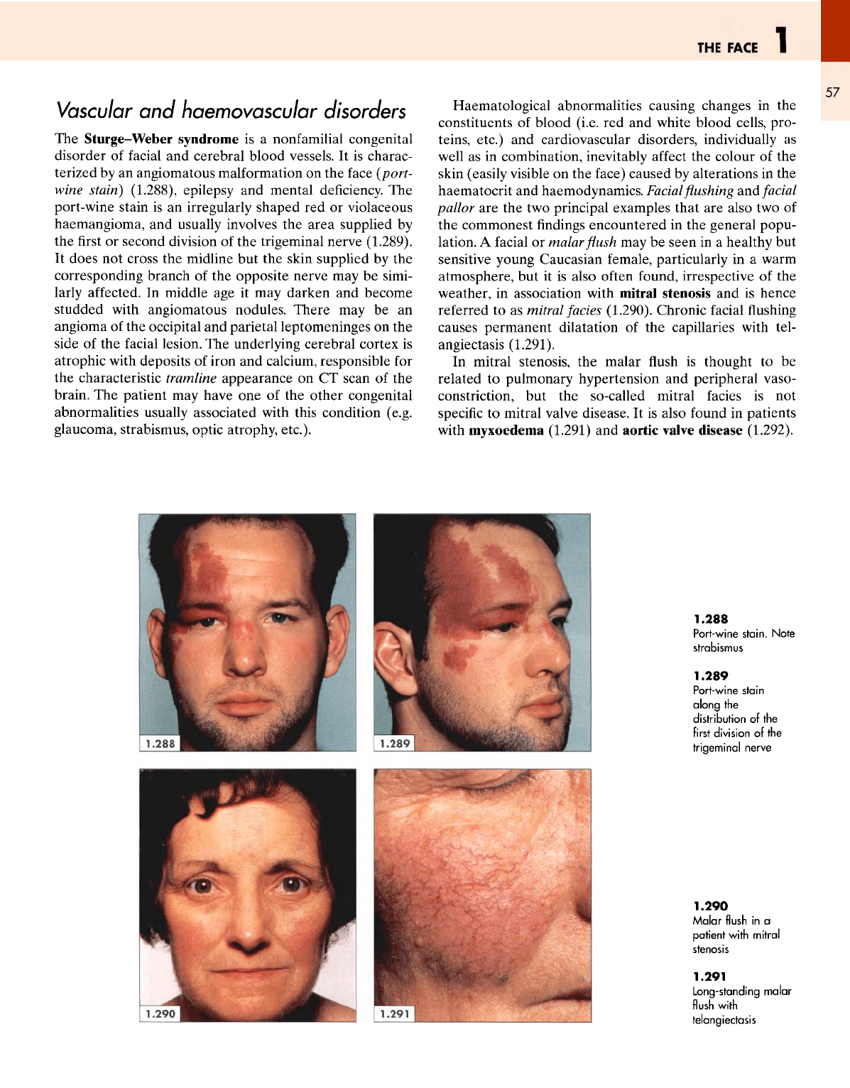
The
Sturge-Weber syndrome
is a
nonfamilial congenital
disorder
of
facial
and
cerebral blood vessels.
It is
charac-
terized
by an
angiomatous
malformation
on the
face
(port-
wine
stain)
(1.288),
epilepsy
and
mental deficiency.
The
port-wine stain
is an
irregularly shaped
red or
violaceous
haemangioma,
and
usually involves
the
area supplied
by
the first or
second division
of the
trigeminal nerve
(1.289).
It
does
not
cross
the
midline
but the
skin supplied
by the
corresponding branch
of the
opposite
nerve
may be
simi-
larly
affected.
In
middle
age it may
darken
and
become
studded with angiomatous nodules.
There
may be an
angioma
of the
occipital
and
parietal leptomeninges
on the
side
of the
facial
lesion.
The
underlying cerebral cortex
is
atrophic with deposits
of
iron
and
calcium, responsible
for
the
characteristic tramline appearance
on CT
scan
of the
brain.
The
patient
may
have
one of the
other congenital
abnormalities usually associated with this condition (e.g.
glaucoma,
strabismus,
optic
atrophy, etc.).
Haematological abnormalities causing changes
in the
constituents
of
blood (i.e.
red and
white blood cells, pro-
teins,
etc.)
and
cardiovascular disorders, individually
as
well
as in
combination, inevitably
affect
the
colour
of the
skin (easily visible
on the
face) caused
by
alterations
in the
haematocrit
and
haemodynamics.
Facial
flushing and
facial
pallor
are the two
principal examples that
are
also
two of
the
commonest
findings
encountered
in the
general popu-
lation.
A
facial
or
malar
flush may be
seen
in a
healthy
but
sensitive young Caucasian female, particularly
in a
warm
atmosphere,
but it is
also
often
found, irrespective
of the
weather,
in
association with mitral
stenosis
and is
hence
referred
to as
mitral fades (1.290). Chronic
facial
flushing
causes permanent dilatation
of the
capillaries with
tel-
angiectasis
(1.291).
In
mitral stenosis,
the
malar
flush is
thought
to be
related
to
pulmonary hypertension
and
peripheral vaso-
constriction,
but the
so-called mitral facies
is not
specific
to
mitral valve disease.
It is
also found
in
patients
with
myxoedema
(1.291)
and
aortic valve disease (1.292).
1.288
Port-wine
stain,
strabismus
Note
1.289
Port-wine
stain
along
the
distribution
of the
first
division
of the
trigeminal
nerve
1.290
Malar
flush
in a
patient
with
mitral
stenosis
1.291
Long-standing
malar
flush
with
telangiectasis
1
ATLAS
OF
CLINICAL
DIAGNOSIS
58
A
dusky
red
face (1.293)
is
often seen
in
patients with
polycythaemia
rubra vera although
not all
patients with
facial
plethora have polycythaemia. These patients
may be
asymptomatic
or may
complain
of
headaches, dizziness
or
visual
symptoms.
On
questioning they
may
admit
to
having
night sweats
and
generalized pruritus made worse
by a
warm
bath.
In
addition
to
facial
plethora, clinical exami-
nation
may
reveal retinal vein distension
and a
palpable
spleen.
At the
other
extreme,
pallor
is a
dependable mani-
festation
of
either
a low
volume,
low
perfusion state
or of
anaemia
(1.294).
In
either event, pallor
as a
clinical sign
should
be
confirmed
by
inspecting
the
mucous membranes,
particularly
by
looking
at the
guttering between
the
palpe-
bral
and
bulbar conjunctiva (see 3.24).
The
signs associated with hyperlipidaemias
and
abnor-
mal
hyperproteinaemias
are
discussed elsewhere (pp.
50,
110,192).
Diagnostic considerations
The
diagnosis
of
various skin lesions
is
dependent essen-
tially
on
familiarity
with their appearance which
can be
1.292
Long-standing
malar
flush
with
telangiectasis
in a
butterfly
distribution
(aortic
valve disease)
1.293
Polycythemia
rubra
vera:
diffuse
plethora
1.294
Pallor
1.295
Psoriasis:
well-
defined
plaques
with
silvery-white
scales
1.296
Psoriasis:
plaques
on
the
knees
1.297
Familial
hypercholesterolaemia:
tuberous xanthomata
1.298
Psoriasis:
pitting
of
the
nails
THE
FACE
1
59
gained
by
looking
at as
many pictures
and
patients
as
pos-
sible.
Any
clinician
who
takes
the
trouble
to
study
and
understand
the
unique
pattern
of a
lesion
in an
atlas
will
find
it
easier
to
identify
it
when
it is
seen
on a
patient.
When studying
a
lesion, particular attention should
be
paid
to its
various components such
as its
size, shape, base,
edges, surface,
the
state
of the
surrounding skin,
and its
areas
of
predilection.
For
example, even part
of a
lesion
without
its
anatomical relationship
can be
recognized
as
psoriasis
by the
presence
of
sharply
defined,
keratotic,
silvery-white
plaques
on
erythematous skin (1.295).
Its
predilection
for
extensor surfaces (1.296,
see
also 9.61)
is
well known,
as is
that
of
tuberous
xanthomata
(1.297),
yet
the two can
hardly
be
confused with each other since both
lesions have
strikingly
different
appearances.
The
knowl-
edgeable clinician
will
also look
for
other
familiar
lesions
known
to be
caused
by the
suspected disease such
as the
pitting
of the
nails
in
psoriasis (1.298).
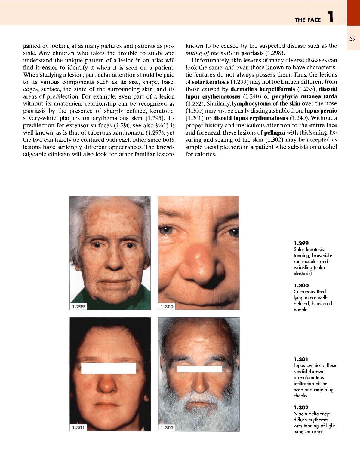
Unfortunately, skin
lesions
of
many
diverse
diseases
can
look
the
same,
and
even those known
to
have characteris-
tic
features
do not
always possess them. Thus,
the
lesions
of
solar keratosis (1.299)
may not
look much
different
from
those caused
by
dermatitis herpetiformis (1.235), discoid
lupus
erythematosus (1.240)
or
porphyria cutanea tarda
(1.252). Similarly, lymphocytoma
of the
skin over
the
nose
(1.300)
may not be
easily distinguishable
from
lupus pernio
(1.301)
or
discoid lupus erythematosus (1.240). Without
a
proper history
and
meticulous attention
to the
entire
face
and
forehead,
these
lesions
of
pellagra with thickening,
fis-
suring
and
scaling
of the
skin (1.302)
may be
accepted
as
simple
facial
plethora
in a
patient
who
subsists
on
alcohol
for
calories.
1.299
Solar keratosis:
tanning,
brownish-
red
macules
and
wrinkling
(solar
elastosis)
1.300
Cutaneous
B-cell
lymphoma:
well-
defined,
bluish-red
nodule
1.301
Lupus
pernio:
diffuse
reddish-brown
granulomatous
infiltration
of the
nose
and
adjoining
cheeks
1.302
Niacin
deficiency:
diffuse
erythema
with
tanning
of
light-
exposed areas
1
ATLAS
OF
CLINICAL
DIAGNOSIS
60
It is
clear that many disorders
of the
skin, like those
of
any
other system, cannot
be
diagnosed without
a
proper
and
comprehensive clinical assessment. Whenever
the
lesions
look
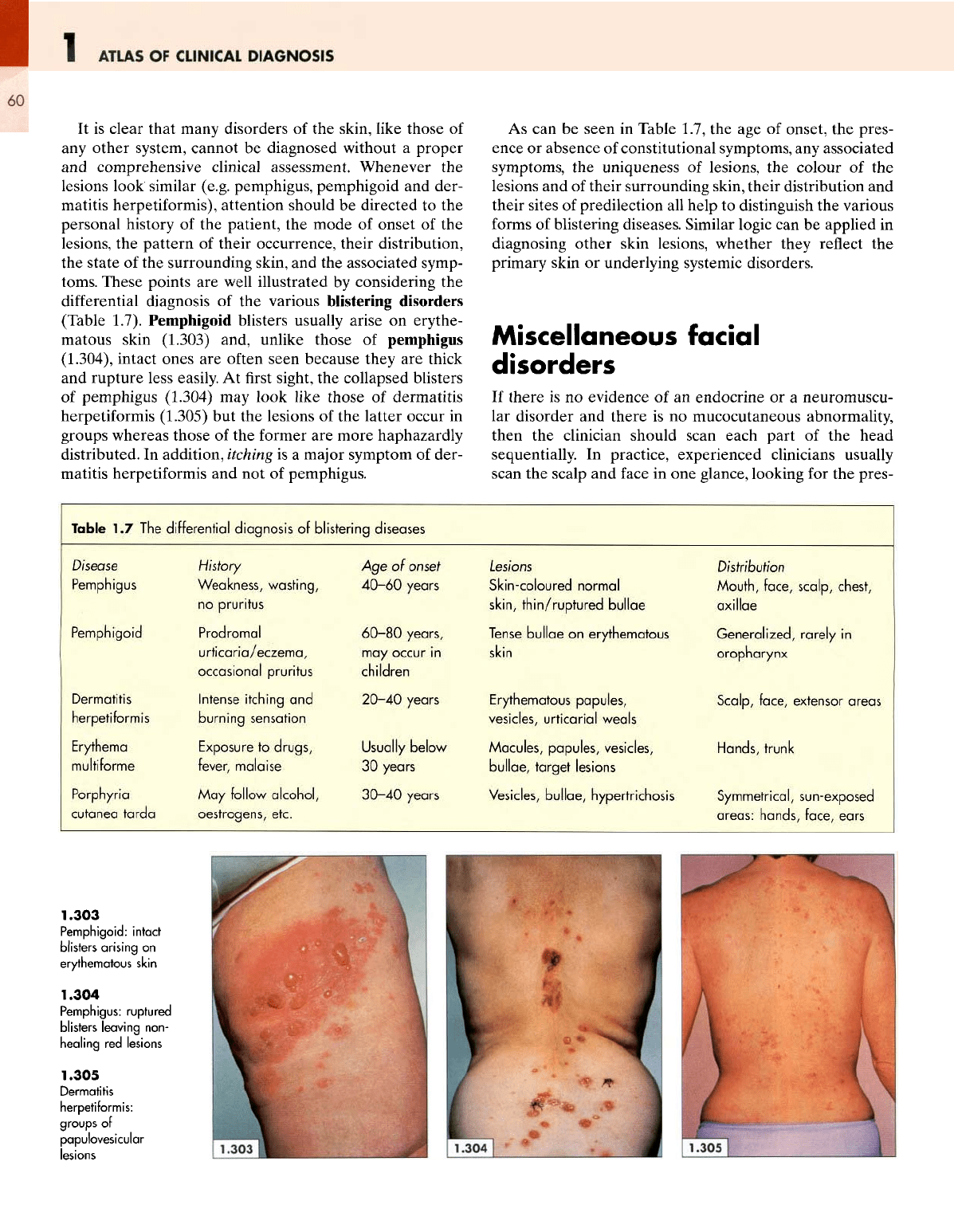
similar (e.g. pemphigus, pemphigoid
and
der-
matitis
herpetiformis), attention should
be
directed
to the
personal history
of the
patient,
the
mode
of
onset
of the
lesions,
the
pattern
of
their occurrence, their distribution,
the
state
of the
surrounding skin,
and the
associated
symp-
toms.
These points
are
well illustrated
by
considering
the
differential
diagnosis
of the
various blistering disorders
(Table
1.7).
Pemphigoid blisters usually arise
on
erythe-
matous skin (1.303)
and,
unlike those
of
pemphigus
(1.304), intact ones
are
often seen because they
are
thick
and
rupture less easily.
At first
sight,
the
collapsed blisters
of
pemphigus
(1.304)
may
look
like
those
of
dermatitis
herpetiformis
(1.305)
but the
lesions
of the
latter occur
in
groups whereas those
of the
former
are
more haphazardly
distributed.
In
addition, itching
is a
major symptom
of
der-
matitis
herpetiformis
and not of
pemphigus.
As can be
seen
in
Table
1.7,
the age of
onset,
the
pres-
ence
or
absence
of
constitutional symptoms,
any
associated
symptoms,
the
uniqueness
of
lesions,
the
colour
of the
lesions
and of
their surrounding skin, their distribution
and
their sites
of
predilection
all
help
to
distinguish
the
various
forms
of
blistering diseases. Similar logic
can be
applied
in
diagnosing other skin lesions, whether they
reflect
the
primary skin
or
underlying systemic disorders.
Miscellaneous
facial
disorders
If
there
is no
evidence
of an
endocrine
or a
neuromuscu-
lar
disorder
and
there
is no
mucocutaneous abnormality,
then
the
clinician should scan each part
of the
head
sequentially.
In
practice, experienced clinicians usually
scan
the
scalp
and
face
in one
glance, looking
for the
pres-
Table
1.7 The d
Disease
Pemphigus
Pemphigoid
Dermatitis
herpetiformis
Erythema
multi
forme
Porphyria
cutanea tarda
fferential
diagnosis
of
blistering
diseases
History
Weakness, wasting,
no
pruritus
Prodromal
urticaria/eczema,
occasional pruritus
Intense
itching
and
burning
sensation
Exposure
to
drugs,
fever,
malaise
May
follow
alcohol,
oestrogens,
etc.
Age of
onset
40-60
years
60-80
years,
may
occur
in
children
20-40
years
Usually below
30
years
30-40
years
Lesions
Skin-coloured normal
skin,
thin/ruptured
bullae
Tense
bullae
on
erythematous
skin
Erythematous
papules,
vesicles,
urticarial
weals
Macules, papules, vesicles,
bullae,
target lesions
Vesicles,
bullae,
hypertrichosis
Distribution
Mouth,
face, scalp,
chest,
axillae
Generalized,
rarely
in
oropharynx
Scalp, face, extensor areas
Hands, trunk
Symmetrical, sun-exposed
areas:
hands, face, ears
1.303
Pemphigoid:
intact
blisters
arising
on
erythematous skin
1.304
Pemphigus:
ruptured
blisters
leaving
non-
healing
red
lesions
1.305
Dermatitis
herpetiformis:
groups
of
papulovesicular
lesions
THE
FACE
1
61
ence
or
absence
of
abnormalities
in all the
major
groups.
Since
the
objective
is
that
nothing must
be
missed,
the
observer should
follow
a set
pattern, taking into account
all
the
groups without
the
need
to
exclude
one
group
before
looking
for the
lesions
from
the
other group.
It
is
beyond
any
argument that
a
clinician
familiar
with
the
appearances
of the
typical facies
of
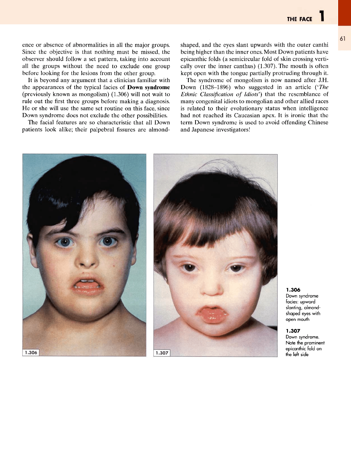
Down syndrome
(previously
known
as
mongolism) (1.306)
will
not
wait
to
rule
out the first
three groups before making
a
diagnosis.
He or she
will
use the
same
set
routine
on
this
face,
since
Down syndrome does
not
exclude
the
other possibilities.
The
facial
features
are so
characteristic that
all
Down
patients look alike; their palpebral
fissures are
almond-
shaped,
and the
eyes slant upwards with
the
outer
canthi
being higher
than
the
inner
ones.
Most
Down patients have
epicanthic
folds
(a
semicircular
fold
of
skin crossing verti-
cally
over
the
inner
can
thus) (1.307).
The
mouth
is
often
kept open with
the
tongue partially protruding through
it.
The
syndrome
of
mongolism
is now
named
after
J.H.
Down
(1828-1896)
who
suggested
in an
article
('The
Ethnic
Classification
of
Idiots']
that
the
resemblance
of
many
congenital idiots
to
mongolian
and
other allied races
is
related
to
their evolutionary status when intelligence
had
not
reached
its
Caucasian apex.
It is
ironic that
the
term Down syndrome
is
used
to
avoid
offending
Chinese
and
Japanese investigators!
1.306
Down
syndrome
facies:
upward
slanting,
almond-
shaped eyes
with
open
mouth
1.307
Down
syndrome.
Note
the
prominent
epicanthic
fold
on
the
left side