Ortiz de Montellano Paul R.(Ed.) Cytochrome P450. Structure, Mechanism, and Biochemistry
Подождите немного. Документ загружается.

Inhibition of Cytochrome P450 Enzymes
275
such as myoglobin that have an imidazole as the
fifth iron ligand.
The likelihood that alkyl radicals, like their aryl
counterparts, bind to the iron before shifting to the
nitrogen is supported by the observation that the
type II complexes formed between alkyldiazenes
and P450 in the absence of oxygen are converted,
in the presence of limited amounts of oxygen,
to complexes with an absorption maximum at
~480 nm characteristic of iron-carbon a-bonded
complexes^^^'
^^^.
Furthermore, alkyl diazene-iron
tetraphenylporphyrin complexes can be prepared
under anaerobic conditions^^"^. However, the
alkyl-iron complexes are much less stable and less
well characterized than the aryl-iron complexes
and their involvement in heme A^-alkylation
reactions remains to be demonstrated.
In addition, some aryl hydrazines, notably dihy-
dralazine, also inactivate P450 via protein modifi-
cation in a mechanism-based process^^^. Thus,
dihydralazine has been shown to inactivate rat liver
microsomal CYP1A2, -2C11 and -3A, but not
CYP2B1 or -lAl^^^, and also inactivates human
liver CYP1A2 and -3A4, but not CYP2C9326 jy^^^
inactivation appears to involve irreversible binding
of a reactive metabolite to the P450 protein in a
process that is not affected by co-incubation with
GSH (5
mM)^^^.
The generation of irreversible
dihydralazine-protein adducts in vivo and their
subsequent immunoproteasomal processing into
antigenic P450 peptides could possibly account for
the detection of CYPlA2-reactive anti-liver micro-
somal (anti-LM) autoantibodies, in the sera of
patients with dihydralazine-induced immunoaller-
gic hepatitis^^^'
^^^.
3.3.5. Other N-N Functions
The P450 heme is iV-alkylated or 7V-arylated
by reactive intermediates formed when it oxidizes
1-aminoaryltriazoles, 2,3-bis(carbethoxy)-2,3-
diazabicyclo[2.2.0]hex-5-ene, and the sydnones.
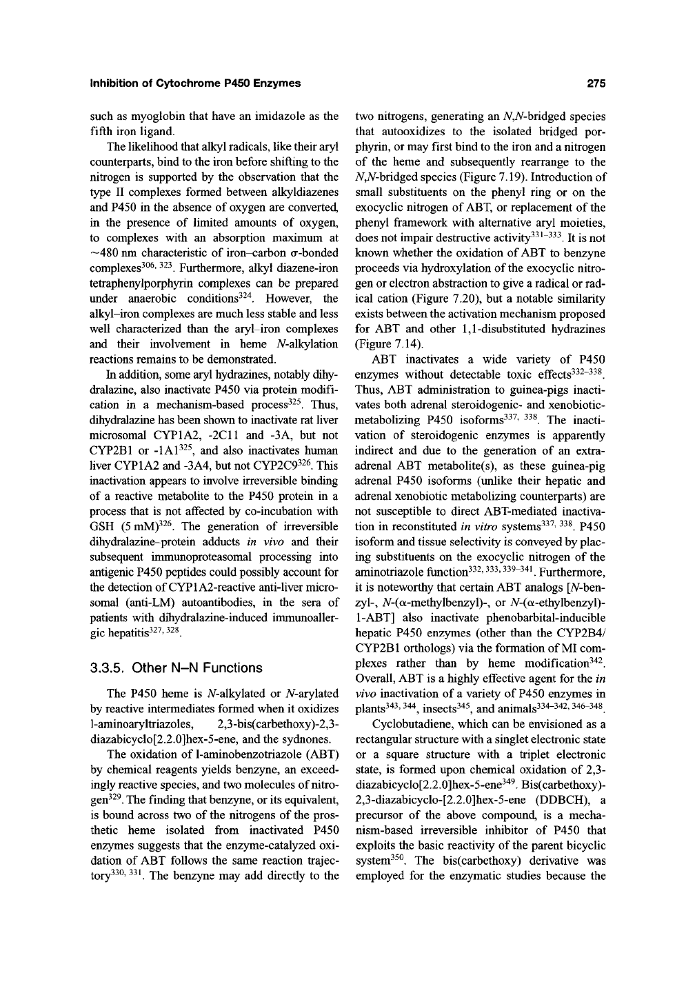
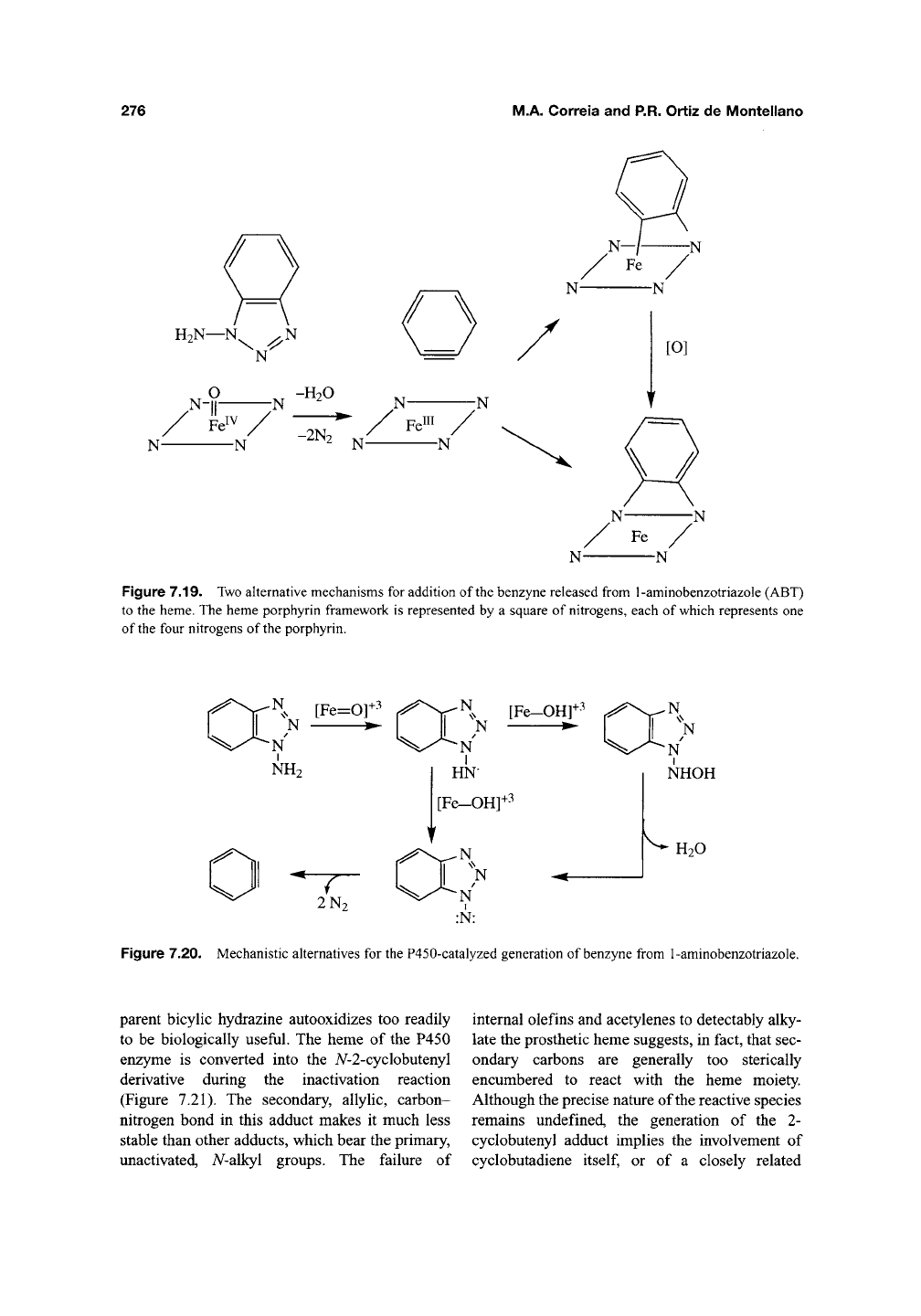
The oxidation of 1-aminobenzotriazole (ABT)
by chemical reagents yields benzyne, an exceed-
ingly reactive species, and two molecules of
nitro-
gen^^^.
The finding that benzyne, or its equivalent,
is bound across two of the nitrogens of the pros-
thetic heme isolated fi-om inactivated P450
enzymes suggests that the enzyme-catalyzed oxi-
dation of ABT follows the same reaction trajec-
^Qj^330,331
jY^Q
benzyne may add directly to the
two nitrogens, generating an N,N-bhdgQd species
that autooxidizes to the isolated bridged por-
phyrin, or may first bind to the iron and a nitrogen
of the heme and subsequently rearrange to the
A^,A^-bridged species (Figure 7.19). Introduction of
small substituents on the phenyl ring or on the
exocyclic nitrogen of ABT, or replacement of the
phenyl fi'amework with alternative aryl moieties,
does not impair destructive activity^^^"^^^. It is not
known whether the oxidation of ABT to benzyne
proceeds via hydroxylation of the exocyclic nitro-
gen or electron abstraction to give a radical or rad-
ical cation (Figure 7.20), but a notable similarity
exists between the activation mechanism proposed
for ABT and other
1,1-disubstituted
hydrazines
(Figure 7.14).
ABT inactivates a wide variety of P450
enzymes without detectable toxic eflfects^^^"^^^.
Thus,
ABT administration to guinea-pigs inacti-
vates both adrenal steroidogenic- and xenobiotic-
metabolizing P450 isoforms^^^' ^^^. The inacti-
vation of steroidogenic enzymes is apparently
indirect and due to the generation of an extra-
adrenal ABT metabolite(s), as these guinea-pig
adrenal P450 isoforms (unlike their hepatic and
adrenal xenobiotic metabolizing counterparts) are
not susceptible to direct ABT-mediated inactiva-
tion in reconstituted in vitro systems^^^'
^^^.
P450
isoform and tissue selectivity is conveyed by plac-
ing substituents on the exocyclic nitrogen of the
aminotriazole
ftinction^^^'
^^^' 339-341
Furthermore,
it is noteworthy that certain ABT analogs [A^-ben-
zyl-,
7V-(a-methylbenzyl)-, or iV-(a-ethylbenzyl)-
1-ABT]
also inactivate phenobarbital-inducible
hepatic P450 enzymes (other than the CYP2B4/
CYP2B1 orthologs) via the formation of MI com-
plexes rather than by heme modification^^^
Overall, ABT is a highly effective agent for the in
vivo inactivation of a variety of P450 enzymes in
plants343,344^ insects345, and animals334-342,346-348
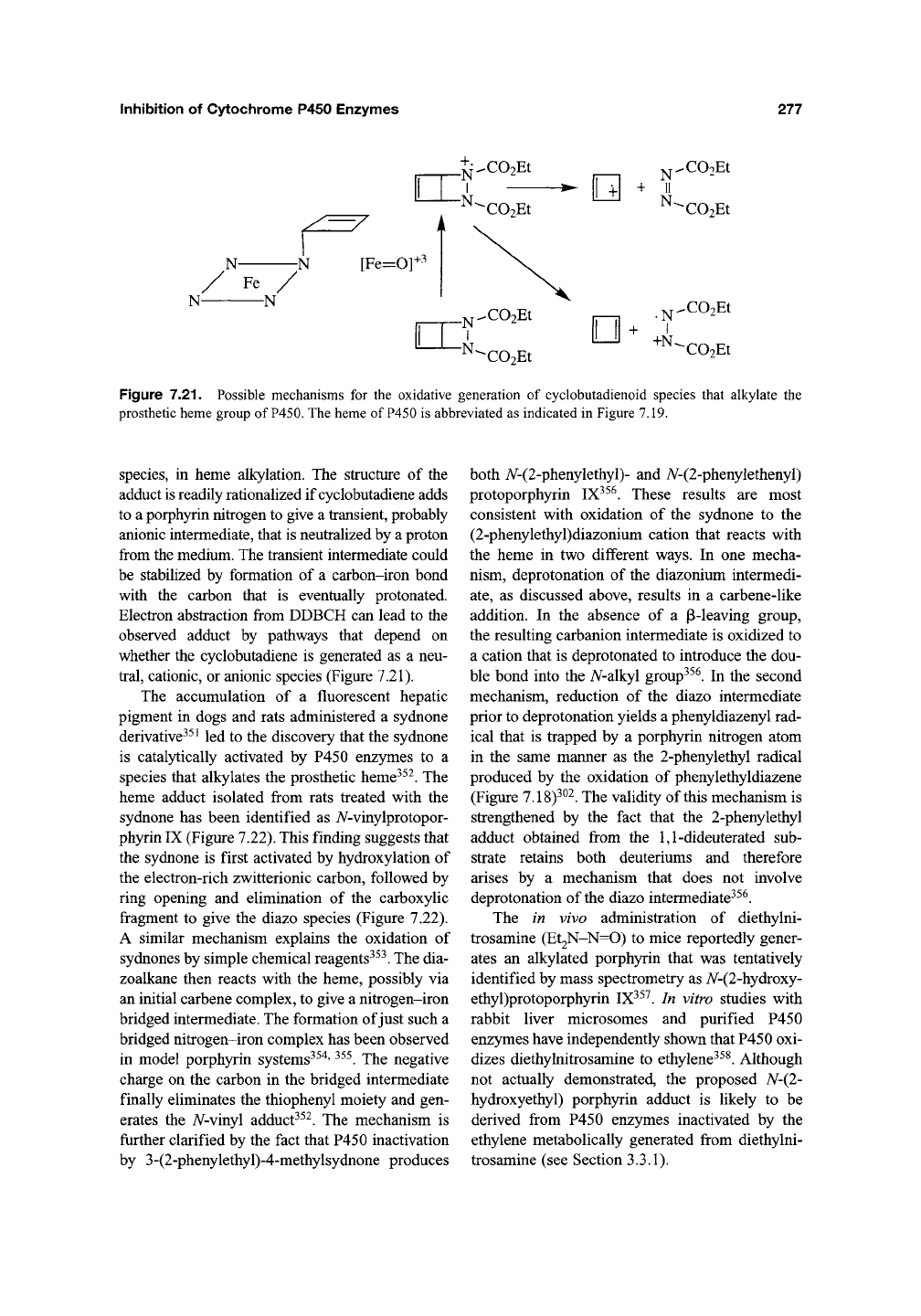
Cyclobutadiene, which can be envisioned as a
rectangular structure with a singlet electronic state
or a square structure with a triplet electronic
state,
is formed upon chemical oxidation of 2,3-
diazabicyclo[2.2.0]hex-5-ene34^. Bis(carbethoxy)-
2,3-diazabicyclo-[2.2.0]hex-5-ene (DDBCH), a
precursor of the above compound, is a mecha-
nism-based irreversible inhibitor of P450 that
exploits the basic reactivity of the parent bicyclic
system^^^. The bis(carbethoxy) derivative was
employed for the enzymatic studies because the
276
M.A. Correia and P.R. Ortiz de Montellano
/ \
/ \
H2N—N^ ^N
N
N-jl N -"-° N N
N N
Fe^
III
-2N,
^: ^-
Figure 7.19. Two alternative mechanisms for addition of the benzyne released from 1-aminobenzotriazole (ABT)
to the heme. The heme porphyrin framework is represented by a square of nitrogens, each of which represents one
of the four nitrogens of the porphyrin.
-^m+3
N [Fe=0]^
N •
N
NH2
N
[Fe-OH]
N
N
I
HN
1+3
[Fe-OH]-'^
"7^
2N2
N
]
N
I
:N:
N
N
NHOH
k
H2O
Figure 7.20. Mechanistic alternatives for the P450-catalyzed generation of benzyne from 1-aminobenzotriazole.
parent bicylic hydrazine autooxidizes too readily
to be biologically useful. The heme of the P450
enzyme is converted into the 7V-2-cyclobutenyl
derivative during the inactivation reaction
(Figure 7.21). The secondary, allylic, carbon-
nitrogen bond in this adduct makes it much less
stable than other adducts, which bear the primary,
unactivated, 7V-alkyl groups. The failure of
internal olefins and acetylenes to detectably alky-
late the prosthetic heme suggests, in fact, that sec-
ondary carbons are generally too sterically
encumbered to react with the heme moiety.
Although the precise nature of the reactive species
remains undefined, the generation of the 2-
cyclobutenyl adduct implies the involvement of
cyclobutadiene
itself,
or of a closely related
Inhibition of Cytochrome P450 Enzymes 277
u
II
I
C02Et
^COoEt
C02Et
XOoEt
C02Et
Figure 7.21. Possible mechanisms for the oxidative generation of cyclobutadienoid species that alkylate the
prosthetic heme group of
P450.
The heme of P450 is abbreviated as indicated in Figure 7.19.
species, in heme alkylation. The structure of the
adduct is readily rationalized if cyclobutadiene adds
to a porphyrin nitrogen to give a transient, probably
anionic intermediate, that is neutralized by a proton
from the medium. The transient intermediate could
be stabilized by formation of a carbon-iron bond
with the carbon that is eventually protonated.
Electron abstraction from DDBCH can lead to the
observed adduct by pathways that depend on
whether the cyclobutadiene is generated as a neu-
tral,
cationic, or anionic species (Figure 7.21).
The accumulation of a fluorescent hepatic
pigment in dogs and rats administered a sydnone
derivatives^
^
led to the discovery that the sydnone
is catal)^ically activated by P450 enzymes to a
species that alkylates the prosthetic heme^^^. The
heme adduct isolated from rats treated with the
sydnone has been identified as A/-vinylprotopor-
phyrin IX (Figure 7.22). This finding suggests that
the sydnone is first activated by hydroxylation of
the electron-rich zwitterionic carbon, followed by
ring opening and elimination of the carboxylic
fragment to give the diazo species (Figure 7.22).
A similar mechanism explains the oxidation of
sydnones by simple chemical reagents^^^. The dia-
zoalkane then reacts with the heme, possibly via
an initial carbene complex, to give a nitrogen-iron
bridged intermediate. The formation of just such a
bridged nitrogen-iron complex has been observed
in model porphyrin systems^^"^'
^^^.
The negative
charge on the carbon in the bridged intermediate
finally eliminates the thiophenyl moiety and gen-
erates the A^-vinyl adduct^^^. The mechanism is
fiirther clarified by the fact that P450 inactivation
by 3-(2-phenylethyl)-4-methylsydnone produces
both A^-(2-phenylethyl)- and 7V-(2-phenylethenyl)
protoporphyrin IX^^^. These results are most
consistent with oxidation of the sydnone to the
(2-phenylethyl)diazonium cation that reacts with
the heme in two different ways. In one mecha-
nism, deprotonation of the diazonium intermedi-
ate,
as discussed above, results in a carbene-like
addition. In the absence of a p-leaving group,
the resulting carbanion intermediate is oxidized to
a cation that is deprotonated to introduce the dou-
ble bond into the N-a\ky\ group^^^. In the second
mechanism, reduction of the diazo intermediate
prior to deprotonation yields a phenyldiazenyl rad-
ical that is trapped by a porph3Tin nitrogen atom
in the same manner as the 2-phenylethyl radical
produced by the oxidation of phenylethyldiazene
(Figure 7.18)^^^. The validity of this mechanism is
strengthened by the fact that the 2-phenylethyl
adduct obtained from the 1,1-dideuterated sub-
strate retains both deuteriums and therefore
arises by a mechanism that does not involve
deprotonation of
the
diazo intermediate^^^.
The in vivo administration of diethylni-
trosamine (Et2N-N=0) to mice reportedly gener-
ates an alkylated porphyrin that was tentatively
identified by mass spectrometry as iV-(2-hydroxy-
ethyl)protoporphyrin IX^^^. In vitro studies with
rabbit liver microsomes and purified P450
enzymes have independently shown that P450 oxi-
dizes diethylnitrosamine to ethylene^^^. Although
not actually demonstrated, the proposed N-(2-
hydroxyethyl) porphyrin adduct is likely to be
derived from P450 enzymes inactivated by the
ethylene metabolically generated from diethylni-
trosamine (see Section 3.3.1).
278
M.A. Correia and P.R. Ortiz de Montellano
RCHov. +
Me.
QH
[Fe=0]3-'
C>
N-
N-
Fe
N-
-N
RCHo
R.
~N.
1
R
—N R = CH2SPh N-/-^N
N N N^^ N
R = CHsPh
R
-N
Figure 7.22. Mechanism proposed for the oxidation of sydnones to reactive intermediates that add to
the prosthetic heme group of P450. The structures of the A^-alkylporphyrins isolated from rats treated with the
3-(2-phenykhioethyl)- and 3-(2-phenylethyl) sydnones are shown.
3.3.6. Other Functionalities
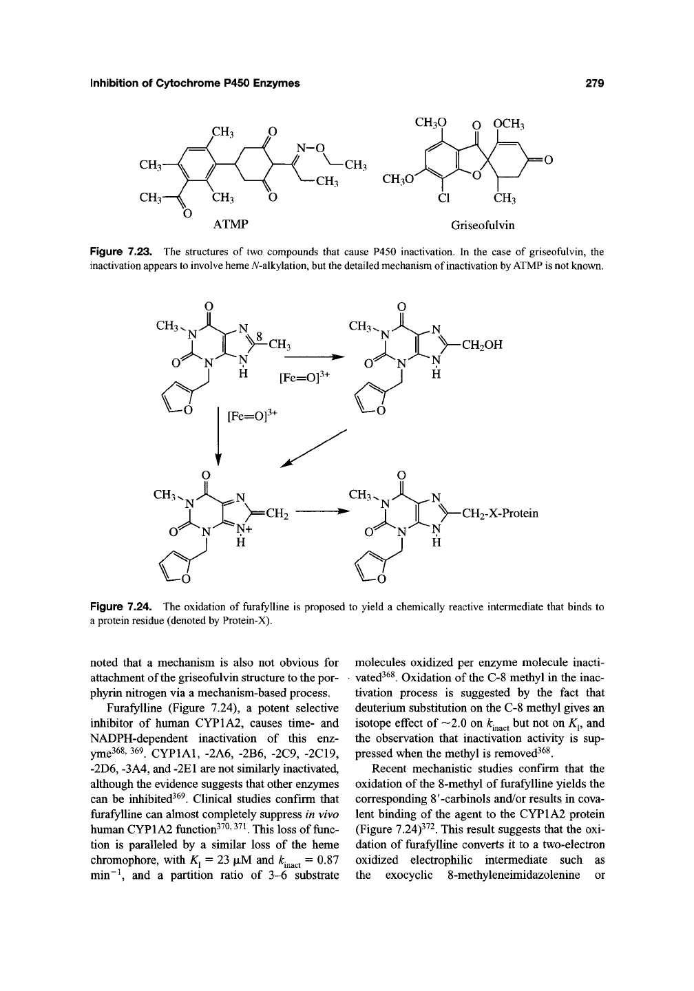
The herbicide l-[4-(3-acetyl-2,4,6-trimethyl-
phenyl)-2,6-cyclohexanedionyl]-0-ethyl propi-
onaldehyde oxime (ATMP) (Figure 7.23) causes
hepatic protoporphyria in the mouse, albeit not
in other species, and this derangement of the
porphyrin biosynthetic pathway appears to be
linked to P450 inactivation via a heme modifica-
tion mechanism^^^. A pigment tentatively identi-
fied as A/-methylprotoporphyrin IX by HPLC
analysis has been isolated from mice treated
with ATMP, and its formation has been shown
to be decreased by pretreatment with the P450
inhibitors SKF 525A and piperonyl butoxide, con-
sistent with a P450-dependent process^^^.
Replacement of the ethyl on the oxime carbon
with a propyl group suppresses the porphyrino-
genic activity of the analog, presumably by pre-
venting heme alkylation. Nothing further is
known about the mechanism by which ATMP
mediates the P450-dependent formation of an
A^-alkyl (possibly iV-methyl) protoporphyrin IX
adduct.
Treatment of mice with griseofulvin
(Figure 7.23), an agent long known to cause
hepatic porphyrias, has been reported to cause the
catalysis-dependent destruction of P450 and the
formation of
a
green pigment^^^. In order to char-
acterize the pigment, the A^-alkyl group was trans-
ferred to an amine in a copper-mediated reaction
and the alkyl amine was analyzed by mass
spectrometry. The results suggested that N-
methylprotoporphyrin IX was a minor product and
a porphyrin with most of the griseofulvin struc-
ture bound to the nitrogen of pyrrole ring C of the
heme was the major product^^^"^^^. The NMR
spectrum of the latter adduct confirms that it is
an iV-alkylated porphyrin with the griseofulvin
structure attached to the nitrogen of either pyrrole
ring C or
D^^"^.
The process appears to be species-
specific because, at most, only very low levels of
pigment have been observed with rats or chicken
embryos^^^"^^^. It is difficult to postulate a mech-
anism for the formation of TV-methyl heme from
griseofulvin, particularly as an identical pigment
is reportedly present in lower amounts in the
livers of control mice^^^' ^^^. The presence of an
endogenous A/-alkylporphyrin in mice would
clearly be of high interest, but more definitive evi-
dence on its origin is required before the signifi-
cance of these results can be evaluated. It is to be
Inhibition of Cytochrome P450 Enzymes
279
CH3O o OCH3
CH3
CH3O
ATMP
CI CH3
Griseofulvin
Figure 7.23. The structures of two compounds that cause P450 inactivation. In the case of griseofulvin, the
inactivation appears
to
involve
heme
A^-alkylation,
but
the
detailed mechanism of inactivation
by ATMP is
not known.
I I >^CH3
CH
[Fe=0]^
CH2OH
CH3
^ ''^'^\^^ N
I |[ y-CH2-X-Protein
H
Figure 7.24. The oxidation of furafylline is proposed to yield a chemically reactive intermediate that binds to
a protein residue (denoted by Protein-X).
noted that a mechanism is also not obvious for
attachment of the griseofulvin structure to the por-
phyrin nitrogen via a mechanism-based process.
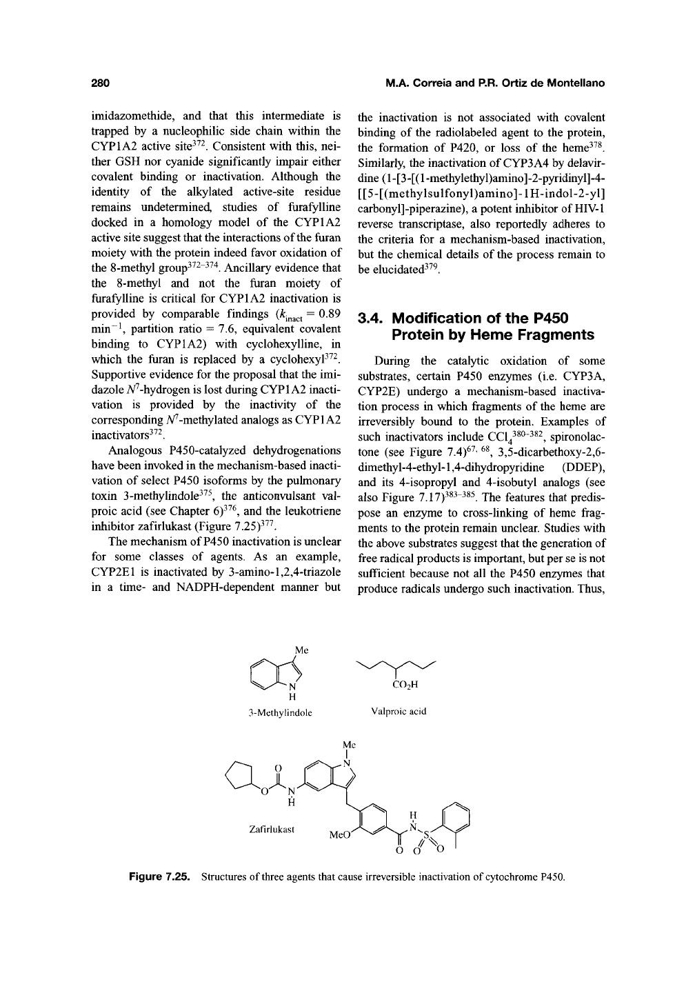
Furafylline (Figure 7.24), a potent selective
inhibitor of human CYP1A2, causes time- and
NADPH-dependent inactivation of this enz-
y^e368,369 CYPIAI, -2A6, -2B6, -2C9, -2C19,
-2D6,
-3A4, and
-2E1
are not similarly inactivated,
although the evidence suggests that other enzymes
can be inhibited^^^. Clinical studies confirm that
furafylline can almost completely suppress in vivo
human CYP1A2 function^^^'
^'^K
This loss of
func-
tion is paralleled by a similar loss of the heme
chromophore, with K^ = 23 fjiM and
A:.^^^^
= 0.87
min~^ and a partition ratio of 3-6 substrate
molecules oxidized per enzyme molecule inacti-
vated^^^. Oxidation of
the
C-8 methyl in the inac-
tivation process is suggested by the fact that
deuterium substitution on the C-8 methyl gives an
isotope effect of —2.0 on
k^^^^^
but not on
K^,
and
the observation that inactivation activity is sup-
pressed when the methyl is removed^^^.
Recent mechanistic studies confirm that the
oxidation of the 8-methyl of furafylline yields the
corresponding 8'-carbinols and/or results in cova-
lent binding of the agent to the CYP1A2 protein
(Figure 7.24)^^^. This result suggests that the oxi-
dation of furafylline converts it to a two-electron
oxidized electrophilic intermediate such as
the exocyclic 8-methyleneimidazolenine or
280
M.A. Correia and P.R. Ortiz de Montellano
imidazomethide, and that this intermediate is
trapped by a nucleophilic side chain within the
CYP1A2 active site^^^. Consistent with this, nei-
ther GSH nor cyanide significantly impair either
covalent binding or inactivation. Although the
identity of the alkylated active-site residue
remains undetermined, studies of furafylline
docked in a homology model of the CYP1A2
active site suggest that the interactions of the furan
moiety with the protein indeed favor oxidation of
the
8-methyl
group^^^"^'^'^. Ancillary evidence that
the
8-methyl
and not the furan moiety of
furafylline is critical for CYP1A2 inactivation is
provided by comparable findings
{h^^
0.89
min~\ partition ratio = 7.6, equivalent covalent
binding to CYP1A2) with cyclohexylline, in
which the furan is replaced by a cyclohexyP^^.
Supportive evidence for the proposal that the imi-
dazole A^^-hydrogen is lost during CYPl A2 inacti-
vation is provided by the inactivity of the
corresponding A^^-methylated analogs as CYPl A2
inactivators^^^.
Analogous P450-catalyzed dehydrogenations
have been invoked in the mechanism-based inacti-
vation of select P450 isoforms by the pulmonary
toxin 3-methylindole^^^, the anticonvulsant val-
proic acid (see Chapter
6)^^^,
and the leukotriene
inhibitor zafirlukast (Figure 7.25)^^^.
The mechanism of P450 inactivation is unclear
for some classes of agents. As an example,
CYP2E1 is inactivated by 3-amino-l,2,4-triazole
in a time- and NADPH-dependent manner but
the inactivation is not associated with covalent
binding of the radiolabeled agent to the protein,
the formation of P420, or loss of the heme^^^.
Similarly, the inactivation of CYP3A4 by delavir-
dine (1 - [3 - [(1 -methylethy l)amino] -2-pyridinyl]
-4-
[[5-[(methylsulfonyl)amino]-lH-indol-2-yl]
carbonyl]-piperazine), a potent inhibitor of HIV-1
reverse transcriptase, also reportedly adheres to
the criteria for a mechanism-based inactivation,
but the chemical details of the process remain to
be elucidated^^^.
3.4. Modification of the P450
Protein by Heme Fragments
During the catalytic oxidation of some
substrates, certain P450 enzymes (i.e. CYP3A,
CYP2E) undergo a mechanism-based inactiva-
tion process in which fragments of the heme are
irreversibly bound to the protein. Examples of
such inactivators include CCl^^^^"^^^, spironolac-
tone (see Figure 7.4)^^' ^^, 3,5-dicarbethoxy-2,6-
dimethyl-4-ethyl-1,4-dihydropyridine (DDEP),
and its 4-isopropyl and 4-isobutyl analogs (see
also Figure 7.17)^^^"^^^. The features that predis-
pose an enzyme to cross-linking of heme frag-
ments to the protein remain unclear. Studies with
the above substrates suggest that the generation of
free radical products is important, but per se is not
sufficient because not all the P450 enzymes that
produce radicals undergo such inactivation. Thus,
ai
Zafirlukast
Figure 7.25. Structures of three agents that cause irreversible inactivation of
cytochrome
P450.
Inhibition of Cytochrome P450 Enzymes
281
during the one-electron oxidation of DDEP,
CYP2C6 and -2C11 undergo heme A^-ethylation,
whereas the CYP3A enzymes predominantly
incur cross-linking of heme fragments to the pro-
tein^^^'
^^^.
Furthermore, studies with DDEP and
its analogs with a secondary carbon attached to
the 4-position (4-isopropyl and 4-isobutyl) reveal
that the reaction outcome is largely dictated by the
P450 active-site structure rather than by the inabil-
ity of the inactivator to iV-alkylate the heme^^^.
Thus,
DDEP, which can form iV-ethyl porphyrins,
and the 4-isopropyl and 4-isobutyl analogs that
cannot, exhibit comparable extents of heme
destruction and heme fragment cross-linking.
Cross-linking of heme fragments to the protein is
thus not merely the result of a defective or ineffi-
cient heme A/-alkylation process. The fact that
spironolactone inactivates hepatic CYP3A
enzymes via heme fragment cross-linking^^'
^^
but
inactivates adrenal P450 enzymes by direct pro-
tein modification^^ frirther demonstrates that the
binding of heme fragments to the protein is iso-
form-specific. Conceivably, the propensity of the
CYP3A enzymes to undergo heme fragment
cross-linking is related to their unusually large
and, given their ability to accommodate large sub-
strates such as cyclosporin, macrolide antibiotics,
and FK506, as well as small substrates such as
DDEP, highly flexible active sites. Their active
sites may therefore exhibit an unusual degree of
substrate mobility and/or water content (Chapter
10).
Regardless of the mechanism, it is clear that
CYP3A active sites are particularly susceptible to
inactivation by heme fragment cross-linking. A
related process is mediated by peroxides such as
H2O2 and cumene hydroperoxide that partially
degrade the prosthetic heme to soluble monopyr-
role and dipyrrole fragments^^^' 386-388 ^^ these
reactions, the bulk of the heme is fragmented to
products that irreversibly bind to the pro-
tein382-39i.
Myoglobin and hemoglobin have been
employed as models in efforts to elucidate this
unusual heme degradation process, but it now
appears that these hemoproteins are not good
models for the P450 reaction392-395 j^ie Rfi^'
mediated oxidation of myoglobin results in the
covalent binding of its heme via either the a or
^-meso carbon or one of the vinyl groups to
Tyrl03^^^.
In contrast, the reduction of CCI4
or CBrClg by myoglobin results in covalent
attachment of the heme via one of its vinyl groups
to
His93^^'*'
^^^.
In both of these processes, as well
as during the hemoglobin-mediated reduction of
CBrCl3^^^
the cross-linked heme retains its Soret
absorption maximum (at —405 nm) and is thus
bound to the protein without substantial structural
disruption of its chromophore. Furthermore, 7-
meso alkylated heme adducts without appreciable
heme-protein cross-linking are observed during
the myoglobin-mediated oxidative metabolism of
alkylhydrazines^^^. In contrast, the cross-linking
of heme fragments to protein observed in the P450
reactions with H2O2, cumene hydroperoxide,
DDEP, and spironolactone involves complete loss
of the heme chromophore and therefore major
structural disruption of the heme skeleton^^' ^^^'
386-391 jjjjg heme degradation also occurs if the
cumene hydroperoxide-mediated inactivation is
carried out under anaerobic conditions, albeit at
a considerably slower rate, implying a role for
molecular O2 in this process^^^' ^^^.
Attempts to elucidate this process have focused
on cumene hydroperoxide-inactivated [^"^CJ-heme-
labeled CYP3A23, -3A4, and -261^89, 390
Proteolytic digestion with lysyl endopeptidase-C
of the [^"^CJ-heme-modified P450enzymes, cou-
pled with HPLC-mapping of the [^"^CJ-heme-
modified peptides, Tricine-SDS-PAGE, elec-
troblotting, microEdman degradation/amino
acid sequencing, and electrospray ionization mass
spectrometry (ESIMS), have located the specific
sites modified by the heme fragments within the
active sites of the P450 enzymes^^^' ^^^. Speci-
fically, the labeled peptide in CYP3A23 encom-
passes residues 287-330, and in CYP2B1 residues
434^66.
Sequence alignment of CYP3A23
and -2B1 with bacterial CYPlOl, -102,-107 and
-108 reveal that the [^'^CJ-heme-fragment-
modified peptide in CYP3A23 corresponds to the
bacterial I-helix^^^"^^3 j^is domain contains the
conserved Thr, which in the crystal structure of
CYPlOl is known to interact both with the sub-
strate and the heme-bound
O2
and to be part of the
active site (Chapter
3)^^^^^^.
On the other hand,
the labeled CYP2B1 region corresponds to the
bacterial L-helix that provides the conserved
Cys thiolate ligand, and thus is also within the
active site. However, until recently, the structure
of the attached heme-derived fragments remained
uncharacterized, largely because of their highly
labile nature under the experimental conditions
282 M.A. Correia and P.R. Ortiz de Montellano
required for isolation and structural analysis of
the modified P450 peptides.
Optimization of the methodology combined
with the larger peptide amounts available through
the use of recombinant [^"^CJ-heme-labeled
CYP3A4, enabled the structural characterization
not only of the CYP3A4 peptide targets, but
also of the protein modifying heme-fragments^^^.
The combined structural analyses identified three
major heme-modified CYP3A4 peptides com-
prised of residues
354-371,
372-386, and
429-450. Sequence alignments and homology
modeling of CYP3A4 reveal that the 354-371 and
429-450 peptides correspond to the K-region
and helix L/Cys region respectively, of P450.
Several residues in these peptides are within 5 A
of the heme and thus within striking distance.
Differential LC-ESMS analyses of the native and
heme-modified peptide fragments provided
molecular masses of «302, 314, and 197 for the
heme-modifying species, corresponding to the
deformylated and formylated A-D/B-C ring
dipyrroles and the monopyrrole 2-formyl hema-
tinic acid^^^. The precise amino acid residues
modified in these peptides remain to be identified.
Nevertheless, these findings suggest that the per-
oxidative inactivation of CYP3A4 (and presum-
ably other P450 enzymes) involves rupture of its
tetrapyrrolic skeleton along its a-7 and/or (3-8
axes to yield reactive heme fragments that modify
residues in their immediate proximity.
It is instructive that in this process, HCOOH
(rather than CO) is the major product, and that
together with minor amounts of CO and CO2,
it stoichiometrically accounts for the oxidative
loss of two heme me^o-carbons^^^' ^^^' 397-399 j^
contrast, peroxidative heme degradation in model
systems yields considerably larger quantities of
soluble dipyrrolic species [hydroxylated and nonhy-
droxylated propentdyopents and HCOOH] as major
products^^^' 397^00 Approximately 15^(^20% of
the prosthetic heme loss after NADPH-induced
oxidative uncoupling can be traced to soluble
mono- and dipyrrolic products in incubations of
purified CYP2BP^^. However, this fraction drops
to —2.5% (comprised largely of hematinic acid,
with traces of methylvinylmaleimide and propent-
dyopents) in CYP3A-enriched rat liver microso-
mal incubations with DDEP or cumene
hydroperoxide^^^. Accordingly, in liver microso-
mal or purified P450 incubations, the bulk of the
heme-derived species appear to irreversibly mod-
ify the CYP3A proteins.
The chemical nature of the heme fragment-
protein adduct remains to be elucidated. In princi-
ple,
it could entail a Schiff-base between a protein
NH2-group and the formyl group of a hydroxy-
dipyrrolic fragment. Alternatively, it could involve
attack by a suitable nucleophilic protein moiety on
the 2-formylated dipyrrole. The possibility of a
protein adduct with the heme vinyl also exists,
even though no vinyl-modified dipyrrolic species
have been detected in model heme degradation
systems.
Both in vivo and in vitro, cross-linking of heme
fragments to the CYP2E1 and -3 A proteins targets
them for proteasomal degradation by the 20S
or ubiquitin-dependent 26S
species^^^'
^01-405
^^^
basal levels of microsomal heme fragment-cross-
linked P450 proteins detected after [^'^C]labeling
of the P450 heme moiety in vivo indicate that
P450 heme-modification probably occurs physio-
logically, possibly as a result of futile oxidative
cycling of the enzymes. Furthermore, the extent of
this cross-linking is increased considerably after
CYP3A is induced by dexamethasone and pheno-
barbitaP^^'"^^^. Since this endogenous post-transla-
tional modification targets the P450 proteins for
proteolytic degradation, it could serve as a deter-
minant of their normal physiological turnover. Not
surprisingly, suppression of P450 futile oxidative
cycling by blocking the P450 heme iron with
TAO or isosafrole, or interrupting the electron
flow through chemical or genetic impairment of
P450 reductase, results in protein stabilization
and consequent "induction" of CYP1A2, -2E1,
and -3A246-249^
3.5- Other Modes of P450
Heme Degradation and
Protein Denaturation
The inactivation of CYP2B4 by aldehydes such
as citral (an a,p unsaturated terpenoid aldehyde),
and other aromatic aldehydes (cinnamaldehyde,
benzaldehyde, and 3-phenylpropionaldehyde) is
accompanied by bleaching of the heme chro-
mophore that is not prevented by catalase, super-
oxide dismutase, epoxide hydrolase, GSH, or
ascorbic acid"^^^'
'^^^.
The corresponding
k^^^^^
val-
ues revealed that saturated aldehydes are generally
Inhibition of Cytochrome P450 Enzymes
283
more inhibitory than their afi unsaturated coun-
terparts, and primary aldehydes are more potent
inactivators than the structurally related secondary
and tertiary aldehydes^^^. Studies with wild-type
CYP2B4 and its T302A mutant, including meas-
urements of the deuterium isotope effects, rates of
inactivation, and rates of product formation sug-
gest that aldehyde-mediated CYP2B4 inactivation
involves deformylation of the aldehyde. In the
inactivation of CYP2B4 by 3-phenylpropionalde-
hyde, a heme adduct is formed with a molecular
weight equal to that of native heme plus 104 mass
units,
in agreement with loss of the carbonyl
group from the original aldehyde. P450 inactiva-
tion by aldehydes has been proposed to involve
homolytic cleavage of a peroxyhemiacetal inter-
mediate to yield formic acid and an alkyl radical
that adds to the heme moiety'*^^. The heme adduct
obtained in similar reactions of the F87G mutant
of CYP108 (P450BM3) has been fully character-
ized by NMR and has been shown to involve
addition of the decarbonylated substrate radi-
cal specifically to the y-meso position of the
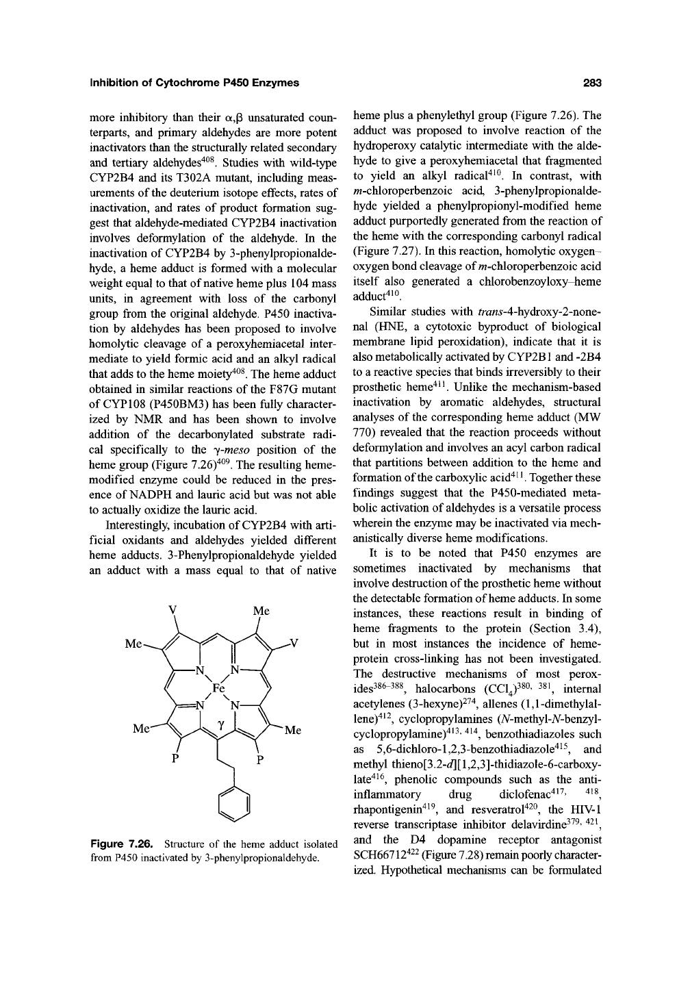
heme group (Figure 7.26)"^^^. The resulting heme-
modified enzyme could be reduced in the pres-
ence of NADPH and lauric acid but was not able
to actually oxidize the lauric acid.
Interestingly, incubation of CYP2B4 with arti-
ficial oxidants and aldehydes yielded different
heme adducts. 3-Phenylpropionaldehyde yielded
an adduct with a mass equal to that of native
Figure 7.26. Structure of the heme adduct isolated
from P450 inactivated by 3-phenylpropionaldehyde.
heme plus a phenylethyl group (Figure 7.26). The
adduct was proposed to involve reaction of the
hydroperoxy catalytic intermediate with the alde-
hyde to give a peroxyhemiacetal that fragmented
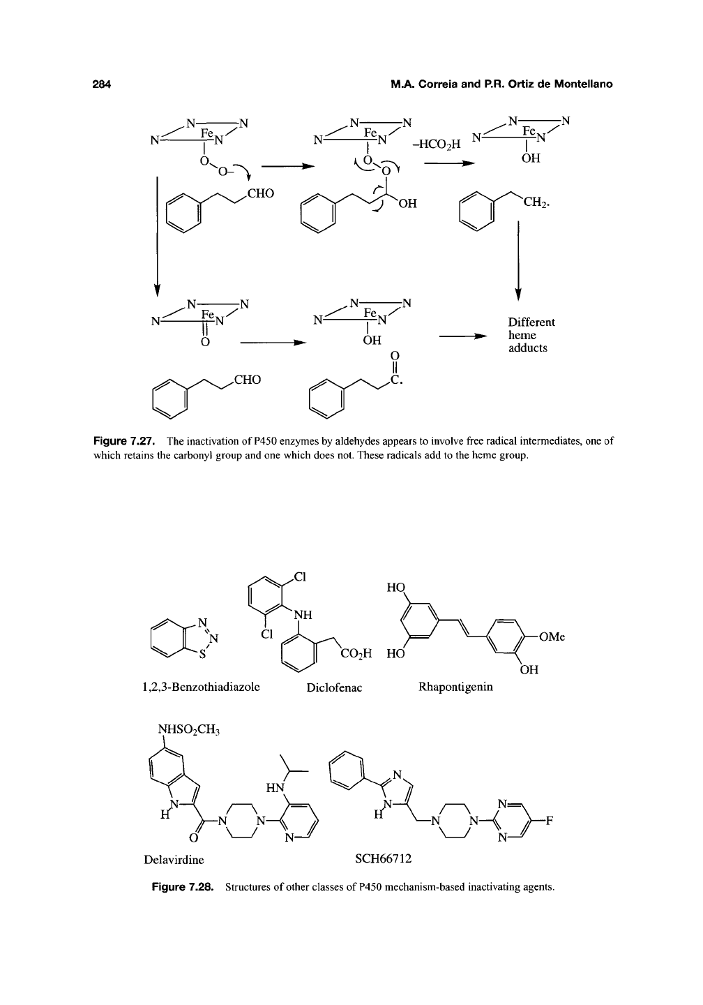
to yield an alkyl radical^^^. In contrast, with
w-chloroperbenzoic acid, 3-phenylpropionalde-
hyde yielded a phenylpropionyl-modified heme
adduct purportedly generated from the reaction of
the heme with the corresponding carbonyl radical
(Figure 7.27). In this reaction, homolytic oxygen-
oxygen bond cleavage of m-chloroperbenzoic acid
itself also generated a chlorobenzoyloxy-heme
adduct^io.
Similar studies with fra«5'-4-hydroxy-2-none-
nal (HNE, a cytotoxic byproduct of biological
membrane lipid peroxidation), indicate that it is
also metabolically activated by CYP2B1 and -2B4
to a reactive species that binds irreversibly to their
prosthetic heme'^^^ Unlike the mechanism-based
inactivation by aromatic aldehydes, structural
analyses of the corresponding heme adduct (MW
770) revealed that the reaction proceeds without
deformylation and involves an acyl carbon radical
that partitions between addition to the heme and
formation of the carboxylic acid'^^^ Together these
findings suggest that the P450-mediated meta-
bolic activation of aldehydes is a versatile process
wherein the enzyme may be inactivated via mech-
anistically diverse heme modifications.
It is to be noted that P450 enzymes are
sometimes inactivated by mechanisms that
involve destruction of the prosthetic heme without
the detectable formation of heme adducts. In some
instances, these reactions result in binding of
heme fragments to the protein (Section 3.4),
but in most instances the incidence of heme-
protein cross-linking has not been investigated.
The destructive mechanisms of most perox-
ides386-388^ halocarbons (CCl4)380' 38i^ internal
acetylenes (3-hexyne)2^'^, allenes (1,1-dimethylal-
lene)"^^^,
cyclopropylamines (7V-methyl-A^-benzyl-
cyclopropylamine)^^^'
^^^,
benzothiadiazoles such
as 5,6-dichloro-l,2,3-benzothiadiazole^^^, and
methyl thieno[3.2-d][ 1,2,3]-thidiazole-6-carboxy-
late"^^^,
phenolic compounds such as the anti-
inflammatory drug diclofenac"^^^' '^^^,
rhapontigenin"^^^, and resveratrol"^^^, the HIV-1
reverse transcriptase inhibitor delavirdine^^^' ^^^,
and the D4 dopamine receptor antagonist
SCH66712422 (Figure 7.28) remain poorly character-
ized. Hypothetical mechanisms can be formulated
284
M.A. Correia and P.R. Ortiz de Montellano
N-
7N
N
^ -HC02H ^^
,N-
Fe y
7N
OH
CH2.
Different
^- heme
adducts
Figure 7.27. The inactivation of
P450
enzymes by aldehydes appears to involve free radical intermediates, one of
which retains the carbonyl group and one which does not. These radicals add to the heme group.
N
N
-S'
I II
CO2H
HO
1,2,3-Benzothiadiazole Diclofenac Rhapontigenin
OMe
OH
NHSO2CH3
Delavirdine SCH66712
Figure 7.28. Structures of other classes of P450 mechanism-based inactivating agents.