Townsend Courtney M.Jr., Evers B. Mark. Atlas of General Surgical Techniques: Expert Consult
Подождите немного. Документ загружается.

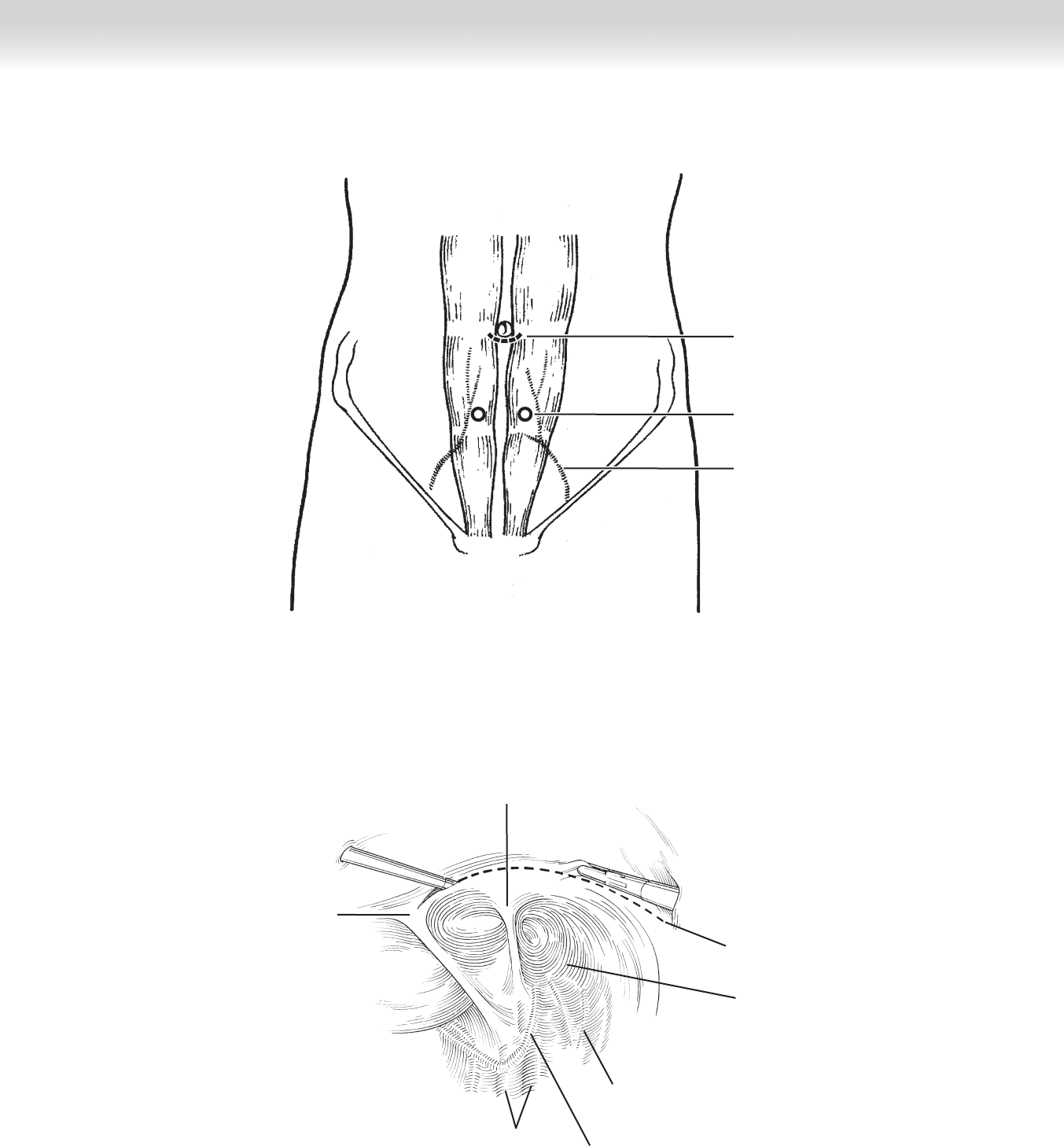
CHAPTER 77 • Inguinal Hernias in Infants and Small Children 839
External oblique
aponeurosis
Ilioinguinal nerve
External
inguinal
ring
FIGURE 77 –2
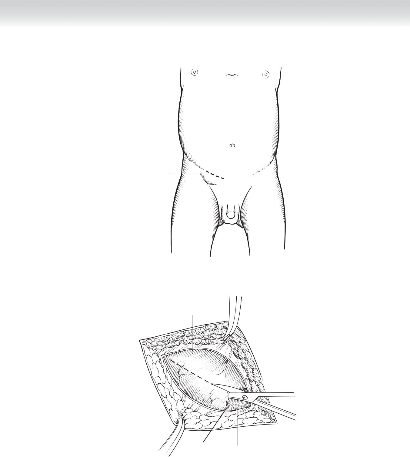
Incision
FIGURE 77 –1

840 Section XI • Hernias
2. DISSECTION
◆ The cremasteric fi bers that surround the spermatic cord are bluntly separated. Be aware that
use of electrocautery in the vicinity of the spermatic vessels or the vas deferens is very haz-
ardous, because transmitted heat or electrical current may damage these structures and may
even result in testicular loss. The hernia sac will be found on the anteromedial aspect of the
spermatic cord (Figure 77-3). Gentle blunt dissection is used to separate the hernia sac
from the spermatic vessels and the vas deferens, avoiding direct manipulation of the latter
(Figure 77-4). These structures must be positively identifi ed before proceeding with the
rest of the operation. Once the hernia sac has been separated from the vas deferens and the
spermatic vessels, the hernia sac is divided between hemostats in its midcourse after it is
ensured that there are no other tissues inside the sac and that there are no sliding compo-
nents making part of the wall of the sac. I fi nd it helpful to place the cord structures within
a vessel loop for gentle traction to avoid injuries. The operation proceeds with dissection of
the proximal portion of the hernia sac up to the level of the internal inguinal ring, where it
is suture ligated with nonabsorbable suture and excised (Figure 77-5).
If you wish to perform a diagnostic laparoscopy, a short 5-mm trocar is introduced through
the sac and secured with a 3-0 Vicryl tie. Pneumoperitoneum is created with a maximum
pressure of 4-8 mm Hg. The patient is placed in the Trendelenburg position, and the table is
tilted toward the surgeon. A 120° telescope is introduced to inspect the contralateral inguinal
ring. After this is done, the trocar is removed, the pneumoperitoneum evacuated, and the
ligation of the sac completed.
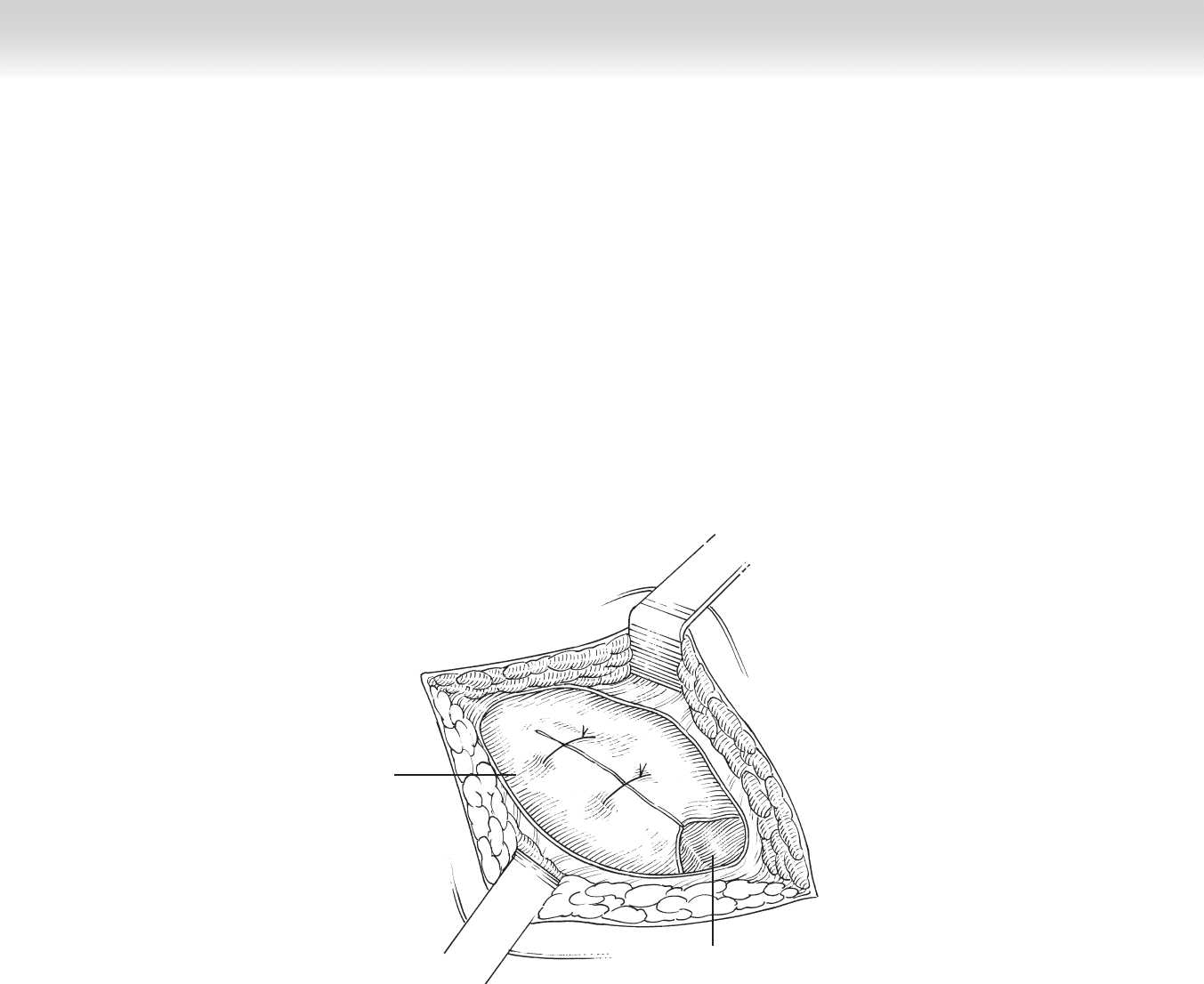
In most cases, high ligation of the hernia sac is suffi cient treatment for an inguinal hernia
in a child. The distal portion of the sac is opened widely; no attempts are made to remove the
sac because this may result in devascularization of the testicle. In patients in whom the fl oor
of the inguinal canal is weak, repair may be performed using the Bassini technique by ap-
proximating the internal oblique muscle to the shelving edge of the inguinal ligament with
two to three interrupted stitches. The most medial stitch approximates the internal oblique
muscle (or the conjoint tendon when present) to the pubic spine. If a hydrocele is present,
the tunica vaginalis is opened and the fl uid is evacuated. The testicle can be brought back
down into the scrotum by gentle caudad traction of the scrotal skin, which will pull the tes-
ticle down along with the gubernaculum testis.
CHAPTER 77 • Inguinal Hernias in Infants and Small Children 841
Spermatic
vessels
Vas deferens
FIGURE 77 –5
Cremaster muscle and
deep spermatic fascia
Spermatic
vessels
Hernia sac
Vas deferens
FIGURE 77 –4
Cremaster muscle and
deep spermatic fascia
Hernia sac
FIGURE 77 –3
842 Section XI • Hernias
3. CLOSING
◆ The external oblique aponeurosis is closed with interrupted fi ne absorbable suture, making
sure that the external inguinal ring does not constrict the cord structures (Figure 77-6).
Scarpa’s fascia is closed with interrupted fi ne absorbable suture, and the skin is closed with
a running fi ne absorbable monofi lament subcuticular suture. The skin is dressed with
adhesive strips.
External
inguinal
ring
External
oblique
aponeurosis
FIGURE 77 –6

CHAPTER 77 • Inguinal Hernias in Infants and Small Children 843
STEP 4: POSTOPERATIVE CARE
◆ Most patients will have either a caudal block, an ilioinguinal block, or subcutaneous infi ltra-
tion of the incision with local anesthetic in the operating room for postoperative pain con-
trol. An oral analgesic such as acetaminophen is prescribed to be given every 4 to 6 hours on
the fi rst postoperative day and then administered only as needed. Children are allowed to
bathe normally 24 hours after the operation and can resume full activity after 2 weeks.
◆ Although rare, the most common complications are wound infections and hematomas. Injury
to the vas deferens, epididymis, or spermatic vessels and hernia recurrence are reported in up
to 1% of cases.
STEP 5: PEARLS AND PITFALLS
◆ Operating immediately after manual reduction of an incarcerated inguinal hernia in a child
is technically diffi cult and fraught with complications, because the hernia sac is edematous
and friable, and the structures of the cord are not easily identifi able. A period of 24 hours
to allow some of the edema to subside is advisable.
◆ As a general rule, no structures should be divided until both the spermatic vessels and the
vas deferens have been positively identifi ed and placed within a vessel loop.
◆ Use of electrocautery in the vicinity of the spermatic cord is discouraged, because arcs of
electrical current may result in thrombosis of the spermatic vessels and loss of the testicle.
SELECTED REFERENCES
1. Weber TR, Tracy TF, Keller MS: Groin hernias and hydroceles. In Ashcraft KW, Holcomb GW,
Murphy JP (eds): Pediatric Surgery, 4th ed. Philadelphia, Elsevier Saunders, 2005, pp 697-705.
2. Engum SA, Grosfeld JL: Hernias in children. In Spitz L, Coran AG (eds): Operative Pediatric Surgery,
6th ed. London, Edward Arnold, 2006, pp 237-244.

844
STEP 1: SURGICAL ANATOMY
◆ A thorough understanding of the preperitoneal space and important structures of the ret-
roperitoneal space and inguinal canal is prerequisite to attempting laparoscopic inguinal
hernia repair (Figure 78-1).
STEP 2: PREOPERATIVE CONSIDERATIONS
INDICATIONS
◆ A laparoscopic approach to inguinal hernias is indicated for any indirect, direct, or femoral
hernia but is particularly suited to bilateral hernias and recurrences from anterior repairs.
◆ Larger hernias, especially with scrotal extension, can make the laparoscopic approach much
more diffi cult.
PREPARATION
◆ A urinary catheter is inserted for bladder decompression.
◆ The patient is placed in the supine Trendelenburg position with the arms padded and
tucked.
CHAPTER
78
Laparoscopic Inguinal
Hernia Repair
Michael D. Trahan
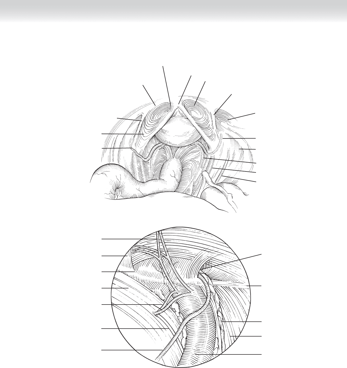
CHAPTER 78 • Laparoscopic Inguinal Hernia Repair 845
Left lateral umbilical fold
with inferior epigastric
vessels
Left deep
inguinal ring
Vas deferens
A
Iliac nerve
Iliac vessels
Right ureter
Lateral cutaneous
nerves of thigh
Vas deferens
Deep inguinal ring
indirect inguinal hernia
Inferior epigastric vessels with
right lateral umbilical fold
Right direct inguinal hernia
Median umbilical fold
bladder
Left medial umbilical
fold
Left direct inguinal hernia
B
Epigastric vessels
Rectus muscles
Conjoined tendon
Cooper's ligament
Obturator vessels
Obturator nerve
Vas deferens
Anterior crus of
internal ring
Iliopubic tract
Internal spermatic
vessels
Psoas muscles
External iliac vessels
Normal Anatomic View
FIGURE 78 –1

846 Section XI • Hernias
STEP 3: OPERATIVE STEPS
1. INCISION
◆ Three ports are used: one 10-mm port and two low-profi le 5-mm ports (Figure 78-2).
2. DISSECTION
◆ Transabdominal preperitoneal (TAPP) repair
◆ An optically guided, bladeless 10-mm port is placed into the peritoneal cavity near the
umbilicus. The two 5-mm ports are placed, guided by internal visualization to avoid the
epigastric vessels.
◆ The peritoneum is incised starting at the medial umbilical fold and proceeding laterally
to or past the anterior superior iliac spine. The incision is made well away from the inter-
nal ring to provide ample tissue to cover the peritoneal defect at the end of the procedure
(Figure 78-3).
◆ The peritoneum is peeled down and bluntly dissected from the underlying structures. An
indirect hernia sac is gently pulled out of the internal ring and dissected free from the
cord structures.
CHAPTER 78 • Laparoscopic Inguinal Hernia Repair 847
10-mm umbilical
port
5-mm paramedian
forceps
Inferior epigastric
artery
FIGURE 78 –2
Peritoneal incision
Iliac vessels
Vas deferens
Lateral cutaneous nerve
of thigh
Internal inguinal ring
and indirect inguinal hernia
Lateral umbilical fold
Medial umbilical
fold
Bladder
FIGURE 78 –3

848 Section XI • Hernias
◆ The margins of a complete dissection are the midline medially, the iliac bone laterally,
and Cooper’s ligament inferiorly (Figure 78-4, A-B).
◆ The fatty contents of a direct hernia defect are reduced and trimmed away as necessary
(Figure 78-4, C).