Berg J.M., Tymoczko J.L., Stryer L. Biochemistry
Подождите немного. Документ загружается.


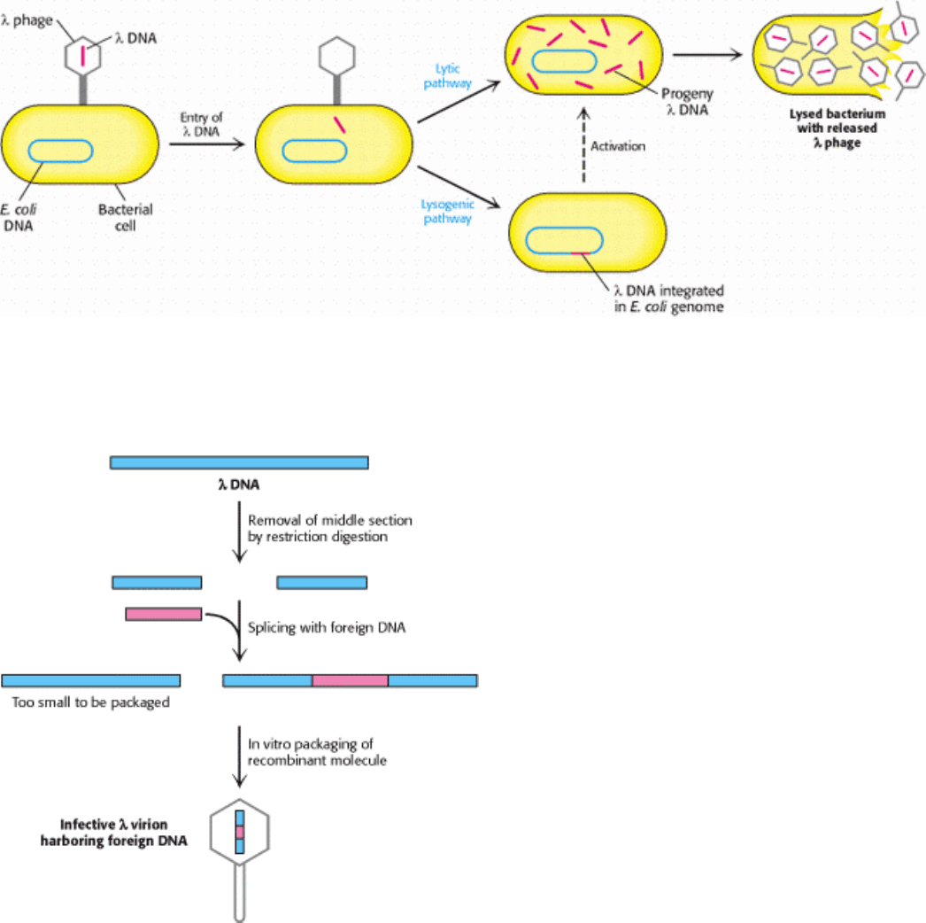
DNA molecule can be removed. The two remaining pieces of DNA (called arms) have a combined length equal to 72%
of a normal genome length. This amount of DNA is too little to be packaged into a λ particle, because only DNA
measuring from 75% to 105% of a normal genome in length can be readily packaged. However, a suitably long DNA
insert (such as 10 kb) between the two ends of λ DNA enables such a recombinant DNA molecule (93% of normal
length) to be packaged. Nearly all infective l particles formed in this way will contain an inserted piece of foreign DNA.
Another advantage of using these modified viruses as vectors is that they enter bacteria much more easily than do
plasmids. Among the variety of λ mutants that have been constructed for use as cloning vectors, one of them, called a
cosmid, is essentially a hybrid of λ phage and a plasmid that can serve as a vector for large DNA inserts (as large as 45
kb).
M13 Phage.
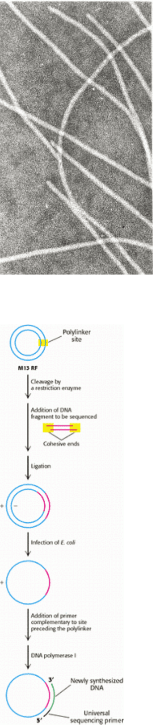
Another very useful vector for cloning DNA, M13 phage is especially useful for sequencing the inserted DNA. This
filamentous virus is 900 nm long and only 9 nm wide (Figure 6.16). Its 6.4-kb single-stranded circle of DNA is protected
by a coat of 2710 identical protein subunits. M13 enters E. coli through the bacterial sex pilus, a protein appendage that
permits the transfer of DNA between bacteria. The single-stranded DNA in the virus particle [called the ( + ) strand] is
replicated through an intermediate circular double-stranded replicative form (RF) containing ( + ) and ( - ) strands. Only
the ( + ) strand is packaged into new virus particles. About a thousand progeny M13 are produced per generation. A
striking feature of M13 is that it does not kill its bacterial host. Consequently, large quantities of M13 can be grown and
easily harvested (1 gram from 10 liters of culture fluid).
An M13 vector is prepared for cloning by cutting its circular RF DNA at a single site with a restriction enzyme. The cut
is made in a polylinker region that contains a series of closely spaced recognition sites for restriction enzymes; only one
of each such sites is present in the vector. A double-stranded foreign DNA fragment produced by cleavage with the same
restriction enzyme is then ligated to the cut vector (Figure 6.17). The foreign DNA can be inserted in two different
orientations because the ends of both DNA molecules are the same. Hence, half the new ( + ) strands packaged into virus
particles will contain one of the strands of the foreign DNA, and half will contain the other strand. Infection of E. coli by
a single virus particle will yield a large amount of single-stranded M13 DNA containing the same strand of the foreign
DNA. DNA cloned into M13 can be easily sequenced. An oligonucleotide that hybridizes adjacent to the polylinker
region is used as a primer for sequencing the insert. This oligomer is called a universal sequencing primer because it can
be used to sequence any insert. M13 is ideal for sequencing but not for long-term propagation of recombinant DNA,
because inserts longer than about 1 kb are not stably maintained.
6.2.3. Specific Genes Can Be Cloned from Digests of Genomic DNA
Ingenious cloning and selection methods have made feasible the isolation of a specific DNA segment several kilobases
long out of a genome containing more than 3×10
6
kb. Let us see how a gene that is present just once in a human genome
can be cloned. A sample containing many molecules of total genomic DNA is first mechanically sheared or partly
digested by restriction enzymes into large fragments (Figure 6.18). This nearly random population of overlapping DNA
fragments is then separated by gel electrophoresis to isolate a set about 15 kb long. Synthetic linkers are attached to the
ends of these fragments, cohesive ends are formed, and the fragments are then inserted into a vector, such as λ phage
DNA, prepared with the same cohesive ends. E. coli bacteria are then infected with these recombinant phages. The
resulting lysate contains fragments of human DNA housed in a sufficiently large number of virus particles to ensure that
nearly the entire genome is represented. These phages constitute a genomic library. Phages can be propagated
indefinitely, and so the library can be used repeatedly over long periods.
This genomic library is then screened to find the very small portion of phages harboring the gene of interest. For the
human genome, a calculation shows that a 99% probability of success requires screening about 500,000 clones; hence, a
very rapid and efficient screening process is essential. Rapid screening can be accomplished by DNA hybridization.
A dilute suspension of the recombinant phages is first plated on a lawn of bacteria (Figure 6.19). Where each phage
particle has landed and infected a bacterium, a plaque containing identical phages develops on the plate. A replica of this

master plate is then made by applying a sheet of nitrocellulose. Infected bacteria and phage DNA released from lysed
cells adhere to the sheet in a pattern of spots corresponding to the plaques. Intact bacteria on this sheet are lysed with
NaOH, which also serves to denature the DNA so that it becomes accessible for hybridization with a
32
P-labeled probe.
The presence of a specific DNA sequence in a single spot on the replica can be detected by using a radioactive
complementary DNA or RNA molecule as a probe. Autoradiography then reveals the positions of spots harboring
recombinant DNA. The corresponding plaques are picked out of the intact master plate and grown. A single investigator
can readily screen a million clones in a day.
This method makes it possible to isolate virtually any gene, provided that a probe is available. How does one obtain a
specific probe? One approach is to start with the corresponding mRNA from cells in which it is abundant. For example,
precursors of red blood cells contain large amounts of mRNA for hemoglobin, and plasma cells are rich in mRNAs for
antibody molecules. The mRNAs from these cells can be fractionated by size to enrich for the one of interest. As will be
described shortly, a DNA complementary to this mRNA can be synthesized in vitro and cloned to produce a highly
specific probe.
Alternatively, a probe for a gene can be prepared if part of the amino acid sequence of the protein encoded by the gene
is known. A problem arises because a given peptide sequence can be encoded by a number of oligonucleotides (Figure
6.20). Thus, for this purpose, peptide sequences containing tryptophan and methionine are preferred, because these
amino acids are specified by a single codon, whereas other amino acid residues have between two and six codons
(Section 5.5.1).
All the DNA sequences (or their complements) that encode the selected peptide sequence are synthesized by the solid-
phase method and made radioactive by phosphorylating their 5
ends with
32
P from [
32
P]-ATP. The replica plate is
exposed to a mixture containing all these probes and autoradiographed to identify clones with a complementary DNA
sequence. Positive clones are then sequenced to determine which ones have a sequence matching that of the protein of
interest. Some of them may contain the desired gene or a significant segment of it.
6.2.4. Long Stretches of DNA Can Be Efficiently Analyzed by Chromosome Walking
A typical genomic DNA library housed in λ phage vectors consists of DNA fragments about 15 kb long. However, many
eukaryotic genes are much longer
for example, the dystrophin gene, which is mutated in Duchenne muscular
dystrophy, is 2000 kb long. How can such long stretches of DNA be analyzed? The development of cosmids helped
because these chimeras of plasmids and λ phages can house 45-kb inserts. Much larger pieces of DNA can now be
propagated in bacterial artificial chromosomes (BACs) or yeast artificial chromosomes (YACs). YACs contain a
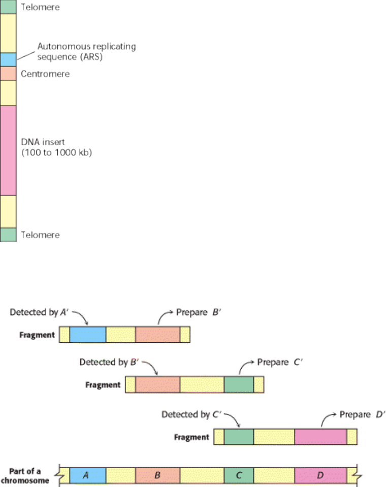
centromere, an autonomous replicating sequence (ARS, where replication begins), a pair of telomeres (normal ends of
eukaryotic chromosomes), selectable marker genes, and a cloning site (Figure 6.21). Genomic DNA is partly digested by
a restriction endonuclease that cuts, on the average, at distant sites. The fragments are then separated by pulsed-field gel
electrophoresis, and the large ones ( ~ 450 kb) are eluted and ligated into YACs. Artificial chromosomes bearing inserts
ranging from 100 to 1000 kb are efficiently replicated in yeast cells.
Equally important in analyzing large genes is the capacity to scan long regions of DNA. The principle technique for this
purpose makes use of overlaps in the library fragments. The fragments in a cosmid or YAC library are produced by
random cleavage of many DNA molecules, and so some of the fragments overlap one another. Suppose that a fragment
containing region A selected by hybridization with a complementary probe A´ also contains region B (Figure 6.22). A
new probe B´ can be prepared by cleaving this fragment between regions A and B and subcloning region B. If the library
is screened again with probe B´, new fragments containing region B will be found. Some will contain a previously
unknown region C. Hence, we now have information about a segment of DNA encompassing regions A, B, and C. This
process of subcloning and rescreening is called chromosome walking. Long stretches of DNA can be analyzed in this
way, provided that each of the new probes is complementary to a unique region.
I. The Molecular Design of Life 6. Exploring Genes 6.2. Recombinant DNA Technology Has Revolutionized All Aspects of Biology
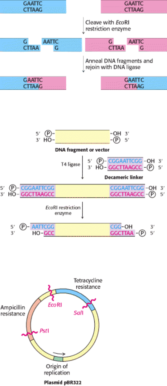
Figure 6.11. Joining of DNA Molecules by the Cohesive-End Method. Two DNA molecules, cleaved with a common
restriction enzyme such as EcoRI, can be ligated to form recombinant molecules.
I. The Molecular Design of Life 6. Exploring Genes 6.2. Recombinant DNA Technology Has Revolutionized All Aspects of Biology
Figure 6.12. Formation of Cohesive Ends. Cohesive ends are formed by the addition and cleavage of a chemically
synthesized linker.
I. The Molecular Design of Life 6. Exploring Genes 6.2. Recombinant DNA Technology Has Revolutionized All Aspects of Biology
Figure 6.13. Genetic Map of the Plasmid PBR322. This plasmid carries two genes for antibiotic resistance. Like all
other plasmids, it is a circular duplex DNA.
I. The Molecular Design of Life 6. Exploring Genes 6.2. Recombinant DNA Technology Has Revolutionized All Aspects of Biology
Figure 6.14. Alternative Infection Modes For λ phage. Lambda phage can multiply within a host and lyse it (lytic
pathway), or its DNA can become integrated into the host genome (lysogenic pathway), where it is dormant until
activated.
I. The Molecular Design of Life 6. Exploring Genes 6.2. Recombinant DNA Technology Has Revolutionized All Aspects of Biology
Figure 6.15. Mutant λ Phage as a Cloning Vector. The packaging process selects DNA molecules that contain an
insert.
I. The Molecular Design of Life 6. Exploring Genes 6.2. Recombinant DNA Technology Has Revolutionized All Aspects of Biology
Figure 6.16. Electron Micrograph of M13 Filamentous Phage. [Courtesy of Dr. Robley Williams.]
I. The Molecular Design of Life 6. Exploring Genes 6.2. Recombinant DNA Technology Has Revolutionized All Aspects of Biology
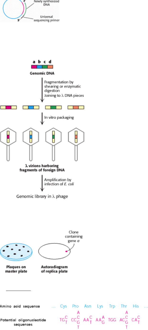
Figure 6.17. M13 Phage DNA, a Cloning and Sequencing Vector. M13 phage DNA is very useful in sequencing
DNA fragments by the dideoxy method. A double-stranded DNA fragment is inserted into M13 RF DNA. Synthesis of
new strand is primed by an oligonucleotide that is complementary to a sequence near the inserted DNA.
I. The Molecular Design of Life 6. Exploring Genes 6.2. Recombinant DNA Technology Has Revolutionized All Aspects of Biology
Figure 6.18. Creation of a Genomic Library. A genomic library can be created from a digest of a whole eukaryotic
genome.
I. The Molecular Design of Life 6. Exploring Genes 6.2. Recombinant DNA Technology Has Revolutionized All Aspects of Biology
Figure 6.19. Screening a Genomic Library for a Specific Gene. Here, a plate is tested from plaques containing gene a
of Figure 6.18.
I. The Molecular Design of Life 6. Exploring Genes 6.2. Recombinant DNA Technology Has Revolutionized All Aspects of Biology
Figure 6.20. Probes Generated from a Protein Sequence. A probe can be generated by synthesizing all possible
oligonucleotides encoding a particular sequence of amino acids. Because of the degeneracy of the genetic code, 256
distinct oligonucleotides must be synthesized to ensure that the probe matching the sequence of seven amino acids is
present.
I. The Molecular Design of Life 6. Exploring Genes 6.2. Recombinant DNA Technology Has Revolutionized All Aspects of Biology
Figure 6.21. Diagram of a Yeast Artificial Chromosome (YAC). DNA inserts as large as 1000 kb can be propagated
in this vector.
I. The Molecular Design of Life 6. Exploring Genes 6.2. Recombinant DNA Technology Has Revolutionized All Aspects of Biology
Figure 6.22. Chromosome Walking. Long regions of unknown DNA can be explored, starting with a known base
sequence, by subcloning and rescreening. New probes are designed on the basis of the DNA sequences that have been
determined.
I. The Molecular Design of Life 6. Exploring Genes
6.3. Manipulating the Genes of Eukaryotes
Eukaryotic genes, in a simplified form, can be introduced into bacteria, and the bacteria can be used as factories to
produce a desired protein product. It is also possible to introduce DNA into higher organisms. In regard to animals, this
ability provides a valuable tool for examining gene action, and it will be the basis of gene therapy. In regard to plants,
introduced genes may make a plant resistant to pests or capable of growing in harsh conditions or able to carry greater
quantities of essential nutrients. The manipulation of eukaryotic genes holds much promise for medical and agricultural
benefits, but it is also the source of controversy.
6.3.1. Complementary DNA Prepared from mRNA Can Be Expressed in Host Cells

How can mammalian DNA be cloned and expressed by E. coli? Recall that most mammalian genes are mosaics of
introns and exons (Section 5.6). These interrupted genes cannot be expressed by bacteria, which lack the machinery to
splice introns out of the primary transcript. However, this difficulty can be circumvented by causing bacteria to take up
recombinant DNA that is complementary to mRNA. For example, proinsulin, a precursor of insulin, is synthesized by
bacteria harboring plasmids that contain DNA complementary to mRNA for proinsulin (Figure 6.23). Indeed, bacteria
produce much of the insulin used today by millions of diabetics.
The key to forming complementary DNA (cDNA) is the enzyme reverse transcriptase. As discussed in Section 5.3.1, a
retrovirus uses this enzyme to form a DNA-RNA hybrid in replicating its genomic RNA. Reverse transcriptase
synthesizes a DNA strand complementary to an RNA template if it is provided with a DNA primer that is base-paired to
the RNA and contains a free 3
-OH group. We can use a simple sequence of linked thymidine [oligo(T)] residues as the
primer. This oligo(T) sequence pairs with the poly(A) sequence at the 3
end of most eukaryotic mRNA molecules
(Section 5.4.4), as shown in Figure 6.24. The reverse transcriptase then synthesizes the rest of the cDNA strand in the
presence of the four deoxyribonucleoside triphosphates. The RNA strand of this RNA-DNA hybrid is subsequently
hydrolyzed by raising the pH. Unlike RNA, DNA is resistant to alkaline hydrolysis. The single-stranded DNA is
converted into double-stranded DNA by creating another primer site. The enzyme terminal transferase adds
nucleotides
for instance, several residues of dG to the 3 end of DNA. Oligo(dC) can bind to dG residues and prime
the synthesis of the second DNA strand. Synthetic linkers can be added to this double-helical DNA for ligation to a
suitable vector. Complementary DNA for all mRNA that a cell contains can be made, inserted into vectors, and then
inserted into bacteria. Such a collection is called a cDNA library.
Complementary DNA molecules can be inserted into vectors that favor their efficient expression in hosts such as E. coli.
Such plasmids or phages are called expression vectors. To maximize transcription, the cDNA is inserted into the vector
in the correct reading frame near a strong bacterial promoter site. In addition, these vectors ensure efficient translation by
encoding a ribosome-binding site on the mRNA near the initiation codon. Clones of cDNA can be screened on the basis
of their capacity to direct the synthesis of a foreign protein in bacteria. A radioactive antibody specific for the protein of
interest can be used to identify colonies of bacteria that harbor the corresponding cDNA vector (Figure 6.25). As
described in Section 6.2.3, spots of bacteria on a replica plate are lysed to release proteins, which bind to an applied
nitrocellulose filter. A
125
I-labeled antibody specific for the protein of interest is added, and autoradiography reveals the
location of the desired colonies on the master plate. This immunochemical screening approach can be used whenever a
protein is expressed and corresponding antibody is available.
6.3.2. Gene-Expression Levels Can Be Comprehensively Examined
Most genes are present in the same quantity in every cell
namely, one copy per haploid cell or two copies per diploid
cell. However, the level at which a gene is expressed, as indicated by mRNA quantities, can vary widely, ranging from
no expression to hundreds of mRNA copies per cell. Gene-expression patterns vary from cell type to cell type,
distinguishing, for example, a muscle cell from a nerve cell. Even within the same cell, gene-expression levels may vary
as the cell responds to changes in physiological circumstances.
Using our knowledge of complete genome sequences, it is now possible to analyze the pattern and level of expression of
all genes in a particular cell or tissue. One of the most powerful methods developed to date for this purpose is based on
hybridization. High-density arrays of oligonucleotides, called DNA microarrays or gene chips, can be constructed either
through lightdirected chemical synthesis carried out with photolithographic microfabrication techniques used in the
semiconductor industry or by placing very small dots of oligonucleotides or cDNAs on a solid support such as a
microscope slide. Fluorescently labeled cDNA is hybridized to the chip to reveal the expression level for each gene,
identifiable by its known location on the chip. (Figure 6.26).
The intensity of the fluorescent spot on the chip reveals the extent of transcription of a particular gene. DNA chips have
been prepared that contain oligonucleotides complementary to all known open reading frames, 6200 in number, within
the yeast genome (Figure 6.27). An analysis of mRNA pools with the use of these chips revealed, for example, that

approximately 50% of all yeast genes are expressed at steady-state levels of between 0.1 and 1.0 mRNA copy per cell.
This method readily detected variations in expression levels displayed by specific genes under different growth
conditions. These tools will continue to grow in power as genome sequencing efforts continue.
6.3.3. New Genes Inserted into Eukaryotic Cells Can Be Efficiently Expressed
Bacteria are ideal hosts for the amplification of DNA molecules. They can also serve as factories for the production of a
wide range of prokaryotic and eukaryotic proteins. However, bacteria lack the necessary enzymes to carry out
posttranslational modifications such as the specific cleavage of polypeptides and the attachment of carbohydrate units.
Thus, many eukaryotic genes can be correctly expressed only in eukaryotic host cells. The introduction of recombinant
DNA molecules into cells of higher organisms can also be a source of insight into how their genes are organized and
expressed. How are genes turned on and off in embryological development? How does a fertilized egg give rise to an
organism with highly differentiated cells that are organized in space and time? These central questions of biology can
now be fruitfully approached by expressing foreign genes in mammalian cells.
Recombinant DNA molecules can be introduced into animal cells in several ways. In one method, foreign DNA
molecules precipitated by calcium phosphate are taken up by animal cells. A small fraction of the imported DNA
becomes stably integrated into the chromosomal DNA. The efficiency of incorporation is low, but the method is useful
because it is easy to apply. In another method, DNA is microinjected into cells. A fine-tipped (0.1- µ m-diameter) glass
micropipet containing a solution of foreign DNA is inserted into a nucleus (Figure 6.28). A skilled investigator can inject
hundreds of cells per hour. About 2% of injected mouse cells are viable and contain the new gene. In a third method,
viruses are used to bring new genes into animal cells. The most effective vectors are retroviruses. As discussed in
Section 5.3.1, retroviruses replicate through DNA intermediates, the reverse of the normal flow of information. A
striking feature of the life cycle of a retrovirus is that the double-helical DNA form of its genome, produced by the action
of reverse transcriptase, becomes randomly incorporated into host chromosomal DNA. This DNA version of the viral
genome, called proviral DNA, can be efficiently expressed by the host cell and replicated along with normal cellular
DNA. Retroviruses do not usually kill their hosts. Foreign genes have been efficiently introduced into mammalian cells
by infecting them with vectors derived from Moloney murine leukemia virus, which can accept inserts as long as 6 kb.
Some genes introduced by this retroviral vector into the genome of a transformed host cell are efficiently expressed.
Two other viral vectors are extensively used. Vaccinia virus, a large DNA-containing virus, replicates in the cytoplasm
of mammalian cells, where it shuts down host-cell protein synthesis. Baculovirus infects insect cells, which can be
conveniently cultured. Insect larvae infected with this virus can serve as efficient protein factories. Vectors based on
these large-genome viruses have been engineered to express DNA inserts efficiently.
6.3.4. Transgenic Animals Harbor and Express Genes That Were Introduced into
Their Germ Lines
Genetically engineered giant mice illustrate the expression of foreign genes in mammalian cells (Figure 6.29). Giant
mice were produced by introducing the gene for rat growth hormone into a fertilized mouse egg. Growth hormone
(somatotropin), a 21-kd protein, is normally synthesized by the pituitary gland. A deficiency of this hormone produces
dwarfism, and an excess leads to gigantism. The gene for rat growth hormone was placed on a plasmid next to the mouse
metallothionein promoter (Figure 6.30). This promoter site is normally located on a chromosome, where it controls the
transcription of metallothionein, a cysteine-rich protein that has high affinity for heavy metals. Metallothionein binds to
and sequesters heavy metals, many of which are toxic for metabolic processes (Section 17.3.2). The synthesis of this
protective protein by the liver is induced by heavy-metal ions such as cadmium. Hence, if mice contain the new gene, its
expression can be initiated by the addition of cadmium to the drinking water.
Several hundred copies of the plasmid containing the promoter and growth-hormone gene were microinjected into the
male pronucleus of a fertilized mouse egg, which was then inserted into the uterus of a foster mother mouse. A number
of mice that developed from such microinjected eggs contained the gene for rat growth hormone, as shown by Southern
blots of their DNA. These transgenic mice, containing multiple copies ( ~ 30 per cell) of the rat growth-hormone gene,

grew much more rapidly than did control mice. In the presence of cadmium, the level of growth hormone in these mice
was 500 times as high as in normal mice, and their body weight at maturity was twice normal. The foreign DNA had
been transcribed and its five introns correctly spliced out to form functional mRNA. These experiments strikingly
demonstrate that a foreign gene under the control of a new promoter site can be integrated and efficiently expressed in
mammalian cells.
6.3.5. Gene Disruption Provides Clues to Gene Function
A gene's function can also be probed by inactivating the gene and looking for resulting abnormalities. Powerful methods
have been developed for accomplishing gene disruption (also called gene knockout) in organisms such as yeast and mice.
These methods rely on the process of homologous recombination. Through this process, regions of strong sequence
similarity exchange segments of DNA. Foreign DNA inserted into a cell thus can disrupt any gene that is at least in part
homologous by exchanging segments (Figure 6.31). Specific genes can be targeted if their nucleotide sequences are
known.
For example, the gene knockout approach has been applied to the genes encoding gene regulatory proteins (also called
transcription factors) that control the differentiation of muscle cells. When both copies of the gene for the regulatory
protein myogenin are disrupted, an animal dies at birth because it lacks functional skeletal muscle. Microscopic
inspection reveals that the tissues from which muscle normally forms contain precursor cells that have failed to
differentiate fully (Figure 6.32). Heterozygous mice containing one normal myogenin gene and one disrupted gene
appear normal, indicating that the level of gene expression is not essential for its function. Analogous studies have
probed the function of many other genes to generate animal models for known human genetic diseases.
6.3.6. Tumor-Inducing Plasmids Can Be Used to Introduce New Genes into Plant Cells
The common soil bacterium Agrobacterium tumefaciens infects plants and introduces foreign genes into plants cells
(Figure 6.33). A lump of tumor tissue called a crown gall grows at the site of infection. Crown galls synthesize opines, a
group of amino acid derivatives that are metabolized by the infecting bacteria. In essence, the metabolism of the plant
cell is diverted to satisfy the highly distinctive appetite of the intruder. Tumor-inducing plasmids (Ti plasmids) that are
carried by Agrobacterium carry instructions for the switch to the tumor state and the synthesis of opines. A small part of
the Ti plasmid becomes integrated into the genome of infected plant cells; this 20-kb segment is called T-DNA
(transferred DNA; Figure 6.34).
Ti plasmid derivatives can be used as vectors to deliver foreign genes into plant cells. First, a segment of foreign DNA is
inserted into the T-DNA region of a small plasmid through the use of restriction enzymes and ligases. This synthetic
plasmid is added to Agrobacterium colonies harboring naturally occurring Ti plasmids. By recombination, Ti plasmids
containing the foreign gene are formed. These Ti vectors hold great promise as tools for exploring the genomes of plant
cells and modifying plants to improve their agricultural value and crop yield. However, they are not suitable for
transforming all types of plants. Ti-plasmid transfer is effective with dicots (broad-leaved plants such as grapes) and a
few kinds of monocots but not with economically important cereal monocots.
Foreign DNA can be introduced into cereal monocots as well as dicots by applying intense electric fields, a technique
called electroporation (Figure 6.35). First, the cellulose wall surrounding plant cells is removed by adding cellulase; this
treatment produces protoplasts, plant cells with exposed plasma membranes. Electric pulses are then applied to a
suspension of protoplasts and plasmid DNA. Because high electric fields make membranes transiently permeable to
large molecules, plasmid DNA molecules enter the cells. The cell wall is then allowed to reform, and the plant cells are
again viable. Maize cells and carrot cells have been stably transformed in this way with the use of plasmid DNA that
includes genes for resistance to antibiotics. Moreover, the transformed cells efficiently express the plasmid DNA.
Electroporation is also an effective means of delivering foreign DNA into animal cells.
The most effective means of transforming plant cells is through the use of "gene guns," or bombardment-mediated
transformation. DNA is coated onto 1- µ m-diameter tungsten pellets, and these microprojectiles are fired at the target