Cook R.A., Stewart B. Colour Atlas of Anatomical Pathology
Подождите немного. Документ загружается.


BONES,
JOINTS
AND
CONNECTIVE
TISSUE
Fig.
10.16
Fig.
10.17
Fig. 10.11
Healed
fracture.
Femur recovered
by
archaeologists from
a
burial about
200
years
ago. There
has
been
a
fracture, which
has
healed with
a
great deal
of
callus
formation.
Fig. 10.12
Multiple
fractures
of the
tibia
and
fibula
which
have
healed
with
very
little
callus
- a
tribute
to
modern
splinting methods
for the
treatment
of
fractures.
Fig. 10.13
Osteoporosis.
F/65.
The
trabecular bone
in the
vertebral
bodies
is
very thin
and the two
lower vertebrae show
the
effects
of
crush fracture
- a
complication
of
this
condition.
Fig. 10.14
Osteoporosis.
M/40. This vertical slice through
the
thoracic spine shows
severe
osteoporosis
of the
vertebrae, with
the
characteristic 'bowing'
or
'curvature'
of the
spine caused
by
collapse
of the
anterior portions
of the
vertebral bodies.
Osteoporosis
of
this
severity
is
more common
in
postmenopausal women.
It is
frequently associated with nerve
pain caused
by
compression
of the
nerves
as
they pass through
the
intervertebral foramina.
The
cause
of the
osteoporosis
was
not
found
in
this case.
Fig. 10.15
Osteoporosis.
X-ray
of the
patient shown
in
Figure
10.14 taken some months before
his
death.
Paget's
disease
James Paget (1814-1899) studied medicine
at St
Bartholomew's
Hospital, London,
and
went
on to
become
a
surgeon
at
that
hospital.
His
name
is
given
to
some
of the
diseases which
he
was
the
first
to
describe. Paget's disease
of
bone
and
Paget's
disease
of the
nipple
are the two
most famous
of
these.
Fig. 10.16 Portrait
of
James Paget displayed
in the
Pathology
Museum
of St
Bartholomew's Hospital, London.
Fig. 10.17 Photographs
of the
first case
of
Paget's disease
of
bone (osteitis deformans), described
by
Paget
in
1876. These
photographs were taken
of a
68-year-old man,
6
months before
he
died.
He
demonstrates
the
features
of
Paget's disease.
The
hats indicate
the
increase
in
size
of his
head between 1844
and
1876.
The
deformation
of the
bones
of the
lower limbs
is
well
illustrated.
211
BONES, JOINTS
AND
CONNECTIVE
TISSUE
Fig.
10.18
Fig.
10.19
Fig.
10.20
Fig.
10.21
212
BONES,
JOINTS
AND
CONNECTIVE
TISSUE
Fig.
10.22
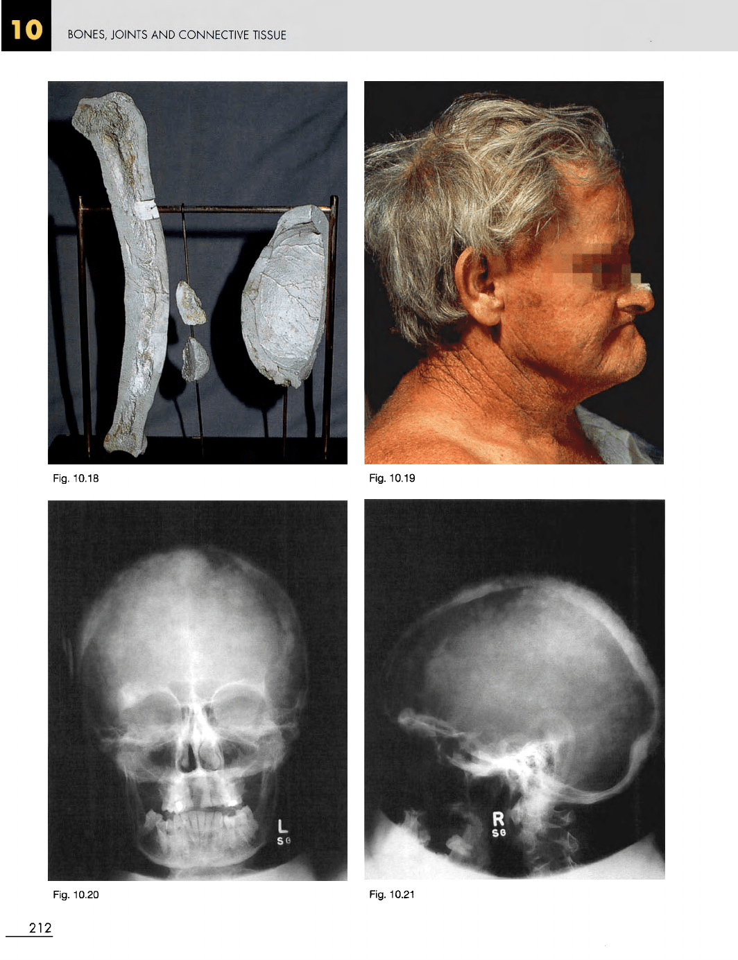
Fig. 10.18 Sample bones from
the
original case
of
Paget's
disease.
They show
the
marked thickening
of the
cortical bone
of
the
tibia,
the
skull
and the
patella.
Fig. 10.19
Paget's
disease.
M/66. Clinical photograph
showing
the
characteristic enlarged head. Patients often
complain
of
headaches.
Fig. 10.20 Anteroposterior
X-ray
of the
skull
of the man
shown
in
Figure 10.19.
Fig. 10.21 Lateral
view
of the
same skull, showing marked
thickening
of the
diploe.
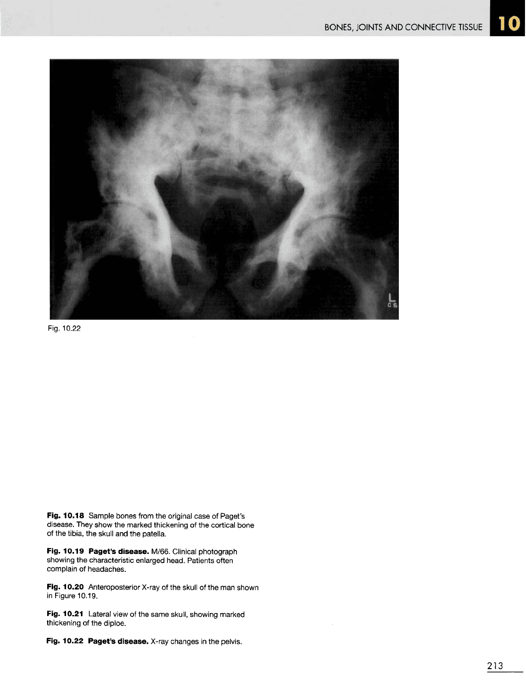
Fig. 10.22
Paget's
disease.
X-ray
changes
in the
pelvis.
213
BONES,
JOINTS
AND
CONNECTIVE
TISSUE
Fig.
10.23
Fig.
10.24
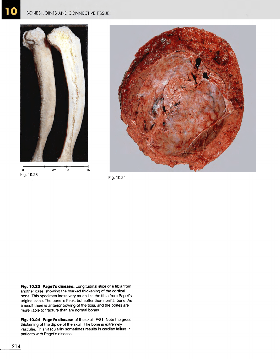
Fig. 10.23
Paget's
disease.
Longitudinal slice
of a
tibia from
another case, showing
the
marked thickening
of the
cortical
bone. This specimen looks very much like
the
tibia from Paget's
original case.
The
bone
is
thick,
but
softer than normal bone.
As
a
result there
is
anterior bowing
of the
tibia,
and the
bones
are
more
liable
to
fracture than
are
normal bones.
Fig. 10.24
Paget's
disease
of the
skull. F/81. Note
the
gross
thickening
of the
diploe
of the
skull.
The
bone
is
extremely
vascular.
This vascularity sometimes results
in
cardiac failure
in
patients with Paget's disease.
214
BONES,
JOINTS
AND
CONNECTIVE
TISSUE
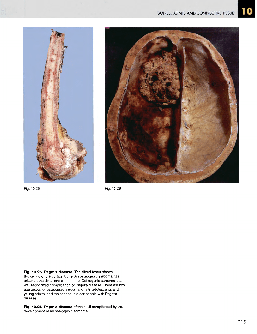
Fig. 10.25
Fig. 10.26
Fig. 10.25 Paget's
disease.
The
sliced femur shows
thickening
of the
cortical
bone.
An
osteogenic
sarcoma
has
arisen
at the
distal
end of the
bone. Osteogenic sarcoma
is a
well
recognized complication
of
Paget's disease. There
are two
age
peaks
for
osteogenic
sarcoma,
one in
adolescents
and
young adults,
and the
second
in
older people with Paget's
disease.
Fig. 10.26
Paget's
disease
of the
skull complicated
by the
development
of an
osteogenic sarcoma.
215
BONES, JOINTS
AND
CONNECTIVE
TISSUE
Fig.
10.27
Fig.
10.28
216

BONES, JOINTS
AND
CONNECTIVE
TISSUE
Fig.
10.30
Fig.
10.29
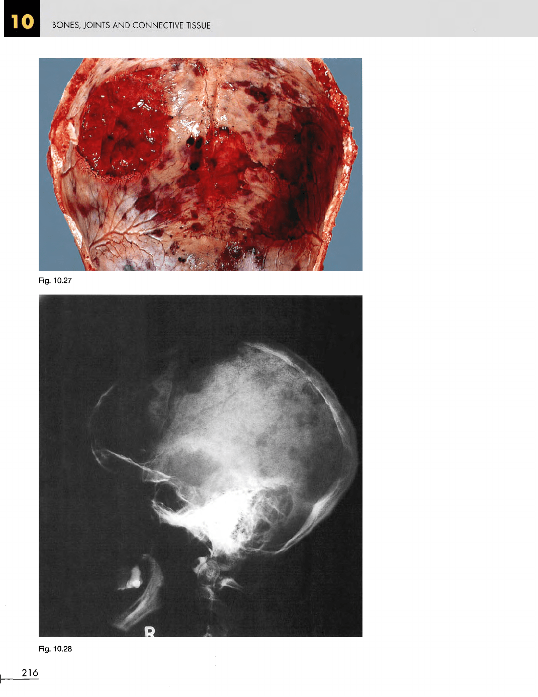
Fig. 10.27 Skull
in
multiple
myeloma. M/39. Multiple,
rounded
red
deposits
of
myeloma
are
present
in the
calvarium.
Fig. 10.28
Multiple
myeloma.
A
lateral X-ray
of the
skull
showing
the
punched-out defects
of
deposits
of
multiple
myeloma.
Fig. 10.29 Vertebral column
in
multiple
myeloma.
The
same
patient
as in
Figure 10.27.
The
myeloma deposits
in the
vertebrae
cause
loss
of
bone
and
consequent crush fractures,
as
demonstrated here.
Fig. 10.30
Multiple
myeloma. Humerus
and
femur
showing
localized areas
of
deposition
of
myeloma
in the
long bones.
217
BONES, JOINTS
AND
CONNECTIVE
TISSUE
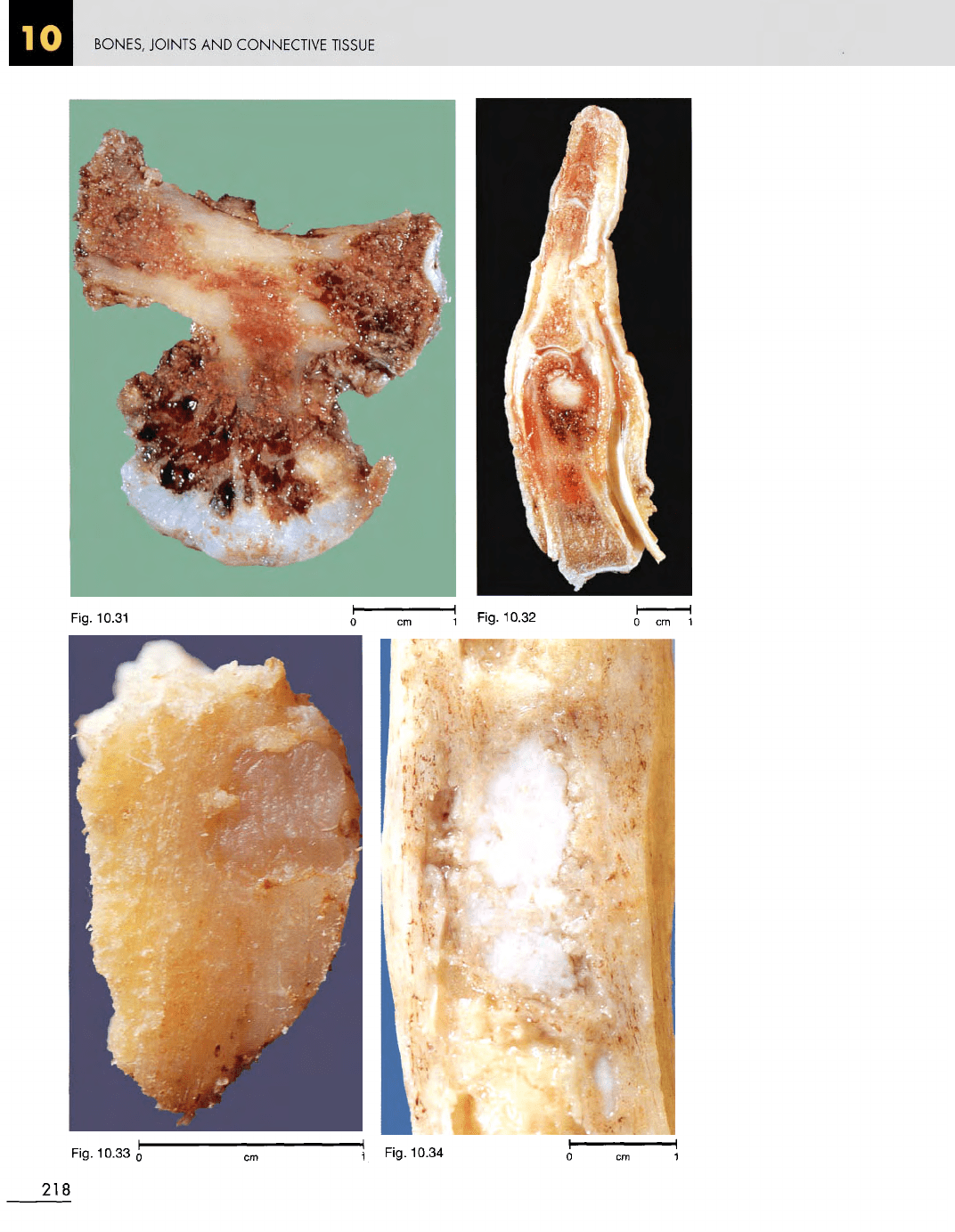
Fig.
10.33
o
\
Fig. 10.34
218
Fig.
10.31
Fig.
10.32
BONES,
JOINTS
AND
CONNECTIVE
TISSUE
Fig.
10.35
Fig. 10.31 Osteochondroma
on a
rib. F/22. This
is a
common benign tumour which usually occurs
in the
region
of
the
epiphyses
of
long bones.
It is
characterized
by
having
a
distinct cartilage cap. Such tumours
are
easily excised.
Fig. 10.32
Osteoid
osteoma
in the
proximal phalanx
of a
finger.
M/30. There
is a
benign, well circumscribed tumour
within
the
medullary cavity.
The
treatment
of
choice
is
local
curettage. Amputation such
as
this
is
overtreatment.
The
commonest site
for an
osteoid osteoma
is the
upper
end of the
tibia.
Fig. 10.33
Benign
chondroma.
M/27. This small cartilage
tumour
was
resected from
the
tibia.
Fig. 10.34 Fibrous dysplasia
in the
medullary
cavity
of
the
midshaft
of the
tibia.
F/14.
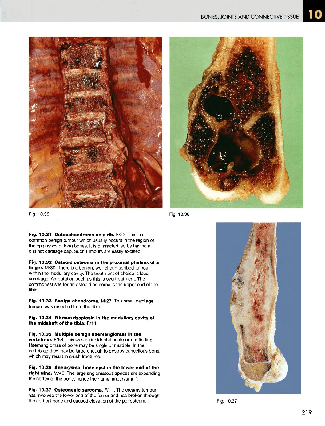
Fig. 10.35
Multiple
benign
haemangiomas
in the
vertebrae.
F/68. This
was an
incidental postmortem finding.
Haemangiomas
of
bone
may be
single
or
multiple.
In the
vertebrae
they
may be
large enough
to
destroy cancellous bone,
which
may
result
in
crush fractures.
Fig. 10.36 Aneurysmal
bone
cyst
in the
lower
end of the
right
ulna.
M/40.
The
large angiomatous spaces
are
expanding
the
cortex
of the
bone, hence
the
name 'aneurysmal'.
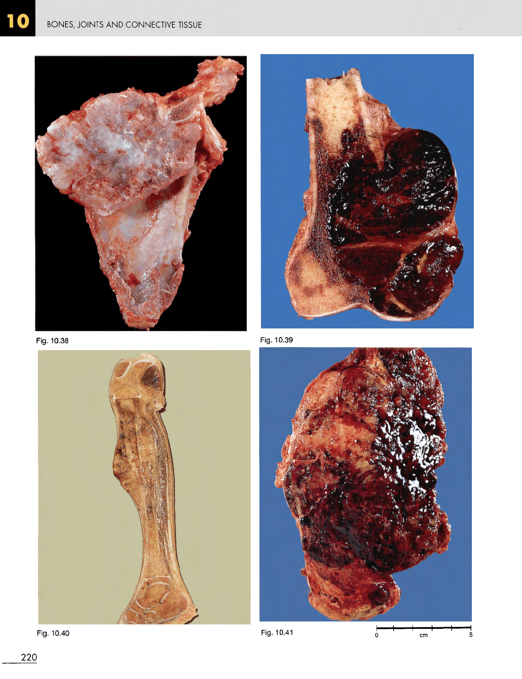
Fig. 10.37
Osteogenic
sarcoma.
F/11.
The
creamy tumour
has
involved
the
lower
end of the
femur
and has
broken through
the
cortical bone
and
caused elevation
of the
periosteum.
Fig. 10.36
Fig.
10.37
219
BONES, JOINTS
AND
CONNECTIVE
TISSUE
Fig.
10.40
Fig.
10.41
220
Fig.
10.38
Fig.
10.39