Cook R.A., Stewart B. Colour Atlas of Anatomical Pathology
Подождите немного. Документ загружается.

ENDOCRINE
SYSTEM
Fig. 9.20
Fig. 9.21
Fig. 9.16
Precocious
puberty.
M/11
months.
Fig. 9.17
Adrenal
cortical
adenoma.
This
was
removed surgically from
the
patient
in
Figure 9.16.
The cut
surface
is
multilobulated
and a
homogeneous
brown colour. This adenoma
was
secreting androgens.
Fig. 9.18 Gynaecomastia. M/5.
Fig. 9.19
Adrenal
cortical
tumour.
This large adrenal tumour
was
removed surgically from
the
patient
in
Figure 9.18.
It was
secreting oestrogens.
The
tumour
was
somewhat adherent
to
adjacent structures
and was
torn during removal.
It is
impossible
to
predict which adrenal tumours will
be
malignant, because
cellular pleomorphism does
not
correlate with their behaviour.
In
spite
of the
local adhesions
and
raggedness
of
this specimen,
the
gynaecomastia subsided
and the
patient
was
alive
and
well
30
years later.
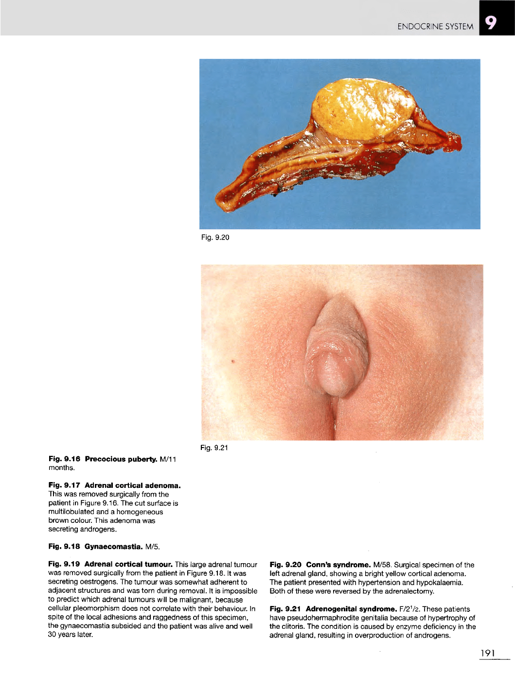
Fig. 9.20 Conn's
syndrome.
M/58. Surgical specimen
of the
left
adrenal gland, showing
a
bright yellow cortical adenoma.
The
patient presented with hypertension
and
hypokalaemia.
Both
of
these were reversed
by the
adrenalectomy.
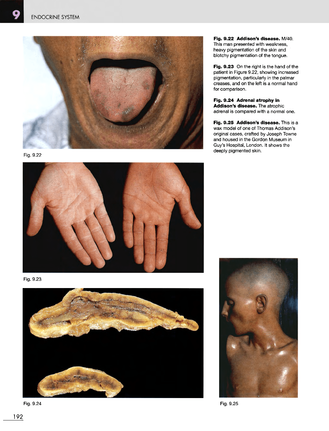
Fig. 9.21
Adrenogenital
syndrome. F/2
1
/2. These patients
have
pseudohermaphrodite genitalia because
of
hypertrophy
of
the
clitoris.
The
condition
is
caused
by
enzyme deficiency
in the
adrenal gland, resulting
in
overproduction
of
androgens.
191
ENDOCRINE
SYSTEM
Fig.
9.22
Fig. 9.23
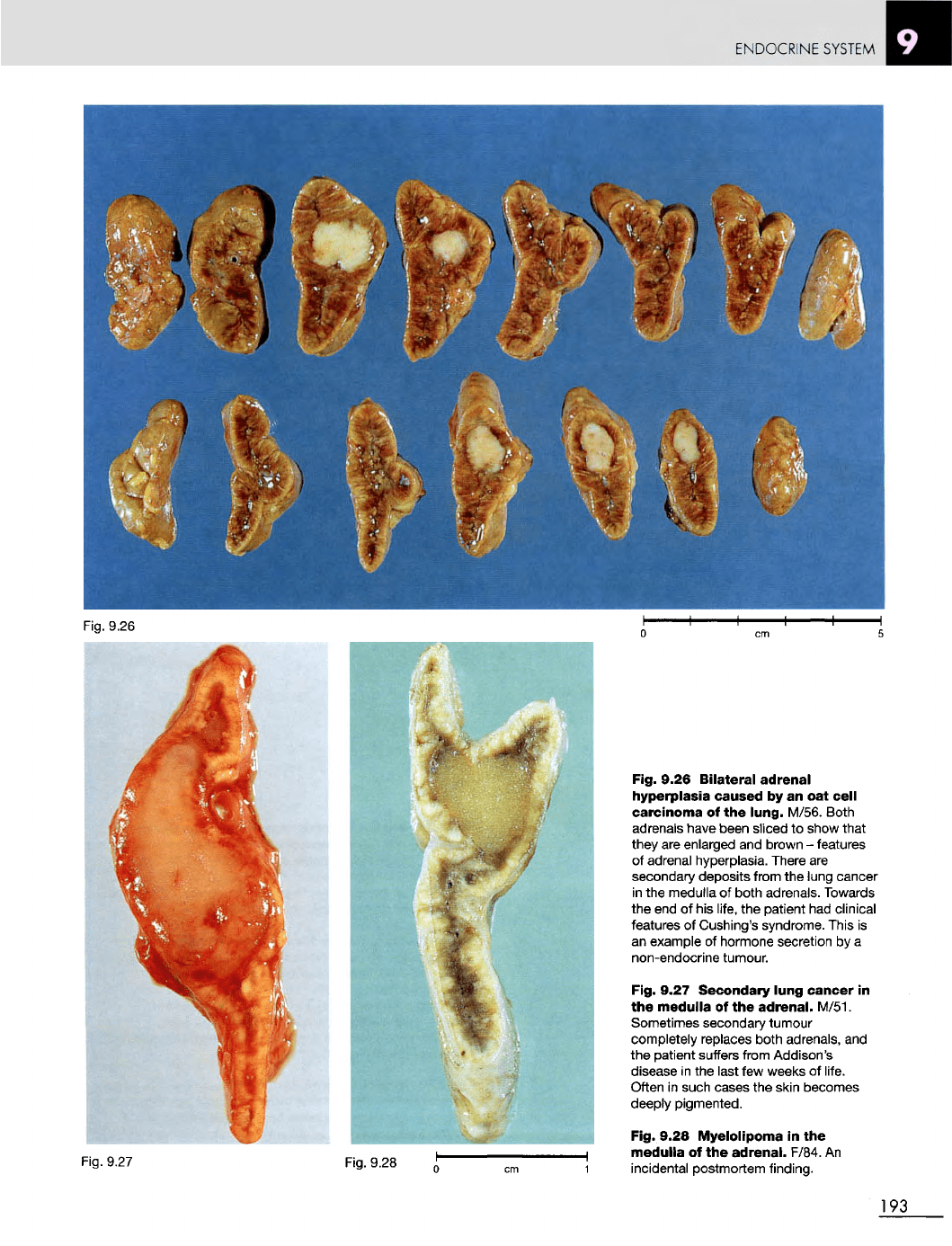
Fig. 9.22 Addison's
disease.
M/40.
This
man
presented
with
weakness,
heavy
pigmentation
of the
skin
and
blotchy pigmentation
of the
tongue.
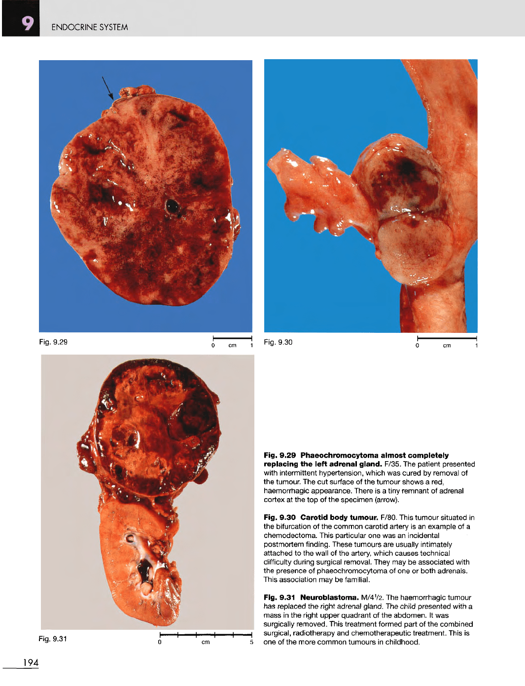
Fig. 9.23
On the
right
is the
hand
of the
patient
in
Figure 9.22, showing increased
pigmentation, particularly
in the
palmar
creases,
and on the
left
is a
normal hand
for
comparison.
Fig. 9.24
Adrenal
atrophy
in
Addison's
disease.
The
atrophic
adrenal
is
compared with
a
normal one.
Fig. 9.25
Addison's
disease.
This
is a
wax
model
of one of
Thomas Addison's
original cases, crafted
by
Joseph
Towne
and
housed
in the
Gordon Museum
in
Guy's
Hospital, London.
It
shows
the
deeply pigmented skin.
Fig.
9.25
Fig.
9.24
192
ENDOCRINE
SYSTEM
Fig. 9.26
Bilateral
adrenal
hyperplasia
caused
by an oat
cell
carcinoma
of the
lung.
M/56. Both
adrenals have been
sliced
to
show that
they
are
enlarged
and
brown
-
features
of
adrenal hyperplasia. There
are
secondary deposits from
the
lung cancer
in
the
medulla
of
both adrenals. Towards
the end of his
life,
the
patient
had
clinical
features
of
Cushing's syndrome. This
is
an
example
of
hormone secretion
by a
non-endocrine tumour.
Fig. 9.27 Secondary lung
cancer
in
the
medulla
of the
adrenal.
M/51.
Sometimes secondary tumour
completely replaces both adrenals,
and
the
patient suffers from Addison's
disease
in the
last
few
weeks
of
life.
Often
in
such cases
the
skin becomes
deeply pigmented.
Fig. 9.28
Myelolipoma
in the
medulla
of the
adrenal.
F/84.
An
incidental postmortem finding.
193
Fig. 9.26
ENDOCRINE
SYSTEM
Fig. 9.31
Fig. 9.29 Phaeochromocytoma
almost
completely
replacing
the
left
adrenal
gland.
F/35.
The
patient presented
with intermittent hypertension, which
was
cured
by
removal
of
the
tumour.
The cut
surface
of the
tumour shows
a
red,
haemorrhagic
appearance. There
is a
tiny remnant
of
adrenal
cortex
at the top of the
specimen
(arrow).
Fig. 9.30
Carotid
body
tumour.
F/80. This tumour situated
in
the
bifurcation
of the
common carotid artery
is an
example
of a
chemodectoma. This particular
one was an
incidental
postmortem
finding.
These tumours
are
usually intimately
attached
to the
wall
of the
artery, which causes technical
difficulty
during surgical removal. They
may be
associated with
the
presence
of
phaeochromocytoma
of one or
both adrenals.
This
association
may be
familial.
Fig. 9.31
Neuroblastoma.
M/4
1
/2.
The
haemorrhagic tumour
has
replaced
the
right
adrenal
gland.
The
child
presented
with
a
mass
in the
right upper quadrant
of the
abdomen.
It was
surgically removed. This treatment formed part
of the
combined
surgical,
radiotherapy
and
chemotherapeutic treatment. This
is
one
of the
more common tumours
in
childhood.
194
Fig.
9.30
Fig 9.29
ENDOCRINE
SYSTEM
Fig.
9.32
Fig. 9.33
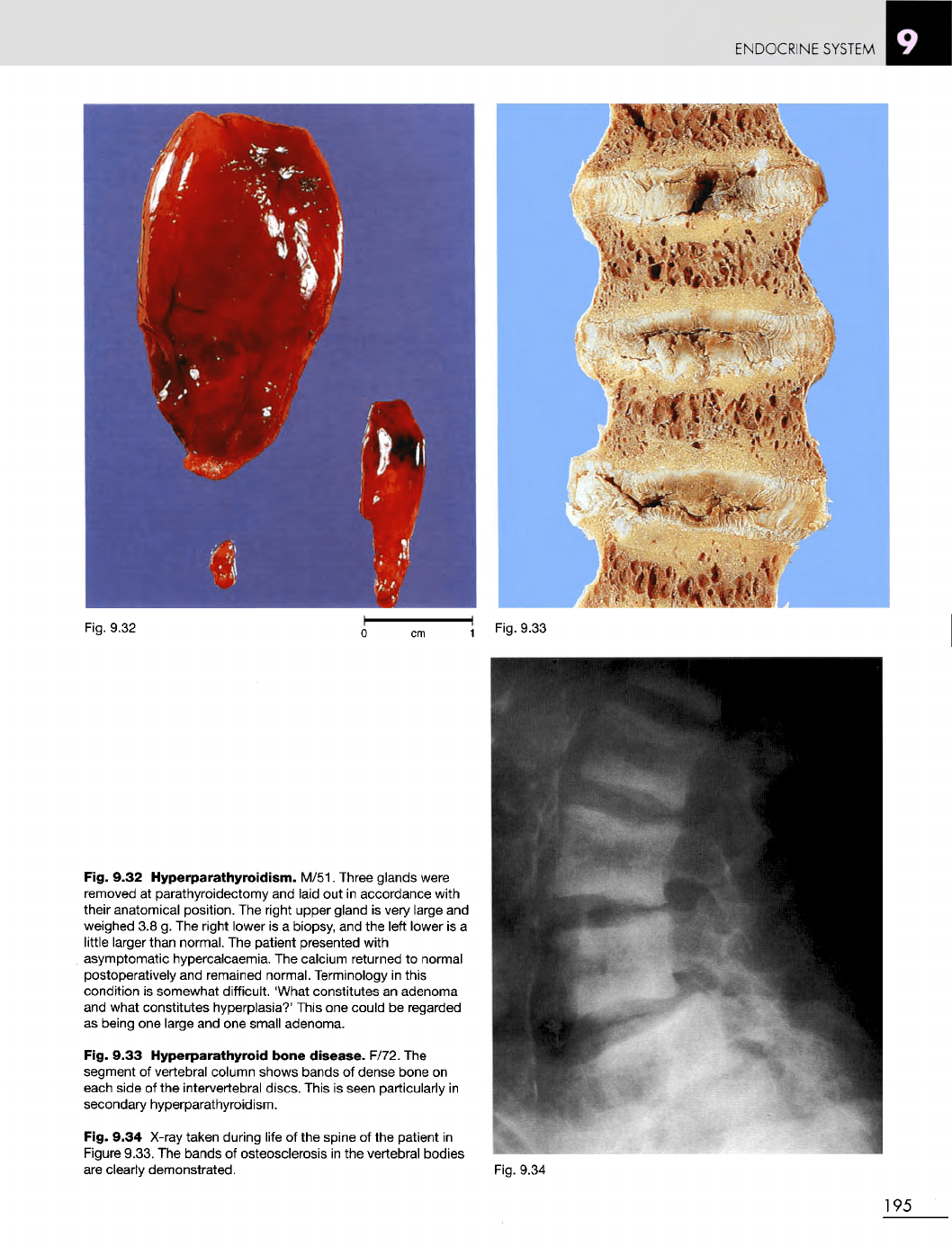
Fig. 9.32
Hyperparathyroidism.
M/51. Three glands were
removed
at
parathyroidectomy
and
laid
out in
accordance with
their
anatomical position.
The
right upper gland
is
very large
and
weighed
3.8 g. The
right lower
is a
biopsy,
and the
left lower
is a
little larger than normal.
The
patient presented with
asymptomatic hypercalcaemia.
The
calcium returned
to
normal
postoperatively
and
remained normal. Terminology
in
this
condition
is
somewhat difficult. 'What constitutes
an
adenoma
and
what constitutes hyperplasia?' This
one
could
be
regarded
as
being
one
large
and one
small adenoma.
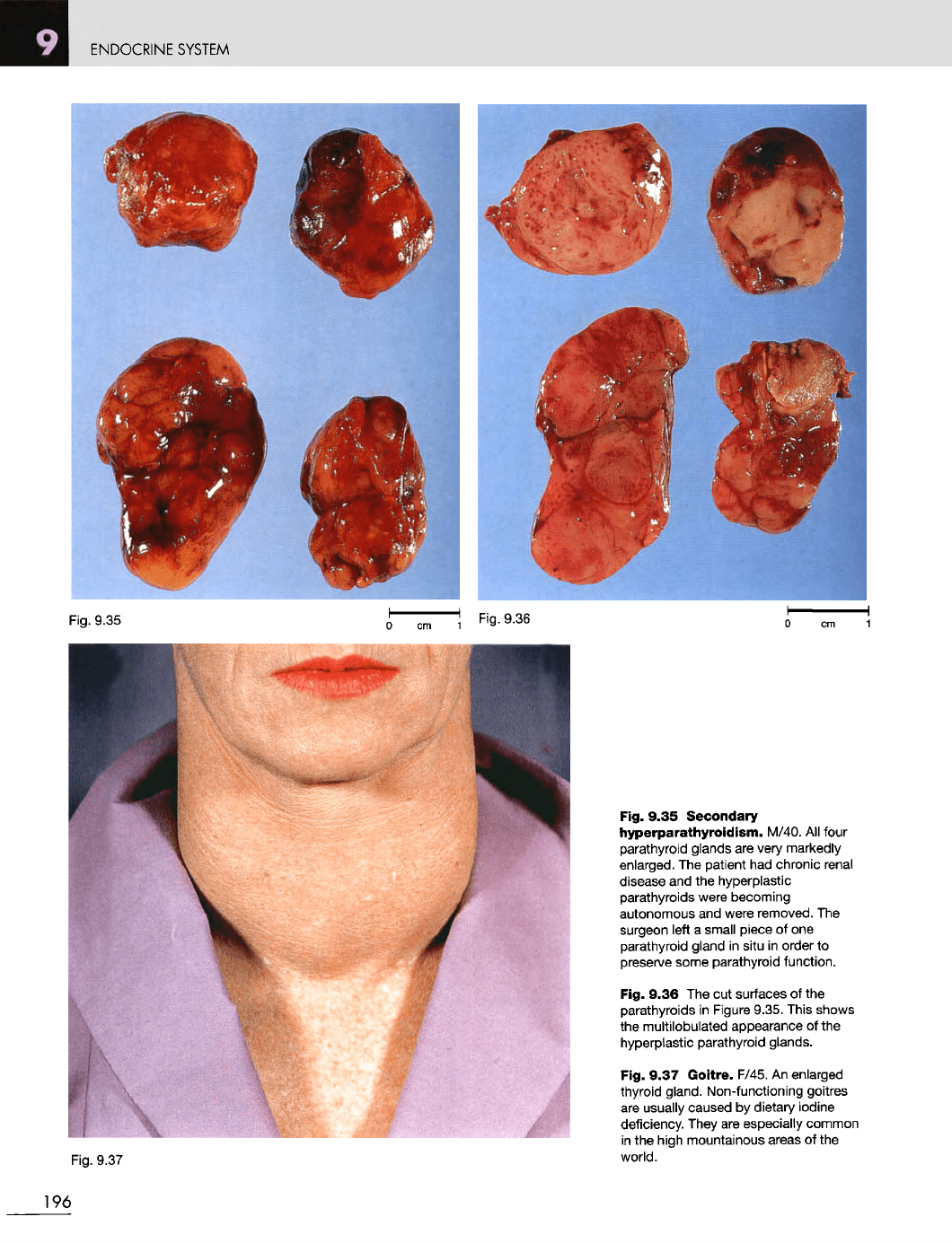
Fig. 9.33
Hyperparathyroid
bone
disease.
F/72.
The
segment
of
vertebral column shows bands
of
dense bone
on
each
side
of the
intervertebral discs. This
is
seen particularly
in
secondary
hyperparathyroidism.
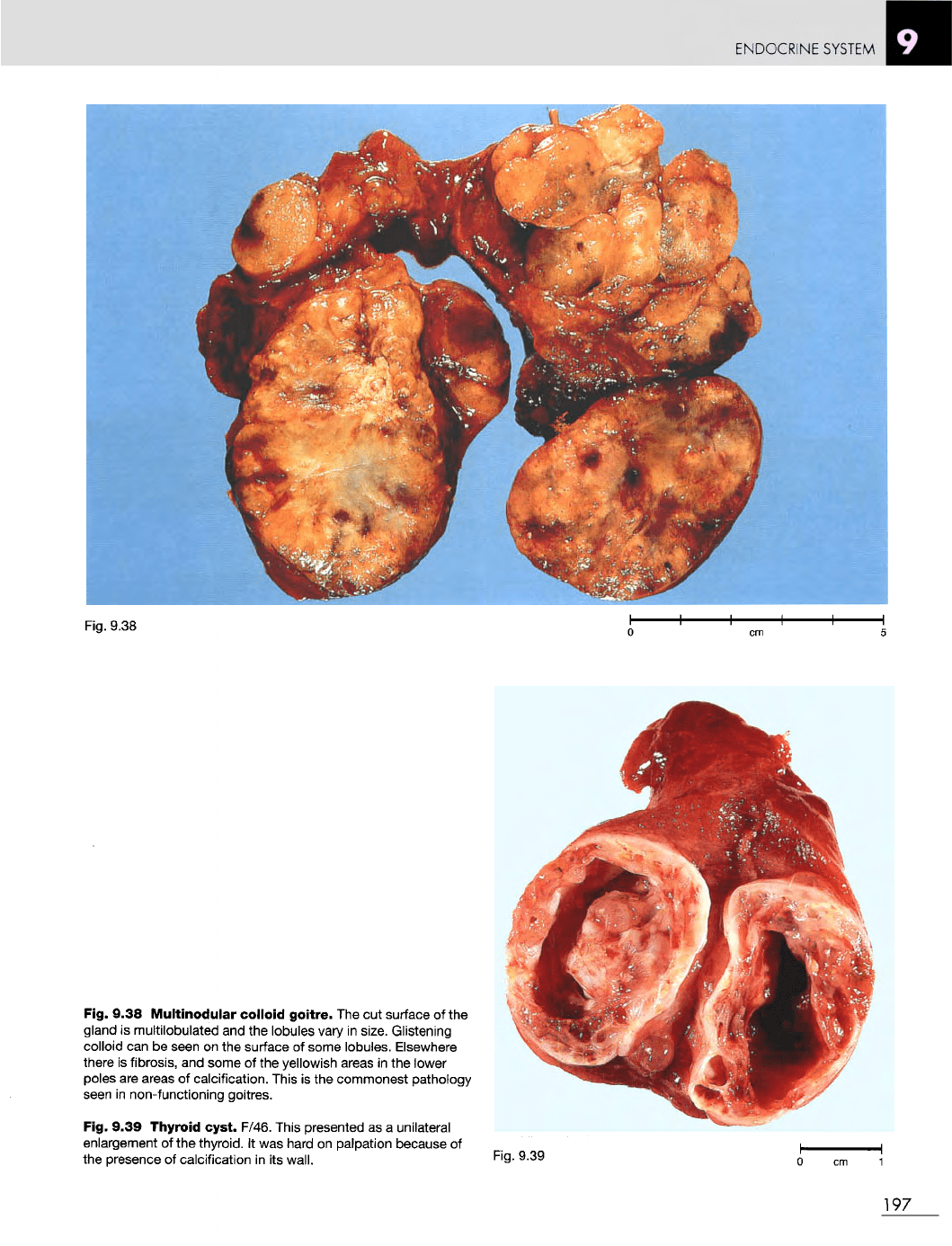
Fig. 9.34
X-ray
taken during
life
of the
spine
of the
patient
in
Figure
9.33.
The
bands
of
osteosclerosis
in the
vertebral bodies
are
clearly demonstrated.
Fig.
9.34
195
ENDOCRINE
SYSTEM
Fig. 9.35
Fig. 9.37
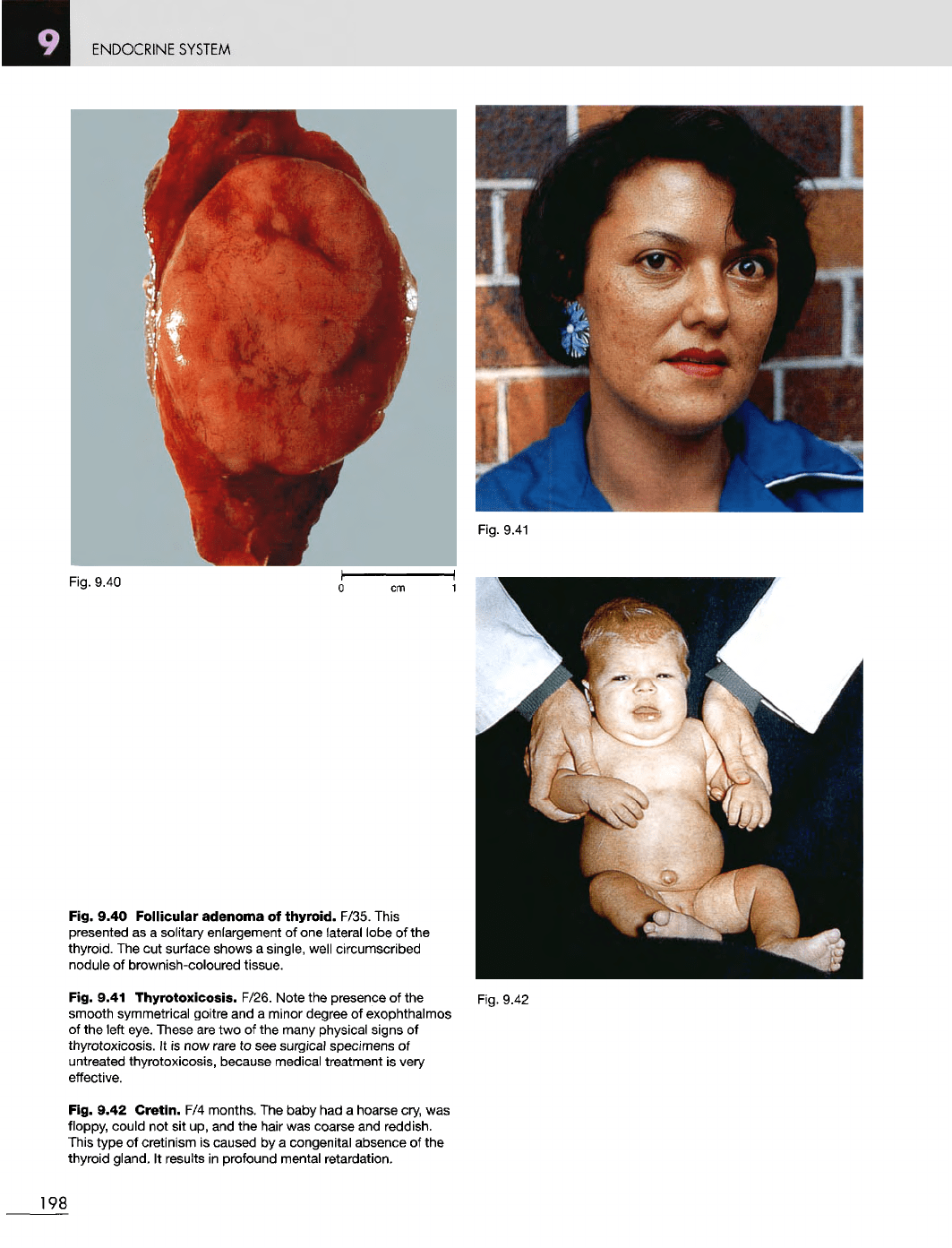
Fig. 9.35
Secondary
hyperparathyroidism.
M/40.
All
four
parathyroid glands
are
very
markedly
enlarged.
The
patient
had
chronic renal
disease
and the
hyperplastic
parathyroids were becoming
autonomous
and
were
removed.
The
surgeon left
a
small piece
of one
parathyroid gland
in
situ
in
order
to
preserve
some parathyroid function.
Fig. 9.36
The cut
surfaces
of the
parathyroids
in
Figure 9.35. This shows
the
multilobulated appearance
of the
hyperplastic parathyroid glands.
Fig. 9.37
Goitre.
F/45.
An
enlarged
thyroid gland. Non-functioning goitres
are
usually caused
by
dietary iodine
deficiency. They
are
especially common
in
the
high mountainous areas
of the
world.
196
Fig.
9.36
ENDOCRINE SYSTEM
Fig.
9.38
Fig. 9.38
Multinodular
colloid
goitre.
The cut
surface
of the
gland
is
multilobulated
and the
lobules
vary
in
size.
Glistening
colloid
can be
seen
on the
surface
of
some lobules. Elsewhere
there
is
fibrosis,
and
some
of the
yellowish areas
in the
lower
poles
are
areas
of
calcification. This
is the
commonest pathology
seen
in
non-functioning goitres.
Fig. 9.39
Thyroid
cyst.
F/46. This presented
as a
unilateral
enlargement
of the
thyroid.
It was
hard
on
palpation because
of
the
presence
of
calcification
in its
wall.
Fig.
9.39
197
ENDOCRINE
SYSTEM
Fig.
9.40
Fig. 9.40
Follicular
adenoma
of
thyroid.
F/35. This
presented
as a
solitary enlargement
of one
lateral lobe
of the
thyroid.
The cut
surface shows
a
single, well circumscribed
nodule
of
brownish-coloured tissue.
Fig. 9.41 Thyrotoxicosis. F/26. Note
the
presence
of the
smooth symmetrical goitre
and a
minor degree
of
exophthalmos
of
the
left eye. These
are two of the
many physical signs
of
thyrotoxicosis.
It is now
rare
to see
surgical specimens
of
untreated thyrotoxicosis, because medical treatment
is
very
effective.
Fig. 9.42
Cretin.
F/4
months.
The
baby
had a
hoarse cry,
was
floppy, could
not sit up, and the
hair
was
coarse
and
reddish.
This
type
of
cretinism
is
caused
by a
congenital absence
of the
thyroid
gland.
It
results
in
profound mental
retardation.
Fig.
9.42
198
Fig.
9.41
ENDOCRINE
SYSTEM
Fig. 9.45
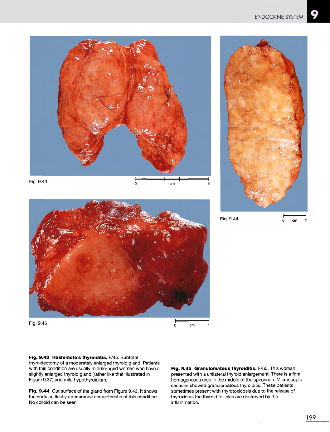
Fig. 9.43 Hashimoto's
thyroiditis.
F/45. Subtotal
thyroidectomy
of a
moderately enlarged thyroid gland. Patients
with this condition
are
usually middle-aged women
who
have
a
slightly
enlarged thyroid gland (rather like that illustrated
in
Figure
9.37)
and
mild hypothyroidism.
Fig. 9.44
Cut
surface
of the
gland from Figure 9.43.
It
shows
the
nodular, fleshy appearance characteristic
of
this condition.
No
colloid
can be
seen.
Fig. 9.45
Granulomatous
thyroiditis.
F/60. This woman
presented with
a
unilateral thyroid enlargement. There
is a
firm,
homogeneous area
in the
middle
of the
specimen. Microscopic
sections showed granulomatous thyroiditis. These patients
sometimes present with thyrotoxicosis
due to the
release
of
thyroxin
as the
thyroid follicles
are
destroyed
by the
inflammation.
199
Fig. 9.44
Fig. 9.43
ENDOCRINE
SYSTEM
Fig.
9.46
Fig.
9.47
Fig.
9.48
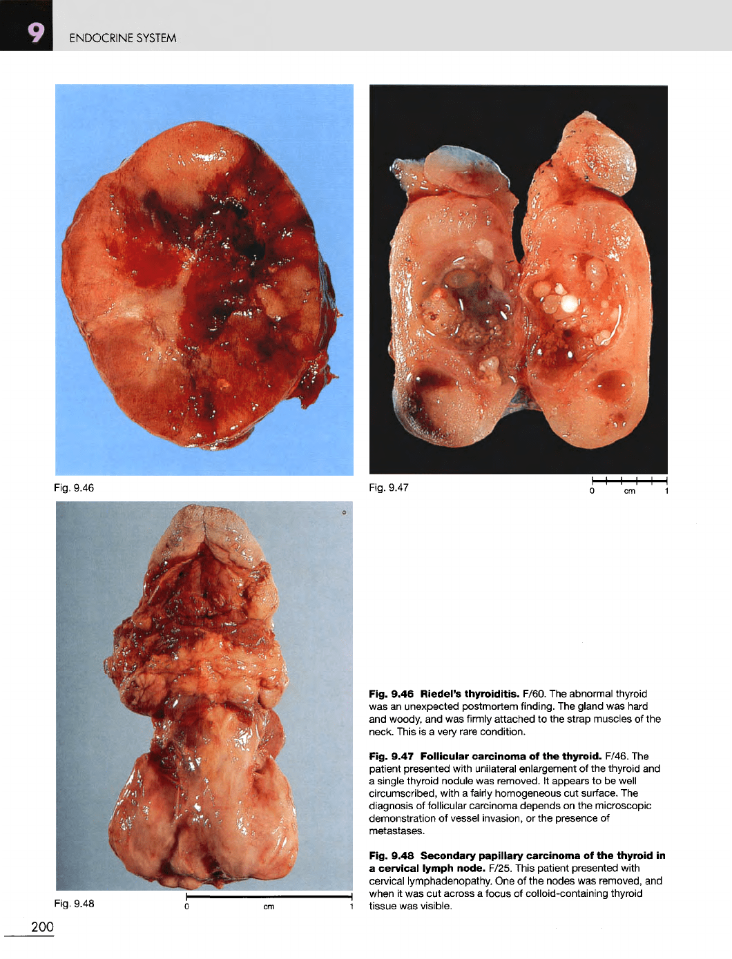
Fig. 9.46 Riedel's
thyroiditis.
F/60.
The
abnormal thyroid
was
an
unexpected postmortem finding.
The
gland
was
hard
and
woody,
and was
firmly
attached
to the
strap muscles
of the
neck. This
is a
very
rare
condition.
Fig. 9.47
Follicular
carcinoma
of the
thyroid.
F/46.
The
patient presented with unilateral enlargement
of the
thyroid
and
a
single thyroid nodule
was
removed.
It
appears
to be
well
circumscribed, with
a
fairly
homogeneous
cut
surface.
The
diagnosis
of
follicular carcinoma depends
on the
microscopic
demonstration
of
vessel invasion,
or the
presence
of
metastases.
Fig. 9.48 Secondary papillary
carcinoma
of the
thyroid
in
a
cervical
lymph node. F/25. This patient presented with
cervical
lymphadenopathy.
One of the
nodes
was
removed,
and
when
it was cut
across
a
focus
of
colloid-containing thyroid
tissue
was
visible.
200