Cook R.A., Stewart B. Colour Atlas of Anatomical Pathology
Подождите немного. Документ загружается.

NERVOUS
SYSTEM
Fig. 11.53
Effects
of
raised
intracranial
pressure
-
Figs
11.50-11.55
Fig. 11.50
Tonsillar
herniation
of the
cerebellum.
The
groove
made
by the
foramen magnum
can be
seen. There
had
been increased intracranial pressure,
and
this forced
the
cerebellar
tonsils
through
the
foramen magnum causing
pressure
on the
brain stem
-
'coning'.
Fig. 11.51
Uncal
herniation.
The
same brain
as in
Figure
11.50.
The
brain
is
viewed
on its
undersurface
and the
left
uncus
is
more prominent than
the
right.
It has
herniated medially,
causing stretching
of the
posterior communicating artery,
and
would have caused stretching
of the
sixth nerve
as
well.
The
tonsillar herniation
of the
cerebellum
is
also apparent.
Fig. 11.52
Multiple
brain-stem
haemorrhages.
Another
complication
of
raised intracranial pressure
and
frequently
the
final
cause
of
death. (Brain-stem haemorrhage
may
also result
from external trauma.)
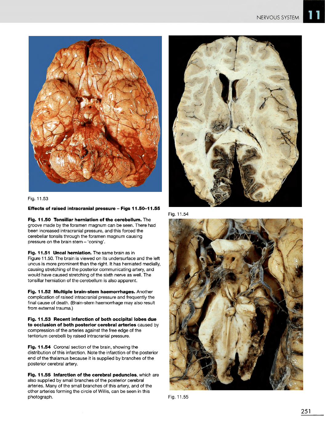
Fig. 11.53
Recent
infarction
of
both
occipital
lobes
due
to
occlusion
of
both
posterior
cerebral
arteries
caused
by
compression
of the
arteries against
the
free
edge
of the
tentorium cerebelli
by
raised intracranial pressure.
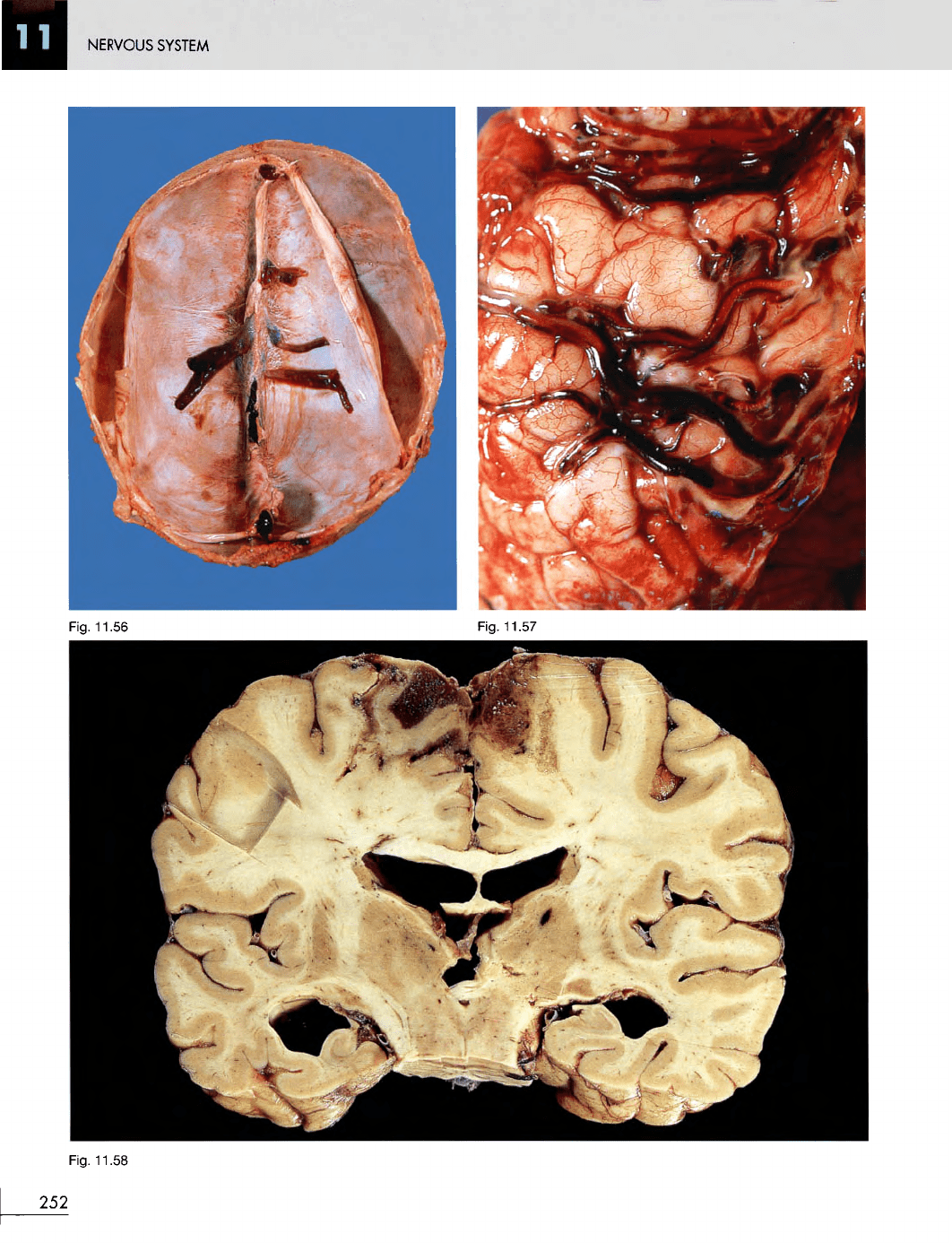
Fig. 11.54 Coronal section
of the
brain, showing
the
distribution
of
this infarction. Note
the
infarction
of the
posterior
end of the
thalamus because
it is
supplied
by
branches
of the
posterior cerebral artery.
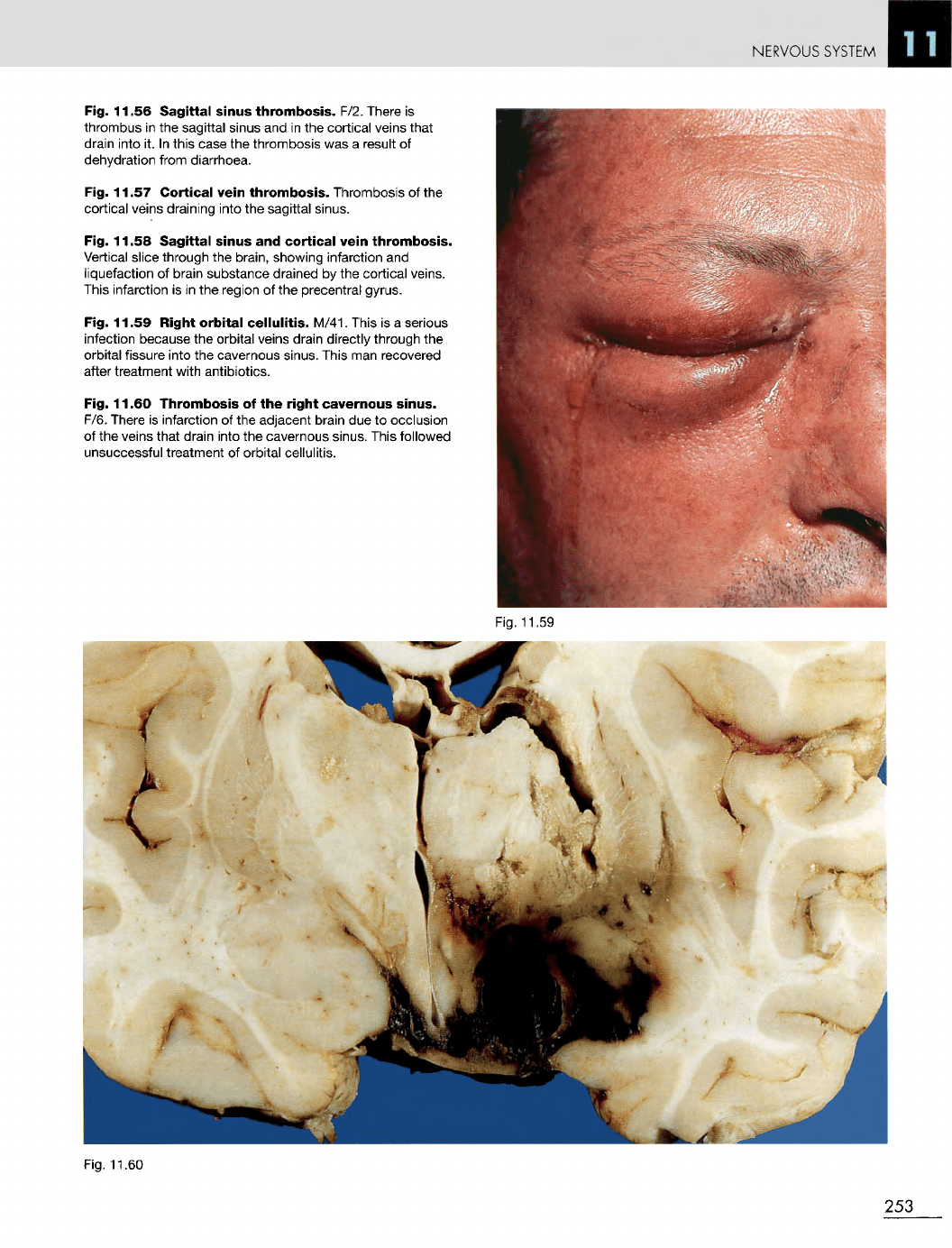
Fig. 11.55
Infarction
of the
cerebral
peduncles,
which
are
also
supplied
by
small branches
of the
posterior cerebral
arteries.
Many
of the
small branches
of
this artery,
and of the
other arteries forming
the
circle
of
Willis,
can be
seen
in
this
photograph.
Fig. 11.54
Fig. 11.55
251
NERVOUS
SYSTEM
Fig. 11.58
252
Fig.
11.56
Fig. 11.57
NERVOUS
SYSTEM
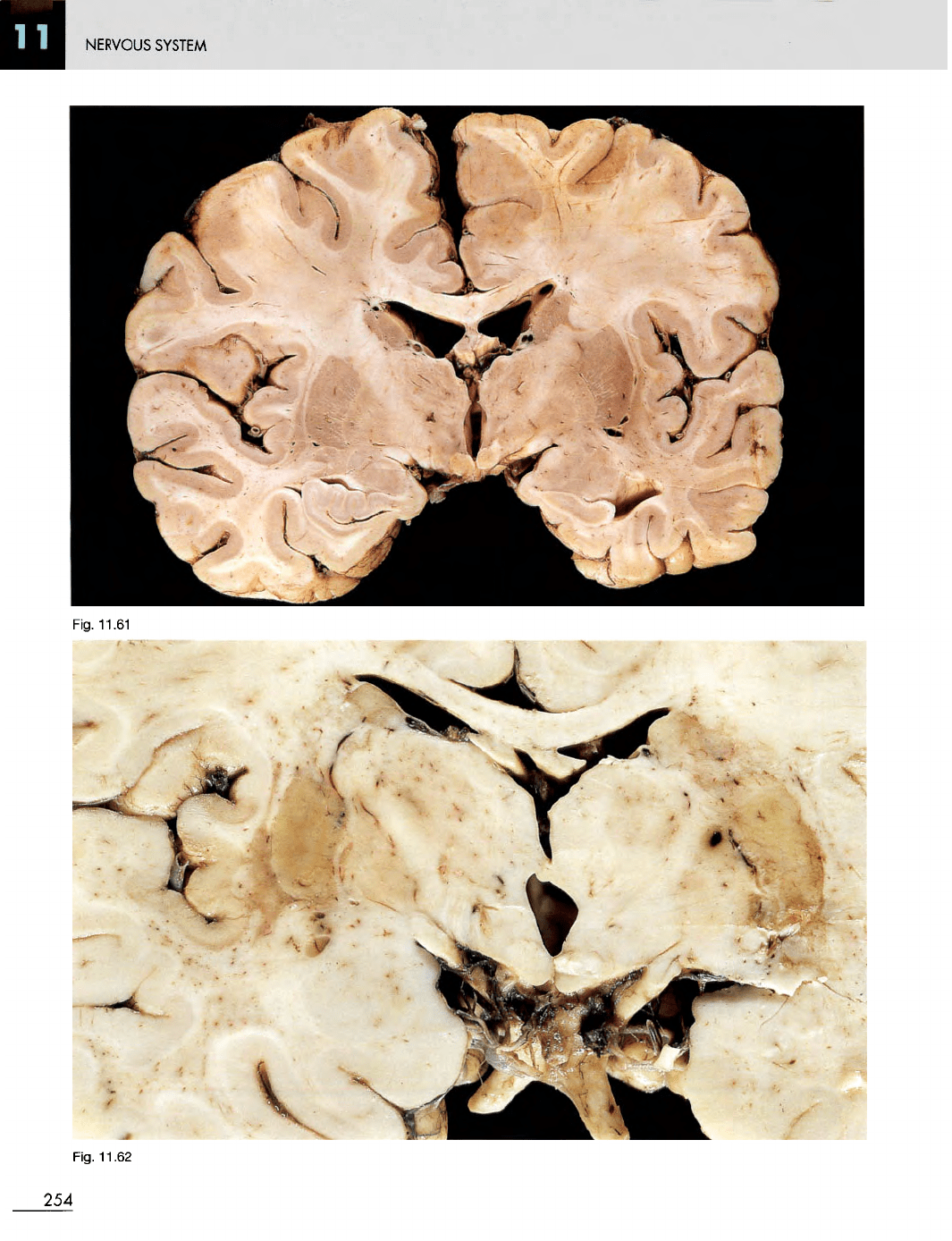
Fig. 11.56
Sagittal
sinus
thrombosis.
F/2. There
is
thrombus
in the
sagittal sinus
and in the
cortical veins that
drain
into
it. In
this case
the
thrombosis
was a
result
of
dehydration from diarrhoea.
Fig. 11.57
Cortical
vein
thrombosis.
Thrombosis
of the
cortical veins draining into
the
sagittal sinus.
Fig. 11.58
Sagittal
sinus
and
cortical
vein
thrombosis.
Vertical
slice through
the
brain, showing infarction
and
liquefaction
of
brain substance drained
by the
cortical veins.
This
infarction
is in the
region
of the
precentral gyrus.
Fig. 11.59
Right
orbital
cellulitis.
M/41. This
is a
serious
infection
because
the
orbital veins drain directly through
the
orbital fissure into
the
cavernous sinus. This
man
recovered
after
treatment with antibiotics.
Fig. 11.60
Thrombosis
of the
right
cavernous
sinus.
F/6. There
is
infarction
of the
adjacent brain
due to
occlusion
of
the
veins that drain into
the
cavernous sinus. This followed
unsuccessful treatment
of
orbital cellulitis.
Fig.
11.59
Fig.
11.60
253
NERVOUS
SYSTEM
254
Fig.
11.61
Fiq.
11.62
NERVOUS
SYSTEM
Fig.
11.63
Fig.
11.64
Fig. 11.61 Normal
brain.
Slice
of
brain showing
the
normal
internal deep cerebral veins running between
the
head
of the
caudate nucleus
and the
thalamus.
Can you
identify
all the
anatomical structures
in
this slice
of
brain?
Fig. 11.62
Deep
cerebral
vein
thrombosis.
F/3. Infarction
of
white matter
in
both hemispheres
in the
region
of the
internal
capsules resulting from thrombosis
of
branches
of the
deep
cerebral
veins. This occurred during
an
episode
of
hypotension
following open heart surgery.
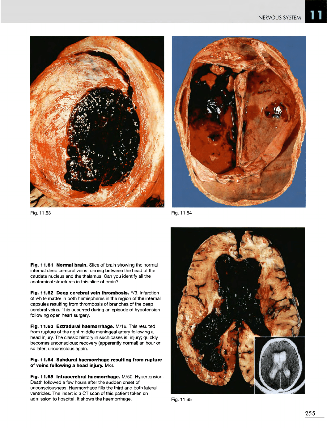
Fig. 11.63 Extradural
haemorrhage.
M/16. This resulted
from
rupture
of the
right middle meningeal artery following
a
head
injury.
The
classic history
in
such cases
is:
injury; quickly
becomes unconscious; recovery (apparently normal)
an
hour
or
so
later; unconscious again.
Fig. 11.64
Subdural
haemorrhage
resulting
from
rupture
of
veins
following
a
head
injury.
M/3.
Fig. 11.65
Intracerebral
haemorrhage.
M/50. Hypertension.
Death followed
a few
hours after
the
sudden onset
of
unconsciousness. Haemorrhage fills
the
third
and
both lateral
ventricles.
The
insert
is a CT
scan
of
this patient taken
on
admission
to
hospital.
It
shows
the
haemorrhage.
Fig. 11.65
255
NERVOUS
SYSTEM
Fig. 11.67
Fig. 11.66
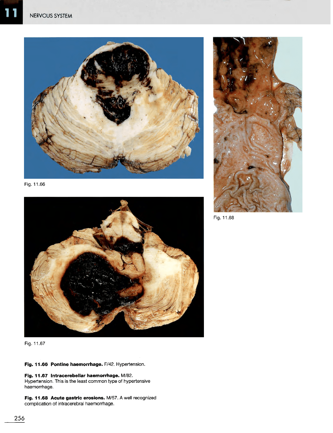
Pontine
haemorrhage.
F/42. Hypertension.
Fig. 11.67
Intracerebellar
haemorrhage.
M/82.
Hypertension. This
is the
least common type
of
hypertensive
haemorrhage.
Fig. 11.68
Acute
gastric
erosions.
M/57.
A
well recognized
complication
of
intracerebral haemorrhage.
256
Fig.
11.66
Fig. 11.68
NERVOUS SYSTEM
Fig. 11.69
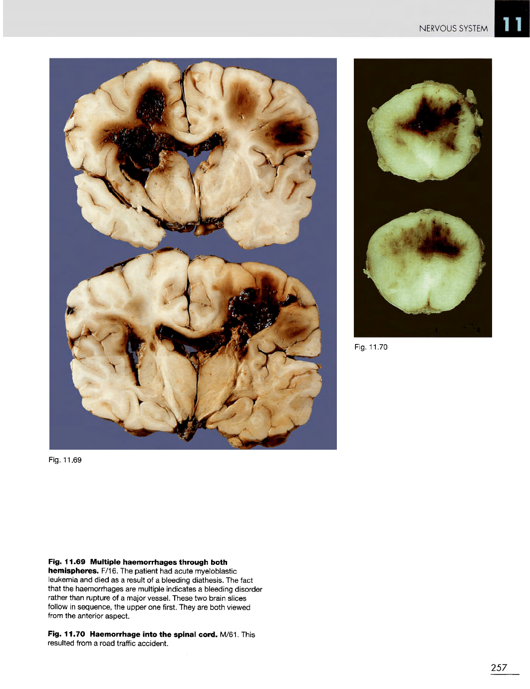
Fig. 11.69
Multiple
haemorrhages
through
both
hemispheres.
F/16.
The
patient
had
acute myeloblastic
leukemia
and
died
as a
result
of a
bleeding diathesis.
The
fact
that
the
haemorrhages
are
multiple indicates
a
bleeding disorder
rather
than rupture
of a
major vessel. These
two
brain slices
follow
in
sequence,
the
upper
one
first. They
are
both viewed
from
the
anterior aspect.
Fig. 11.70 Haemorrhage
into
the
spinal
cord.
M/61. This
resulted from
a
road traffic accident.
257
Fig.
11.70
NERVOUS
SYSTEM
Fig. 11.71
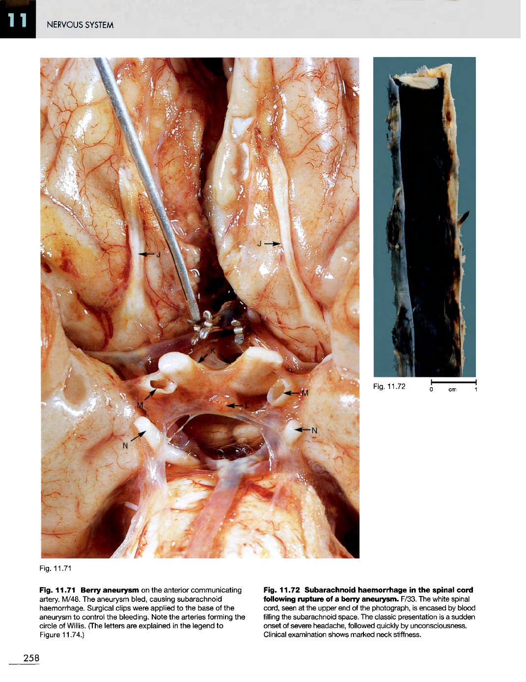
Fig. 11.71 Berry
aneurysm
on the
anterior communicating
artery.
M/48.
The
aneurysm
bled,
causing subarachnoid
haemorrhage. Surgical clips were applied
to the
base
of the
aneurysm
to
control
the
bleeding. Note
the
arteries forming
the
circle
of
Willis. (The letters
are
explained
in the
legend
to
Figure
11.74.)
Fig. 11.72
Subarachnoid
haemorrhage
in the
spinal cord
following
rupture
of a
berry
aneurysm.
F/33.
The
white spinal
cord,
seen
at the
upper
end of the
photograph,
is
encased
by
blood
filling
the
subarachnoid space.
The
classic presentation
is a
sudden
onset
of
severe headache, followed quickly
by
unconsciousness.
Clinical examination shows marked neck stiffness.
258
Fig.
11.72
NERVOUS
SYSTEM
Fig.
11.73
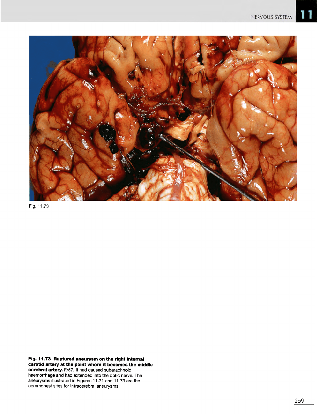
Fig. 11.73 Ruptured aneurysm
on the
right
internal
carotid
artery
at the
point
where
it
becomes
the
middle
cerebral
artery- F/57.
It had
caused subarachnoid
haemorrhage
and had
extended into
the
optic nerve.
The
aneurysms
illustrated
in
Figures 11.71
and
11.73
are the
commonest sites
for
intracerebral aneurysms.
259
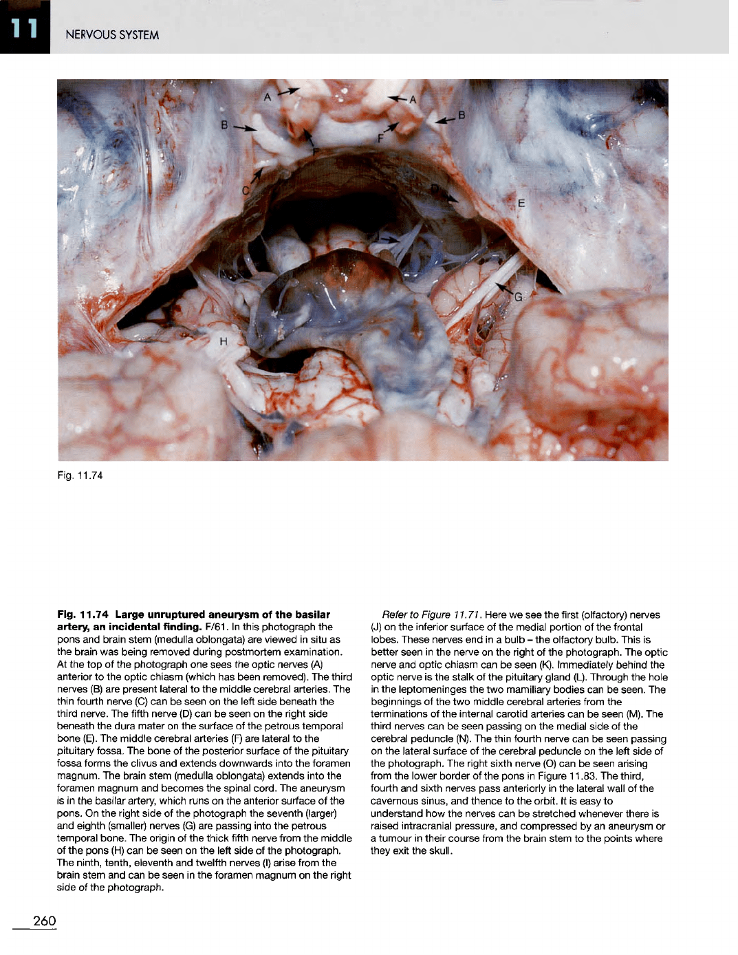
Fig.
11.74
Fig. 11.74
Large
unruptured
aneurysm
of the
basilar
artery,
an
incidental
finding.
F/61.
In
this photograph
the
pons
and
brain stem (medulla oblongata)
are
viewed
in
situ
as
the
brain
was
being removed during postmortem examination.
At
the top of the
photograph
one
sees
the
optic
nerves
(A)
anterior
to the
optic chiasm (which
has
been removed).
The
third
nerves
(B) are
present lateral
to the
middle cerebral arteries.
The
thin fourth nerve
(C) can be
seen
on the
left side beneath
the
third nerve.
The
fifth nerve
(D) can be
seen
on the
right side
beneath
the
dura mater
on the
surface
of the
petrous temporal
bone (E).
The
middle cerebral arteries
(F) are
lateral
to the
pituitary
fossa.
The
bone
of the
posterior
surface
of the
pituitary
fossa forms
the
clivus
and
extends downwards into
the
foramen
magnum.
The
brain stem (medulla oblongata) extends into
the
foramen
magnum
and
becomes
the
spinal cord.
The
aneurysm
is
in the
basilar
artery, which
runs
on the
anterior
surface
of the
pons.
On the
right side
of the
photograph
the
seventh
(larger)
and
eighth (smaller) nerves
(G) are
passing into
the
petrous
temporal bone.
The
origin
of the
thick fifth nerve from
the
middle
of
the
pons
(H) can be
seen
on the
left side
of the
photograph.
The
ninth, tenth, eleventh
and
twelfth nerves
(I)
arise from
the
brain stem
and can be
seen
in the
foramen magnum
on the
right
side
of the
photograph.
Refer
to
Figure
11.71.
Here
we see the
first (olfactory) nerves
(J)
on the
inferior surface
of the
medial portion
of the
frontal
lobes. These nerves
end in a
bulb
- the
olfactory
bulb.
This
is
better seen
in the
nerve
on the
right
of the
photograph.
The
optic
nerve
and
optic
chiasm
can be
seen (K).
Immediately
behind
the
optic nerve
is the
stalk
of the
pituitary gland (L). Through
the
hole
in
the
leptomeninges
the two
mamillary bodies
can be
seen.
The
beginnings
of the two
middle cerebral arteries from
the
terminations
of the
internal carotid arteries
can be
seen (M).
The
third nerves
can be
seen passing
on the
medial side
of the
cerebral peduncle (N).
The
thin fourth nerve
can be
seen passing
on
the
lateral
surface
of the
cerebral
peduncle
on the
left side
of
the
photograph.
The
right sixth
nerve
(O) can be
seen arising
from
the
lower border
of the
pons
in
Figure 11.83.
The
third,
fourth
and
sixth nerves pass anteriorly
in the
lateral wall
of the
cavernous
sinus,
and
thence
to the
orbit.
It is
easy
to
understand
how the
nerves
can be
stretched whenever there
is
raised intracranial pressure,
and
compressed
by an
aneurysm
or
a
tumour
in
their course from
the
brain stem
to the
points where
they exit
the
skull.
260
NERVOUS SYSTEM