Cui Dongmei. Atlas of Histology: with functional and clinical correlations. 1st ed
Подождите немного. Документ загружается.

CHAPTER 13
■
Integumentary System
255
Development of the Skin
SYNOPSIS 13-2 Pathological Terms for the Integumentary System
Acanthosis ■ : Thickening of the stratum spinosum of the epidermis, typically seen in epidermal hyperplasia.
Hypergranulosis
■ : Thickening and prominence of the stratum granulosum of the epidermis, often in response to chronic
mechanical irritation of the skin. Hypergranulosis may also be seen in the declivities of papillary lesions such as verruca
vulgaris, or warts.
Hyperkeratosis
■ : Thickening of the stratum corneum of the epidermis. Orthokeratotic hyperkeratosis refers to hyperkeratosis
without the presence of nuclei.
Papillomatosis
■ : Fingerlike projections from the epidermal surface, often with hyperkeratosis, seen in a variety of conditions
including verruca vulgaris, or warts.
Parakeratosis
■ : A form of hyperkeratosis in which nuclei are retained in the stratum corneum, seen in many conditions
including psoriasis.
Spongiosis
■ : Intercellular edema of the epidermis frequently seen in various etiologies of dermatitis such as allergic contact
dermatitis or irritant dermatitis.
Ulceration
■ : The discontinuity of an epithelial surface including the epidermis or mucous membranes.
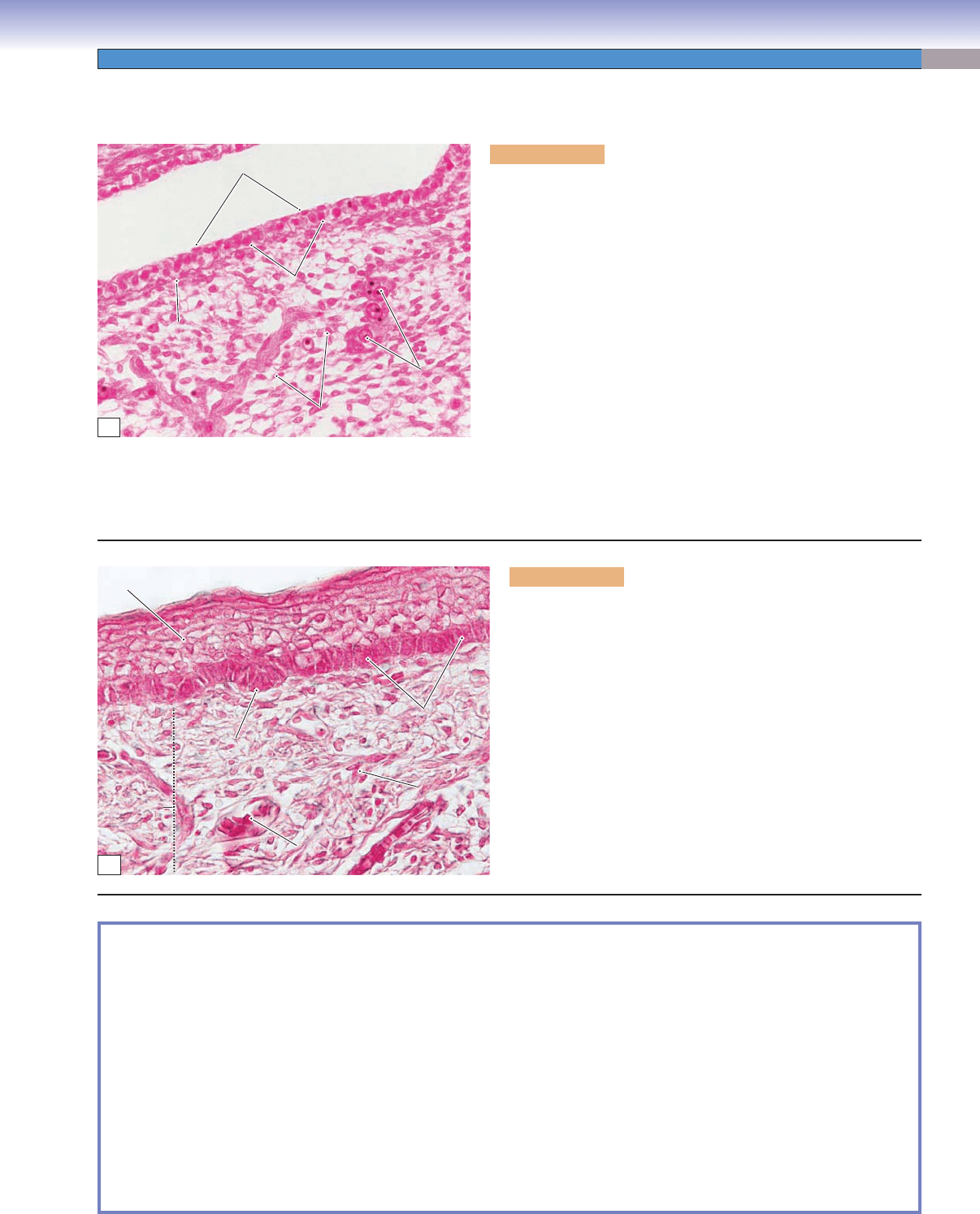
Figure 13-12A. Fetal skin (5–9 weeks). H&E, 331
The two layers (epidermis and dermis) of the skin develop from two
different embryonic tissues. The epidermis develops from the ecto-
derm, and the dermis from the mesoderm. About 4 weeks after con-
ception, the human embryo is covered by a single layer of ectodermal
cells, which are loosely arranged on the basement membrane and
over the mesenchymal tissue. After 5 weeks, the epidermis has two
layers of cells: the superfi cial layer (periderm) and basal layers. At
2 to 3 months, basal cells are dividing rapidly, and the epidermis
becomes several cell layers thick. At the same time, the mesenchyme
differentiates into a more mature connective tissue with blood
vessels. By 4 months, neural crest cells migrate into the basal layer of
the epidermis and differentiate into melanocytes and Merkel cells.
The connective tissue layer beneath the epithelium develops into
the dermis and a deeper layer, the hypodermis. At about 5 months,
the appendages of the skin (hair follicles and glands) start to form.
This section shows an early stage of skin development in an embryo.
There are only two layers of epithelial cells in the epidermis, and fetal
blood vessels are located within the mesenchyme tissue. Nucleated
erythrocytes are shown inside the blood vessels.
Periderm
Periderm
Periderm
Basement
Basement
membrane
membrane
Basement
membrane
Mesenchymal
Mesenchymal
tissue
tissue
Mesenchymal
tissue
Nucleated
Nucleated
erythrocytes
erythrocytes
Nucleated
erythrocytes
Mesenchymal
Mesenchymal
cells
cells
Mesenchymal
cells
Basal layer
Basal layer
Basal layer
A
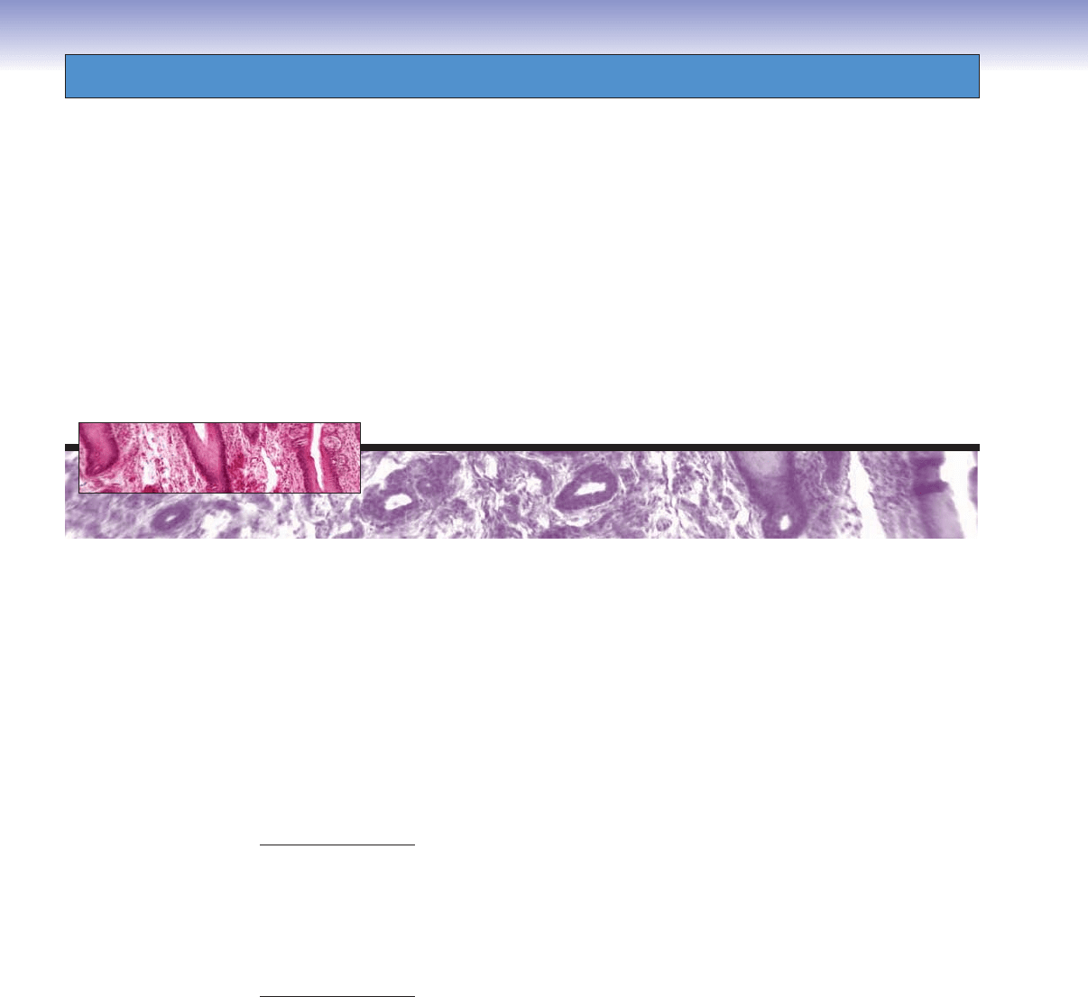
Figure 13-12B. Fetal skin (fi fth month). H&E, 438
This section shows a later stage of fetal skin development. The
epidermis has formed multiple cell layers, and four layers of epi-
dermis can be distinguished to some extent. The basal cells in
the stratum basale layer are highly active and appear as column-
shaped cells. The underlying mesenchymal tissue has differentiated
into connective tissue (dermis). There are many active fi broblasts
in the dermis. An accumulation of basal cells forms a fold called
an epidermal bud, which projects into the dermis. These accu-
mulated cells will interact with the dermis and differentiate into
appendages (hair follicles and glands).
Epidermis
Epidermis
Epidermis
Dermis
Dermis
Dermis
Accumulated
Accumulated
basal cells
basal cells
(epidermal bud)
(epidermal bud)
Accumulated
basal cells
(epidermal bud)
Erythrocytes in
Erythrocytes in
the blood vessels
the blood vessels
Erythrocytes in
the blood vessels
Basal cells
Basal cells
Basal cells
Fibroblasts
Fibroblasts
Fibroblasts
B
CUI_Chap13.indd 255 6/2/2010 8:21:35 AM
14
256
Oral Cavity
Oral Mucosa
Introduction and Key Concepts for Oral Mucosa
Figure 14-1 Overview of the Oral Mucosa and Teeth
Lining Mucosa
Figure 14-2A Overview of the Lip
Figure 14-2B Skin, Lip
Figure 14-2C Vermilion Zone, Lip
Figure 14-2D Labial Mucosa (Lining Mucosa), Lip
Figure 14-3A Buccal Mucosa (Lining Mucosa), Cheek
Figure 14-3B Clinical Correlation: Oral Submucous Fibrosis of the Lip
Table 14-1 Comparison of Lining and Masticatory Mucosae
Masticatory Mucosa
Figure 14-4A Masticatory Mucosa, Gingiva
Figure 14-4B Masticatory Mucosa, Hard Palate
Figure 14-4C Clinical Correlation: Nicotine Stomatitis
Specialized Mucosa
Figure 14-5A Overview of the Tongue
Figure 14-5B Filiform Papillae, Tongue
Figure 14-5C Fungiform Papillae, Tongue
Figure 14-6A Circumvallate Papillae, Tongue
Figure 14-6B Foliate Papillae and Taste Buds, Tongue
Table 14-2 Comparison of Lingual Papillae
Teeth
Introduction and Key Concepts for Teeth
Figure 14-7 Overview of the Teeth
CUI_Chap14.indd 256 6/2/2010 8:22:47 AM

CHAPTER 14
■
Oral Cavity
257
Tooth Development (Odontogenesis)
Figure 14-8 Overview of Tooth Development (Odontogenesis)
Figure 14-9A Bud Stage, Weeks 8–9
Figure 14-9B Cap Stage, Weeks 10–11
Figure 14-10A Bell Stage, Weeks 12–14
Figure 14-10B Bell Stage, Cell Layers
Figure 14-11A Apposition (Crown) Stage, Dentinogenesis
Figure 14-11B Apposition (Crown) Stage, Amelogenesis
Figure 14-12A Tooth Root Development
Figure 14-12B Tooth Eruption
Figure 14-12C Clinical Correlation: Dilaceration
Figure 14-13A Clinical Correlation: Gemination, Incisor
Figure 14-13B Clinical Correlation: Amelogenesis Imperfecta
Figure 14-13C Clinical Correlation: Enamel Pearl
Enamel, Dentin, and Dental Pulp
Figure 14-14A,B Enamel, Tooth
Figure 14-14C Clinical Correlation: Enamel Fluorosis
Figure 14-15A,B Dentin, Tooth
Figure 14-15C Clinical Correlation: Dentinogenesis Imperfecta
Figure 14-16A Clinical Correlation: Vitamin D–Resistant Rickets
Figure 14-16B Clinical Correlation: Dentin Dysplasia
Table 14-3 Dental Hard Tissue
Figure 14-17A,B Dental Pulp
Figure 14-17C Clinical Correlation: Pulp Abscess
Periodontium
Figure 14-18A Acellular Cementum, Cervical Region of Tooth Root
Figure 14-18B Cellular Cementum, Apical Region of Tooth Root
Figure 14-18C Periodontal Ligament and Alveolar Bone
Figure 14-19A Periodontal Ligament and Alveolar Bone, Tooth Root
Figure 14-19B Clinical Correlation: Tooth Ankylosis
Synopsis 14-1 Pathological and Clinical Terms for the Oral Cavity
Oral Mucosa
Introduction and Key Concepts for Oral
Mucosa
The oral cavity refers to the internal part of the mouth and can
be divided into the oral vestibule and the oral cavity proper.
The oral vestibule is the space between the inner lips, cheeks,
and front surface of the teeth. The oral cavity proper is the space
between the upper and lower dental arches, extending from the
inner surface of the teeth to the oropharynx. The structures
inside of the oral cavity include the lips, cheeks, tongue, teeth,
gingiva, palates (hard and soft), salivary glands, and tonsils. The
tonsils are discussed in Chapter 10, “Lymphoid System,” and
salivary glands are discussed in Chapter 16, “Digestive Glands
and Associated Organs.” The structures in the oral cavity are
lined by an oral mucosa, which includes an overlying epithe-
lium and underlying connective tissue. The oral mucosa can be
divided into three types based on differences in the epithelial
covering, organization of the connective tissue, and associated
functions: lining, masticatory, and specialized mucosa.
LINING MUCOSA is covered by nonkeratinized stratifi ed
squamous epithelium with two distinct layers: the stratum basale
and stratum spinosum. The epithelium of the lining mucosa is
similar to the epidermis of the skin, except that it has neither
a stratum corneum nor a stratum lucidum, and the stratum
granulosum is often absent (see Chapter 13, “Integumentary
System,” Figs. 13-2 and 13-3B). The nonkeratinized stratifi ed
epithelium is moistened by saliva. The connective tissues of the
lining mucosa can be divided into the lamina propria and the
submucosa. The lamina propria is a thin layer of loose connec-
tive tissue containing many elastic fi bers and relatively few col-
lagen fi bers. This layer is equivalent to the dermis of the skin
and is located beneath the epithelium. The submucosa is a thick
layer of connective tissue, which contains minor salivary
CUI_Chap14.indd 257 6/2/2010 8:22:53 AM

258
UNIT 3
■
Organ Systems
glands and is attached to the underlying muscle. The lining
mucosa covers the inner oral surfaces of the lips, cheeks, soft
palate, the inferior surface of the tongue, and the fl oor of the
mouth. This type of mucosa is less exposed to abrasion than
the masticatory mucosa. The lining mucosa provides a bar-
rier against the invasion of pathogens and toxic chemicals,
contains receptors for sensations, and serves immunological
functions. The lining mucosa also provides lubrication and
buffering by minor glands in the submucosal layer. Examples
of the lining mucosa include the lip (Fig. 14-2D) and cheek
(Fig. 14-3A).
MASTICATORY MUCOSA is covered by keratinized strati-
fi ed squamous epithelium, which is exposed to signifi cant abra-
sion due to high compression and friction during chewing.
The epithelium of the masticatory mucosa is composed of the
stratum basale, stratum spinosum, stratum granulosum, and
stratum corneum. It has a thick lamina propria that contains a
dense network of collagen fi bers and a few elastic fi bers. This
layer has no submucosa and is directly and fi rmly attached to
the underlying bone. Masticatory mucosa can be found cover-
ing the oral surfaces of the gingiva and the hard palate. Injec-
tion into this area is diffi cult and painful because of its sensitive
periosteum, high collagen density, and fi rm attachment to the
bone. See Figure 14-4A for examples of the gingiva and Figure
14-4B for the hard palate.
SPECIALIZED MUCOSA covers the anterior two thirds of
the tongue and consists of keratinized and nonkeratinized
squamous epithelium and numerous papillae. These papillae
can be classifi ed into four types: fi liform, fungiform, circum-
vallate, and foliate papillae. Most of these papillae have taste
buds. The fi liform papillae are the only papillae without taste
buds; their main function is to aid in mixing food during chew-
ing. The lamina propria (connective tissue) of the specialized
mucosa is attached to the underlying skeletal muscle. These
muscles produce voluntary movement of the tongue and are
innervated by the hypoglossal nerve (cranial nerve [CN] XII).
Lining mucosa covers the inferior surface of the tongue. The
mucosa of the tongue is divided into two parts by a V-shaped
groove called the sulcus terminalis. The anterior two thirds of
the tongue is referred to as the body of the tongue. Its mucosa
is innervated by the facial nerve (CN VII) and the trigeminal
nerve (CN V). The posterior third of the tongue is the base of
the tongue. Its taste buds and mucosa are innervated by the
glossopharyngeal nerve (CN IX). The posterior third of the
tongue contains the lingual tonsils (Fig. 14-5A).
1. Filiform papillae are the smallest and most numerous of the
four types of papillae. They cover almost the entire supe-
rior surface of the anterior two thirds of the tongue and
are packed in rows that parallel the sulcus terminalis. Each
of the papillae appears cone shaped with some branching
processes. Connective tissue forms the central core of each
papilla. Filiform papillae have no taste buds and extend
from the nonkeratinized stratifi ed squamous epithelium.
The surface of the papilla is keratinized and is exposed to a
great deal of abrasion (Fig. 14-5B).
2. Fungiform papillae are less numerous than the fi liform
papillae. They are mushroom shaped and are scattered
among the fi liform papillae (Fig. 14-5C). Fungiform papil-
lae are located at the tip and on the two lateral edges of the
tongue. They are more numerous near the tip of the tongue.
Taste buds are found on the apical surfaces of fungiform
papillae.
3. Circumvallate papillae are large and round with a fl at-
topped cylindrical structure. There are about 10 to 14
papillae arranged in a row along the sulcus terminalis. Each
papilla is surrounded by a deep groove (moat), which forms
a valley around the papilla. Taste buds are found in the lat-
eral walls of each papilla (Fig. 14-6A).
4. Foliate papillae are leafl ike folds with fl at tops and have deep
clefts between the papillae. They are located on the posterior
lateral surface of the tongue. They are more prominent in
some animals (such as rabbits) than in humans. Foliate
papillae contain taste buds in the lateral walls of the papillae
(Fig. 14-6B).
CUI_Chap14.indd 258 6/2/2010 8:22:53 AM
CHAPTER 14
■
Oral Cavity
259
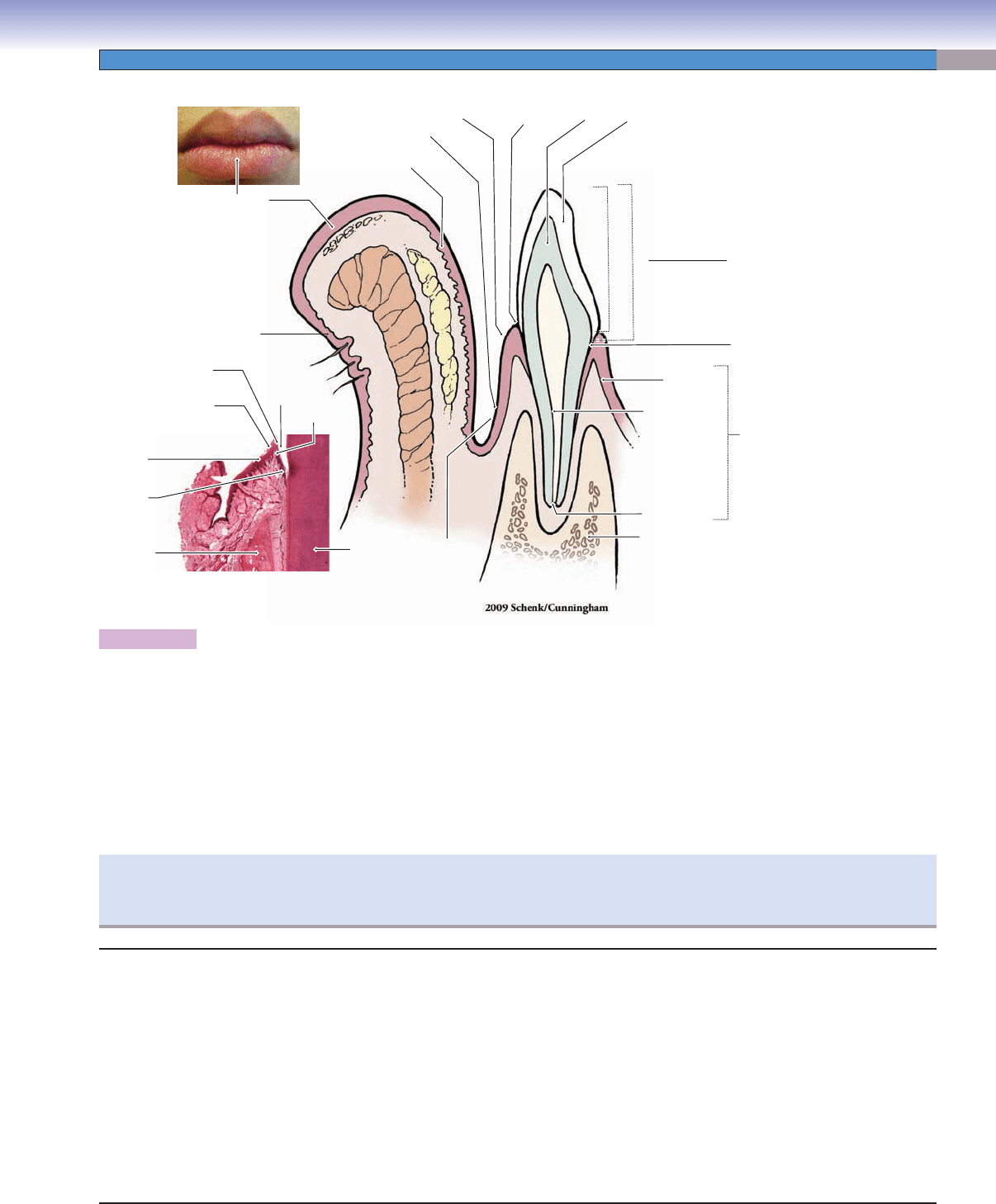
The oral cavity is lined by oral mucosa, which can be divided into masticatory mucosa (gingiva, hard palate), lining mucosa (lips,
soft palate, cheeks, inferior surface of the tongue, fl oor of the mouth), and specialized mucosa (tongue). This illustration represents
the lip and a tooth and the mucosa covering these structures. The lip is covered externally with skin to the vermilion zone (inter-
mediate zone or mucocutaneous junction); the vermilion zone continues to the labial mucosa, which is the lining mucosa on the
internal surface of the lip. The alveolar process of the jaw (containing the tooth roots) is covered by alveolar mucosa (lining mucosa)
and gingiva. The junction between the lining mucosa and gingiva is the mucogingival junction. The tooth can be divided into three
parts: crown, cervix, and root. The crown is the part of the tooth projecting into the oral cavity and has two different defi nitions:
The clinical crown is the part of the crown which is visible in the mouth; the anatomical crown is the part of the tooth covered by
enamel. The root is covered by the gingiva or is inside the bony socket. The region between the crown and root is the cervix (neck).
The gingival sulcus is the space between the free gingiva and the enamel; it is normally 0.5 to 3.0 mm in depth.
Alveolar (lining)
Alveolar (lining)
mucosa
mucosa
Alveolar (lining)
mucosa
Dentin
Dentin
Dentin
Apical foramen
Apical foramen
Apical foramen
Alveolar bone
Alveolar bone
(process)
(process)
Alveolar bone
(process)
Cervix
Root
Crown
Gingiva
Gingiva
Gingiva
Dental pulp
Dental pulp
Dental pulp
A
A
n
n
a
a
to
to
m
m
ic
ic
a
a
l c
l
c
ro
ro
w
w
n
n
Anatomic
a
l crown
Clinical crown
Clinical
crown
Clinical crown
Enamel
Dentin
Labial (lining)
Labial (lining)
mucosa
mucosa
Labial (lining)
mucosa
Dentogingival
junction
Mucogingival
junction
Gingiva
(masticatory mucosa)
Vermilion zone
(intermediate zone
mucocutaneous
junction)
Skin
Enamel
Enamel
space
space
Enamel
space
Sulcus
Sulcus
epithelium
epithelium
Sulcus
epithelium
Free gingiva
Free gingiva
Free gingiva
Gingiva sulcus
Gingiva sulcus
Gingiva sulcus
Gingival
epithelium
Junctional
epithelium
Alveolar
bone
labial sulcus
la
b
ial su
lc
us
Labial sulcus
Figure 14-1. Overview of the oral mucosa and teeth. Lower left, H&E, 18
If the depth of the gingival sulcus is over 3 mm, these spaces are called gingival or periodontal pockets. These pockets represent
an abnormal condition. An accumulation of debris and microbes in the pockets may cause damage to the periodontal ligament
(PDL). For tooth details, see Figure 14-7.
I. Lining mucosa (covering of inner surface of the lips and
cheeks, soft palate, inferior surface of the tongue, and fl oor
of the mouth)
A. Epithelium: nonkeratinized stratifi ed squamous epithelium
B. Lamina propria: connective tissue with many elastic fi bers
and few collagen fi bers
C. Submucosa: connective tissue with minor salivary glands
and their ducts
II. Masticatory mucosa (covering of gingiva and hard palate)
A. Epithelium: keratinized stratifi ed squamous epithelium
B. Lamina propria: connective tissue with few elastic fi bers
and many dense collagen fi bers
C. No submucosa
III. Specialized mucosa (tongue)
A. Filiform papillae: no taste buds
B. Fungiform papillae: taste buds on apical surface of the
papilla—sweet, sour, salty
C. Circumvallate papillae: taste buds in lateral wall of the
papilla—bitter
D. Foliate papillae: taste buds in lateral wall of papilla
Structures of the Oral Mucosa
CUI_Chap14.indd 259 6/2/2010 8:22:53 AM
260
UNIT 3
■
Organ Systems
Lining Mucosa
Figure 14-2A. Overview of the lip, H&E, 7.6
Lining mucosa is a wet mucosa that covers the inside of the mouth
and is lined by nonkeratinized stratifi ed squamous epithelium.
The labial mucosa of the lip is an example of lining mucosa. The
lips are soft, fl exible, and movable; they play important roles in
food intake, speech, and as a sensory organ (e.g., kissing). Lips
can be divided into three regions: (1) thin skin, forming the exter-
nal surface of the lip; (2) vermilion zone, appearing red in color,
also called the intermediate zone or mucocutaneous junction;
and (3) the labial mucosa (lining mucosa), the internal surface
of the lip. The central core of the lip contains the orbicularis oris
(skeletal) muscle, which is innervated by the facial nerve (CN
VII), and contributes to lip movement and facial expressions.
Labial
(lining)
mucosa
Fig.
14-2D
Vermilion
zone
Minor
salivary gland
Fig.
14-2C
Vermilion
zone
Skin
Fig.
14-2B
O
O
rb
rb
ic
ic
u
u
la
la
ris
ris
o
o
ris
ris
m
m
u
u
s
s
c
c
le
le
Orbicularis
oris muscle
Orbicularis
Orbicularis
oris muscle
oris muscle
Orbicularis
oris muscle
A
Sweat
Sweat
glands/ducts
glands/ducts
Sweat
glands/ducts
Sebaceous
Sebaceous
glands
glands
Sebaceous
glands
Sebaceous
Sebaceous
glands
glands
Sebaceous
glands
Sweat
Sweat
glands/ducts
glands/ducts
Sweat
glands/ducts
Hair
follicle
space
Hair
follicle
Epidermis
Dermis
Dermis
Dermis
B
Figure 14-2B. Skin, lip. H&E, 33; inset 44
An example of the skin on the external surface of the lip is shown.
It is covered by keratinized stratifi ed squamous epithelium. The
sebaceous glands in the dermis are associated with hair follicles,
and sweat glands are present. The skin of the lip is like thin skin
elsewhere and can be divided into epidermis and dermis.
C
Ducts of sebaceous-like
Ducts of sebaceous-like
glands
glands
Ducts of sebaceous-like
glands
Fordyce granules/spots
Fordyce granules/spots
ebaceous-like glands
ebaceous-like glands
(s
(s
)
)
Fordyce granules/spots
ebaceous-like glands(s )
Parakeratinized stratified
squamous epithelium
Nonkeratinized stratified
Nonkeratinized stratified
squamous epithelium
squamous epithelium
Nonkeratinized stratified
squamous epithelium
Ducts of minor
Ducts of minor
salivary glands
salivary glands
Ducts of minor
salivary glands
Stratum
Stratum
spinosum
spinosum
Stratum
spinosum
Stratum
Stratum
basale
basale
Stratum
basale
Minor
Minor
salivary
salivary
glands
glands
Minor
salivary
glands
D
Figure 14-2C. Vermilion zone, lip. H&E, 33
The vermilion zone of the lip is covered by parakeratinized strati-
fi ed squamous epithelium. Sebaceous-like glands (Fordyce gran-
ules or spots) may be found in the connective tissue and are not
associated with hair follicles. These glands have ducts that release
their oily product directly onto the surface of the lip. The ver-
milion zone appears red because of many blood vessels near the
surface of the thin and translucent epithelium (Fig. 14-1). This
region can become thick and forms the sucking pad in infants.
Figure 14-2D. Labial mucosa (lining mucosa), lip. H&E, 33
The labial mucosa of the lip is an example of lining mucosa,
which is covered by nonkeratinized stratifi ed squamous epithe-
lium and contains many elastic fi bers; it is very fl exible and can
be stretched. Its submucosa layer contains many minor salivary
glands (mucous glands). The minor salivary glands in the lips are
often called labial glands.
CUI_Chap14.indd 260 6/2/2010 8:22:55 AM
CHAPTER 14
■
Oral Cavity
261
Name of
Mucosa
Epithelium Lamina Propria Submucosa Covering
Region
Special
Features
Clinical Application
Lining
mucosa
Nonkeratinized
stratifi ed
squamous
epithelium
Few collagen
fi bers; many
elastic fi bers
Well-developed
submucosa;
attached
primarily to
underlying muscle
rather than
bone (except for
alveolar mucosa)
Lips, cheeks,
soft palate,
inferior surface
of the tongue,
and fl oor of the
mouth
Thick, soft,
and loose;
fl exible
and can be
stretched
Injections are easy to
make and minimally
painful; if cut, a gap
appears that requires
suturing; infection
spreads quickly
Masticatory
mucosa
Keratinized
stratifi ed
squamous
epithelium
Dense network
of collagen fi bers;
few elastic fi bers
No submucosa
present; lamina
propria is directly
and fi rmly bound
to the bone
Gingiva, hard
palate
Thin, stiff,
dense;
cannot be
stretched
Injections are diffi cult
and painful; if cut,
no gap results and
suturing is not
necessary; infection
spreads slowly
TABLE 14-1 Comparison of Lining and Masticatory Mucosae
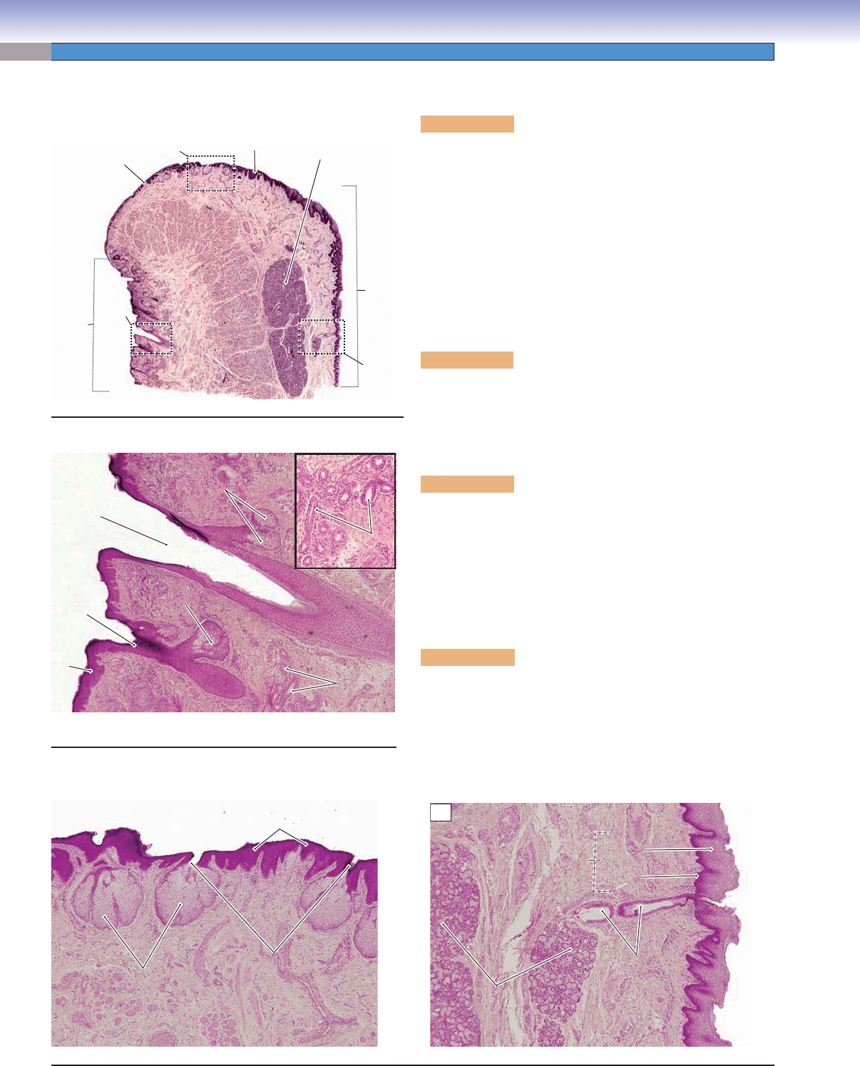
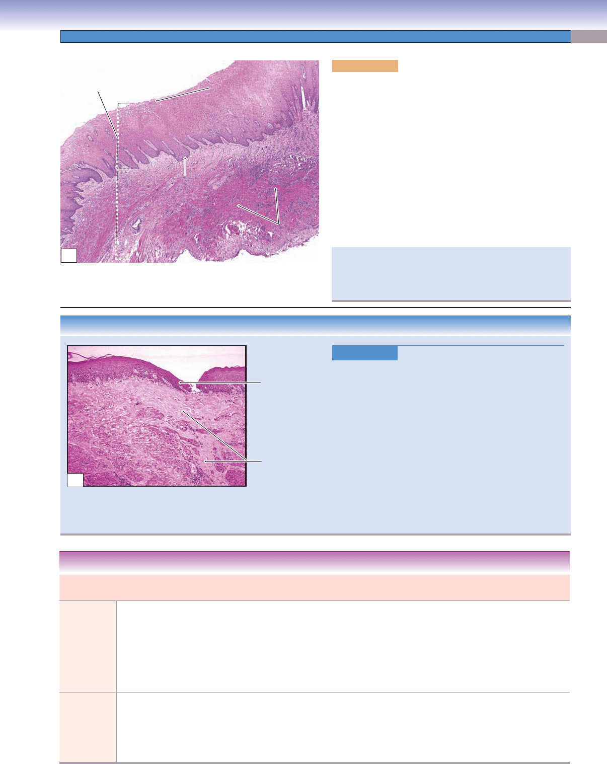
CLINICAL CORRELATION
Figure 14-3B.
Oral Submucous Fibrosis of the Lip.
H&E, 25
Oral submucous fi brosis is a precancerous condition char
-
acterized by a mucosal rigidity due to fi broelastic changes
of the lamina propria and submucosa layers of the lining
mucosa. This causes a progressive diffi culty in opening
the mouth. It affects the buccal mucosa, lips, retromolar
areas, the soft palate, and even the esophagus. Causes of
this condition include the use of chillies and areca nut,
collagen disorders, and autoimmune disorders. Histologi-
cally, it is characterized by atrophic (thinned) epithelium
and increased collagen fi ber formation followed by the
presence of dense collagen fi ber bundles and different
degrees of hyalinization. Prevention and treatments of the
disease include dietary changes and having plastic surgery
to improve the function of the mouth.
Atrophic
epithelium
Increased
collagen fibers
B
Elastic fibers
Elastic fibers
Elastic fibers
Lamina
Lamina
propria
propria
Lamina
propria
Stratum
Stratum
basale
basale
Stratum
basale
Stratum
Stratum
spinosum
spinosum
Stratum
spinosum
Stratum
Stratum
granulosum
granulosum
Stratum
granulosum
Nonkeratinized stratified
squamous epithelium
A
Figure 14-3A. Buccal mucosa (lining mucosa), cheek.
H&E, 16
Each cheek constitutes a lateral wall of the mouth. The inner
surface of the cheeks is lined by lining mucosa known as buccal
mucosa. The buccal mucosa has a nonkeratinized stratifi ed
squamous epithelium with three defi nite layers (stratum basale,
spinosum, and granulosum) with many elastic fi bers in the
lamina propria and minor salivary glands (buccal glands) in the
submucosa layer. This example shows the epithelium and lam-
ina propria of the buccal mucosa, which has a nonkeratinized
stratifi ed squamous epithelium. There are many elastic fi bers in
the lamina propria of the mucosa. Elastic fi bers appear pink and
do not readily stain with H&E stain. Fordyce spots (sebaceous-
like glands) may also be found in the mucosa of the cheek. They
increase with age and are more visible in elderly individuals.
The lingual and inferior alveolar nerves run through the pos-
terior groove of the cheek (between the pterygomandibular
raphe and the ramus of the mandible). This is an important
landmark for local anesthesia injections in the mouth.
CUI_Chap14.indd 261 6/2/2010 8:23:01 AM
262
UNIT 3
■
Organ Systems
Masticatory Mucosa
Alveolar bone
Alveolar bone
Alveolar bone
Mucogingival
Mucogingival
junction
junction
Mucogingival
junction
Enamel space
Enamel space
Enamel space
Junction epithelium
Junction epithelium
Junction epitheliumJunction epithelium
Junction epithelium
Junction epithelium
Sulcus epithelium
Sulcus epithelium
Sulcus epitheliumSulcus epithelium
Sulcus epithelium
Sulcus epithelium
Free
gingiva
A
ttached
gingiva
Alveolar
mucosa
L
L
a
a
m
m
in
in
a
a
p
p
ro
ro
p
p
ria
ria
Lamina propria
Dentin
Dentin
Dentin
Dental
Dental
pulp
pulp
Dental
pulp
Cementum
Cementum
Cementum
A
Figure 14-4A. Masticatory mucosa, gingiva. H&E, 22
Masticatory mucosa covers the gingiva and hard palate; it has a
keratinized stratifi ed squamous epithelium, which is exposed to
more abrasion during chewing than the lining mucosa. Mastica-
tory mucosa lacks a submucosa layer. The lamina propria of the
masticatory mucosa consists of a dense network of collagen fi bers
that are fi rmly attached to the underlying bone. The gingiva (gum)
surrounds the cervix of the tooth and covers the upper part of the
alveolar bone at the tooth root. The gingiva can be divided into
free gingiva and attached gingiva. The superior part of the gingiva
is free gingiva and surrounds, but does not attach to, the cervix of
the tooth. This nonattachment between the sulcus epithelium of the
free gingiva and enamel creates a space called the gingival sulcus, or
free gingival groove, (normally 0.5–3 mm depth). The attached gin-
giva fi rmly attaches to the underlying hard tissues (alveolar bone,
cementum, and edge of the enamel). The gingival-mucosal border
is called the mucogingival junction (see Fig. 14-1), where the epi-
thelium changes from nonkeratinized to keratinized and the color
changes from bright pink (alveolar mucosa) to pale pink (gingiva).
Stratum
Stratum
basale
basale
Stratum
basale
Stratum
Stratum
granulosum
granulosum
Stratum
granulosum
Stratum corneum
Stratum
Stratum
spinosum
spinosum
Stratum
spinosum
Keratinized stratified
squamous epithelium
Lamina
Lamina
propria
propria
Lamina
propria
Collagen
Collagen
fibers
fibers
Collagen
fibers
B
Figure 14-4B. Masticatory mucosa, hard palate. H&E, 35
The hard palate forms the roof of the mouth and is covered
by masticatory mucosa with keratinized stratifi ed squamous
epithelium and a dense lamina propria. It has one more layer
(stratum corneum) than the lining mucosa. The collagen fi bers in
the lamina are thick and dense and fi rmly bind to the periosteum
of the underlying bone. The periosteum consists of dense connec-
tive tissue, which covers the bone and contributes to bone forma-
tion (see Chapter 5, “Cartilage and Bone,” Fig. 5-10B). Most of
the hard palate lacks a submucosal layer. However, the posterior
region near the alveolar process may present a submucosal layer
that contains minor mucous glands. The hard palate signifi cantly
assists in mastication and speech.
Cleft palate is a birth defect in which there is a fi ssure in the hard
palate that is caused by the failure of two parts of the palate
to fuse during facial development. This condition impairs the
quality of speech (an individual is unable to pronounce certain
sounds) and also causes eating problems.
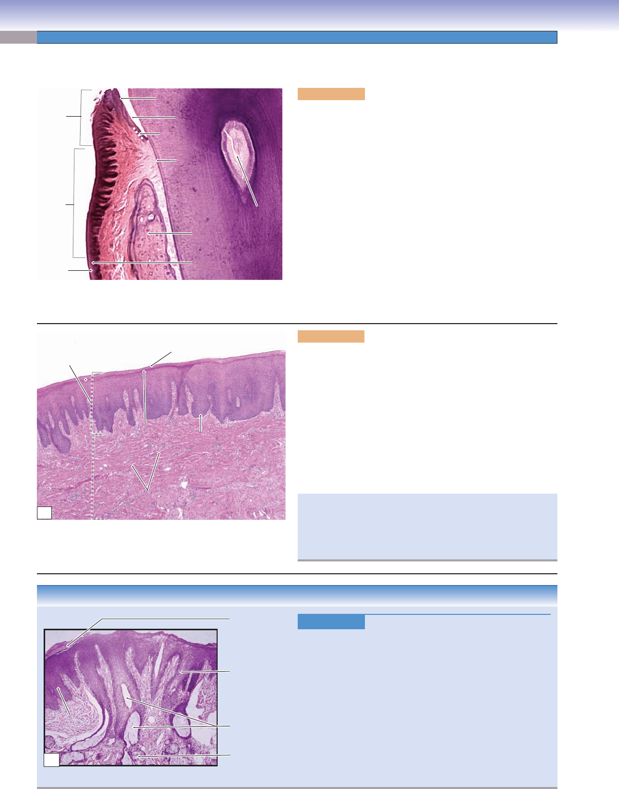
CLINICAL CORRELATION
Acanthosis
Orifice of the
gland ducts
Minor salivary
glands
Hyperkeratosis
Acanthosis
Acanthosis
Acanthosis
C
Figure 14-4C.
Nicotine Stomatitis. H&E, 25
Nicotine stomatitis is a nonprecancerous condition characterized
by a white lesion in the oral mucosa of the hard palate of the
mouth. The causes of this condition are associated with long-
term tobacco smoking, especially pipe smoking, and hot beverage
consumption. The lesion has a white cobblestone or “dried-
mud” appearance because of excessive keratin production. The
hard palate may appear gray or white and contain many papules
that are slightly elevated with red in their center. Histologically,
the squamous mucosa demonstrates hyperkeratosis (thickening
of the stratum corneum) and acanthosis (overgrowth of the
stratum spinosum). Complete smoking cessation usually helps
to diminish and resolve the condition within about two weeks. If
the lesion persists, close monitoring may be necessary.
CUI_Chap14.indd 262 6/2/2010 8:23:03 AM
CHAPTER 14
■
Oral Cavity
263
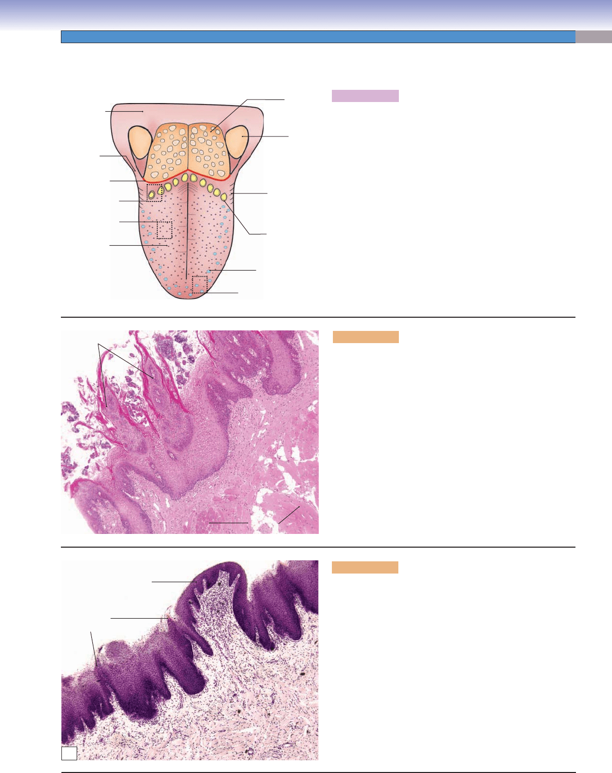
Specialized Mucosa
T. Yang
Foliate
papillae
Circumvallate
papillae
Lingual
tonsil
Palatine
tonsil
Fungiform
papillae
Filiform
papillae
Fig. 14-5B
Fig. 14-6A
Fig. 14-5C
Sulcus
terminalis
Palatoglossus
muscle
Oropharynx
A
Figure 14-5A. Overview of the tongue.
The inferior surface of the tongue and the fl oor of the mouth
are covered by lining mucosa with a nonkeratinized squamous
epithelium. The superior surface of the tongue is covered by
specialized mucosa with numerous projecting papillae includ-
ing fi liform papillae, fungiform papillae, circumvallate papillae,
and foliate papillae. The specialized mucosa is attached to the
underlying skeletal muscle. The tongue has a central core of
skeletal muscle, which controls movements of the tongue and
is innervated by the hypoglossal nerve (CN XII). The base of
the tongue is attached to the fl oor of the mouth. The surface
of the posterior third of the tongue has somatosensory recep-
tors and taste buds that are innervated by the glossopharyn-
geal nerve (CN IX). The anterior two thirds of the tongue has
somatosensory receptors that are innervated by the trigeminal
nerve (CN V), and its taste buds are innervated by the facial
nerve (CN VII). The tongue plays an important role in speech,
taste, and in moving and swallowing (deglutition) food.
Filiform
papillae
Epithelium
Lamina propria
Skeletal
muscle
B
Figure 14-5B. Filiform papillae, tongue. H&E, 35
The fi liform papillae are slender, cone-shaped papillae with
keratinized outer surfaces. They are the most numerous but
smallest in size of the four types of papillae. The fi liform
papillae are often packed in rows and cover the entire supe-
rior surface of the anterior two thirds of the tongue (anterior
to the sulcus terminalis). Each fi liform papilla has a central
connective tissue core with several branches of small papillae.
They are the only papillae that do not have taste buds. The
fi liform papillae’s tips are keratinized, consistent with their
exposure to abrasion during chewing. Because they have no
taste buds, their main functions are to help with chewing and
mixing food. The submucosal layer is absent in the tongue;
the mucosa of the tongue is strongly bound to the underlying
skeletal muscle to allow optimum food bolus control.
C
Fungiform papilla
Filiform
papillae
L
L
a
a
m
m
in
in
a
a
p
p
ro
ro
p
p
ria
ria
E
E
p
p
ith
ith
e
e
liu
liu
m
m
Epithelium
Skeletal
Skeletal
muscle
muscle
Skeletal
muscle
L
L
a
a
m
m
in
in
a
a
p
p
ro
ro
p
p
ria
ria
Lamina propria
Figure 14-5C. Fungiform papillae, tongue. H&E, 44
The fungiform papillae are mushroom shaped and much less
numerous than the fi liform papillae. They tend to be slightly
taller than the fi liform papillae that surround them. Each
fungiform papilla has one to fi ve taste buds on its superior sur-
face. These taste buds are innervated by the chorda tympani
branch of the facial nerve (CN VII), which joins the lingual
branch of the trigeminal nerve (CN V). The fungiform papillae
are covered by nonkeratinized squamous epithelium. They are
distributed at the tip and two sides of the tongue (Fig. 14-5A).
In general, the taste buds have fi ve taste sensations: sweet, bit-
ter, umami, salty, and sour. The salty and sour sensations are
associated with ion channels; the other three taste sensations
are associated with G protein–coupled receptors.
CUI_Chap14.indd 263 6/2/2010 8:23:07 AM
264
UNIT 3
■
Organ Systems
Moat (groove)
Moat (groove)
Moat (groove)
Lateral wall
Lateral wall
(taste buds)
(taste buds)
Lateral wall
(taste buds)
Duct of the
Duct of the
Von Ebner glands
Von Ebner glands
Duct of the
Von Ebner glands
Von Ebner
Von Ebner
glands
glands
Von Ebner
glands
Taste buds
Circumvallate
papilla
A
Figure 14-6A. Circumvallate papillae, tongue. H&E,
34 inset 181
The circumvallate papillae are also called vallate papillae.
They are arranged in a single row, which contains about
10 to 14 papillae that are located immediately anterior to the
sulcus terminalis (Fig. 14-5A). Each circumvallate papilla is
cylindrical in shape and is surrounded by a groove called a
moat. The ducts of the glands of von Ebner (minor serous
salivary glands) open and drain serous products into the
groove; this helps to clear the food debris in the groove and
helps in detection of taste. Most minor salivary glands in the
oral cavity are mucous or mixed glands; von Ebner glands
are the only ones that are pure serous glands. The taste buds
of the circumvallate papillae are located on the lateral walls
of the groove. These taste buds are mainly bitter receptors
and are innervated by the glossopharyngeal nerve (CN IX).
Foliate
papilla
Taste receptor cell
Taste receptor cell
Taste receptor cell
Taste pore
Taste pore
Taste pore
Basal cell
Basal cell
Basal cell
Taste buds
Taste buds
Taste buds
B
Figure 14-6B. Foliate papillae and taste buds, tongue.
H&E, 68 inset 222
The foliate papillae are located on the posterior lateral
surface of the tongue. This type of papilla is not well devel-
oped in humans but is frequently found in animals; illus-
trated here from rabbit tissue.
In general, taste buds are ovular structures embedded
in nonkeratinized stratifi ed squamous epithelium of the
papillae of the tongue. They also can be found in the pal-
ate of the oral cavity and in the mucosa of the oropharynx
and epiglottis. Each taste bud is composed of 40 to 60 elon-
gated taste receptor cells with microvilli at the apical region,
which extend into the taste pore. The basal end of the taste
bud contacts an afferent nerve fi ber. The taste pore is a small
opening that allows taste molecules to contact taste recep-
tor cells. The taste receptor cells have a life span of 1 to
2 weeks, and new taste cells arise from basal cells at the basal
lateral region of the taste bud. The inset shows taste buds.
Name Epithelial Covering Taste Buds Location Main Function
Filiform papillae Keratinized stratifi ed
squamous epithelium
No taste buds Anterior 2/3 of tongue Helps in chewing and
mixing food
Fungiform papillae Nonkeratinized stratifi ed
squamous epithelium
Taste buds located at
the apical surface of
papillae
Tip and two sides of
tongue
Chemoreceptor
detecting taste
Circumvallate papillae Nonkeratinized stratifi ed
squamous epithelium
Taste buds located at
the lateral surface of
papillae
In a V-shaped row just
anterior to the sulcus
terminalis
Chemoreceptor
detecting taste
Foliate papillae Nonkeratinized stratifi ed
squamous epithelium
Taste buds located at
the lateral surface of
papillae
Posterior lateral
surface of the tongue
Chemoreceptor
detecting taste
TABLE 14-2 Comparison of Lingual Papillae
CUI_Chap14.indd 264 6/2/2010 8:23:11 AM