Cui Dongmei. Atlas of Histology: with functional and clinical correlations. 1st ed
Подождите немного. Документ загружается.


CHAPTER 16
■
Digestive Glands and Associated Organs
305
Figure 16-12A Liver Acinus
Figure 16-12B Portal Triad, Liver
Figure 16-12C Clinical Correlation: Alcoholic Fatty Liver (Steatosis)
Figure 16-13A Hepatocytes and Hepatic Sinusoids, Liver
Figure 16-13B Space of Disse, Hepatocyte
Figure 16-14A A Representation of Bile Canaliculi and Hepatocytes
Figure 16-14B Bile Canaliculus, Hepatocytes
Gallbladder
Figure 16-15A Gallbladder
Figure 16-15B Epithelial Cells Lining the Gallbladder
Figure 16-16A Clinical Correlation: Hepatitis C
Figure 16-16B Clinical Correlation: Gallstones
Synopsis 16-2 Pathological and Clinical Terms for the Digestive Glands and Associated Organs
Introduction and Key Concepts
for the Digestive Glands and Associated
Organs
Digestive glands and associated organs include the major
salivary glands, pancreas, liver, and gallbladder. These organs
are located outside the wall of the digestive tract. Their secretory
products are delivered into the digestive tract via a duct system.
Major Salivary Glands
The major salivary glands produce saliva and empty into the
oral cavity. Saliva is 99% water and contains protein, enzymes,
glucose, cholesterol, urea, uric acid, ions (e.g., Na
+
, K
+
, Ca
++
,
HCO
3
-
), and antibacterial agents (lactoferrin, lysozyme, and
IgA). Saliva is produced by both serous and mucous cells in
the salivary glands. It plays important roles in aiding digestion,
lubrication, protection, buffering, wound healing, maintaining
the integrity of the esophagogastric epithelium, perception of
taste, and in hardening of the enamel of the teeth. There are
three major salivary glands: the parotid, submandibular, and
sublingual glands. These are paired glands and have similar
structures of secretory units (acini) and duct systems, including
intralobular ducts (intercalated and striated ducts), interlobular
ducts, lobar ducts, and a main duct (Figs. 16-1 and 16-8B).
1. The parotid gland is the largest of the three major salivary
glands. This gland is surrounded by a connective tissue
capsule. Its secretory unit is composed of only serous cells,
which produce watery proteinaceous fl uid (Figs. 16-3B and
16-4A).
2. The submandibular gland is the second largest salivary
gland. This gland is surrounded by a connective tissue cap-
sule. It is a mixed gland, although the majority of cells are
serous cells. The secretory unit is composed of both serous
and mucous cells. Serous demilunes (serous cell caps on the
mucous cells) are present in the submandibular gland (Figs.
16-5A to 16-7B).
3. The sublingual gland is the smallest salivary gland. This
gland is not surrounded by a connective tissue capsule. This
is also a mixed gland, but is predominately mucous. Acini
that are completely serous are few, but serous demilunes are
commonly present. Striated ducts are not as obvious as in
the other two types of salivary glands (Fig. 16-8A,B).
Pancreas
The pancreas has endocrine and exocrine portions. The
endocrine portion (islets of Langerhans) secretes blood glucose–
regulating hormones (insulin, glucagon, somatostatin, and pan-
creatic polypeptide), which are released into the bloodstream
(see Chapter 17, “Endocrine System”). The exocrine portion
produces pancreatic secretions (juice), which are carried by
pancreatic ducts. Most of these secretions go to the main pan-
creatic duct, which joins the hepatopancreatic ampulla (ampulla
of Vater) and then enters the duodenum through the major duo-
denal papilla (papilla of Vater). The ampulla is surrounded by
smooth muscle called the sphincter of the ampulla (sphincter
of Oddi [Fig. 16-9A]). A small portion of the pancreatic secre-
tion is carried by the accessory pancreatic duct and enters the
duodenum through the minor duodenal papilla. The pancreatic
duct system includes intralobular ducts, interlobular ducts, and
a main duct. The pancreas does not have striated ducts, and
the smallest intralobular ducts are intercalated ducts. The initial
portions of the intercalated ducts are lined by centroacinar cells.
The initial pancreatic secretions (enzymes) are produced by pan-
creatic acinar cells, and a large volume of fl uid (water, sodium,
and bicarbonate) is added by intercalated duct cells. Pancreatic
secretions contain enzymes for digesting proteins, carbohy-
drates, and fats. The major components include water, sodium,
bicarbonate ions, trypsinogen, chymotrypsinogen, procarboxy-
polypeptidase, amylase, lipase, cholesterol esterase, phospho-
lipase, and nucleases. The main enzymes for digesting protein
are trypsinogen, chymotrypsinogen, and procarboxypolypepti-
dase; the main enzyme for digesting carbohydrates is amylase;
and the main enzymes for digesting fat are lipase, cholesterol
esterase, and phospholipase. The nucleases degrade nucleic
acids. When the pancreatic digestive enzymes are fi rst synthe-
sized by the pancreatic cells, they are in an inactive stage. They
become activated after they enter the duodenum and come into
CUI_Chap16.indd 305 6/2/2010 6:42:18 PM

306
UNIT 3
■
Organ Systems
contact with enterokinase in the glycocalyx. The glycocalyx is
an extracellular polymeric material (glycoprotein), which coats
the surface of microvilli in the small intestines (Figs. 16-9A to
16-10B).
Liver
The liver also plays both endocrine and exocrine roles. Its
endocrine role is in synthesizing and releasing plasma proteins,
such as fi brinogen, prothrombin, lipoproteins, and albumins,
into the bloodstream. Its exocrine role is the production of bile.
Bile is important in emulsifying and degrading fat into smaller
molecules and in carrying wastes out of the body. Bile contains
water, bile salts, bilirubin, cholesterol, fatty acids, lecithin, and
electrolytes. Bile is produced by hepatocytes and is collected
by bile canaliculi; it drains into the hepatic duct, then into the
cystic duct, and fi nally enters the gallbladder (Figs. 16-11 and
16-15A). Other functions of the liver include detoxifi cation;
involvement in lipid, carbohydrate, and protein metabolism;
and storage of iron, blood, glycogen, triglycerides (lipid drop-
lets), and vitamins A, D, and B12. The liver is supplied by the
portal veins and the hepatic arteries at the portal triads. The
hepatic arteries carry oxygen-rich blood, and the portal veins
carry nutrient-rich blood to the hepatic sinusoids through their
branches (hepatic arterioles and portal venules). The hepatocytes
are not in direct contact with the blood stream. A small space
between the hepatocyte and the endothelium of the sinusoids
is called the perisinusoidal space or space of Disse. The discon-
tinuous endothelium of the sinusoids allows proteins, nutrients,
wastes, and plasma components (but not blood cells) from the
hepatic sinusoids to enter the space of Disse. Hepatocytes take
up nutrients and transport wastes, such as bilirubin, into the
bile. The central veins collect the exchanged blood from sinu-
soids and drain into the sublobular veins and then into the large
hepatic veins. The basic structure of the liver includes the classic
lobule, the
portal lobule, and the liver acinus (Figs. 16-11 to
16-12A).
Gallbladder
The gallbladder is a pear-shaped, saclike organ closely associ-
ated with the liver. It stores, concentrates, and releases bile.
When the smooth muscle (muscularis) of the gallbladder
contracts, bile is released from the gallbladder through the
cystic duct into the bile duct. This joins the hepatopancreatic
ampulla and drains into the duodenum at the major duode-
nal papilla. The wall of the gallbladder is composed of three
layers: (1) mucosa, which includes epithelium (simple colum-
nar epithelium) and lamina propria; (2) muscularis, which
contains interlacing smooth muscle bundles and contracts in
response to cholecystokinin, which is released by enteroen-
docrine cells in the small intestine (the smooth muscle of the
gallbladder extends from the body into the neck where it is
known as the spiral valve of Heister and is vital in controlling
gallbladder fl ow); and (3) serosa/adventitia, a connective layer
with a lining of mesothelium (serosa) or without mesothelium
(adventitia). Most of the outer surface of the gallbladder is
covered with serosa, which is continuous with the peritoneum
that covers the liver. A small region of the gallbladder is cov-
ered by adventitia, which attaches the gallbladder to the liver
(Fig. 16-15A,B).
CUI_Chap16.indd 306 6/2/2010 6:42:18 PM
CHAPTER 16
■
Digestive Glands and Associated Organs
307
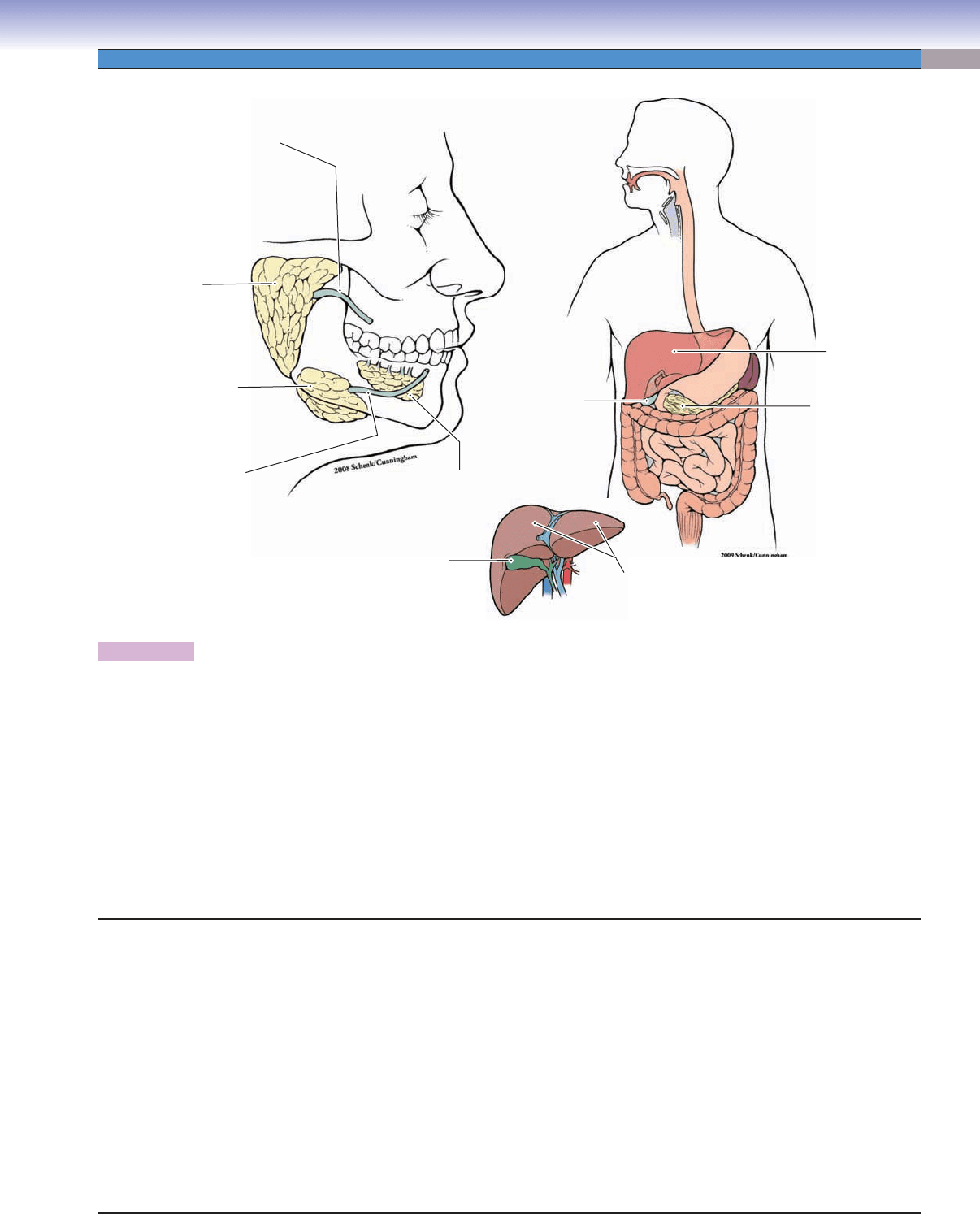
Figure 16-1. Overview of the digestive glands and associated organs.
The major digestive glands include the salivary glands, pancreas, and liver. Although the gallbladder is not a gland, it is closely associated
with the liver and digestive system. The digestive glands and gallbladder are located outside of the digestive tract. Their ducts carry
their products (saliva) into the oral cavity or into the lumen of the duodenum of the small intestine (pancreatic secretions and bile). The
salivary glands include the major salivary glands and the minor salivary glands. The parotid, submandibular, and sublingual glands are
the three major salivary glands. These glands are either serous glands or mixed glands (both serous and mucous). Most of the minor
salivary glands are mucous or mixed glands, with only von Ebner glands being entirely serous glands (see Fig. 14-6A). For details of clas-
sifi cation of the glands see Chapter 3, “Epithelium and Glands.” The pancreas includes an endocrine portion (islets of Langerhans) and
an exocrine portion. The endocrine portion secretes hormones into the bloodstream (see Chapter 17, “Endocrine System”), whereas the
exocrine portion has ducts that carry enzyme products into the lumen of the duodenum (see Figs. 16-9A to 16-10B). The liver produces
bile, which is carried by the bile duct system into the gallbladder (see Figs. 16-11 and 16-15A). The liver also aids in the metabolism of
lipids, carbohydrates, and proteins; stores iron, glycogen, triglycerides, and vitamins A, D, and B12; and detoxifi es certain toxic sub-
stances in the blood. The gallbladder stores, concentrates, and releases bile into the duodenum (see Fig. 16-15A,B).
Parotid
gland
Liver
Liver
Gallbladder
Gallbladder
Pancreas
Main duct of
parotid gland
Submandibular
gland
Sublingual
gland
Main duct of
submandibular
gland
Digestive Glands and Associated Organs
I. Major Salivary Glands
A. Parotid glands (serous)
B. Submandibular glands (mixed, predominately serous)
C. Sublingual glands (mixed, predominately mucous)
II. Pancreas
A. Exocrine portion
B. Endocrine (islets of Langerhans) portion
III. Liver
A. Central artery
B. Portal triad
C. Liver lobules
1. Classic lobule
2. Portal lobule
3. Hepatic acinus
IV. Gallbladder
A. Mucosa
B. Muscularis
C. Serosa
CUI_Chap16.indd 307 6/2/2010 6:42:18 PM
308
UNIT 3
■
Organ Systems
Digestive Glands and Associated Organs with Figure Numbers
Parotid Glands
Figure 16-3B
Figure 16-4A
Figure 16-4B
Figure 16-4C
Submandibular Glands
Figure 16-5A
Figure 16-5B
Figure 16-6A
Figure 16-6B
Figure 16-7A
Figure 16-7B
Sublingual Glands
Figure 16-8A
Figure 16-8B
Figure 16-8C
Pancreas
Figure 16-9A
Figure 16-9B
Figure 16-9C
Figure 16-10A
Figure 16-10B
Liver
Figure 16-11
Figure 16-12A
Figure 16-12B
Figure 16-12C
Figure 16-13A
Figure 16-13B
Figure 16-14A
Figure 16-14B
Figure 16-16A
Gallbladder
Figure 16-15A
Figure 16-15B
Figure 16-16B
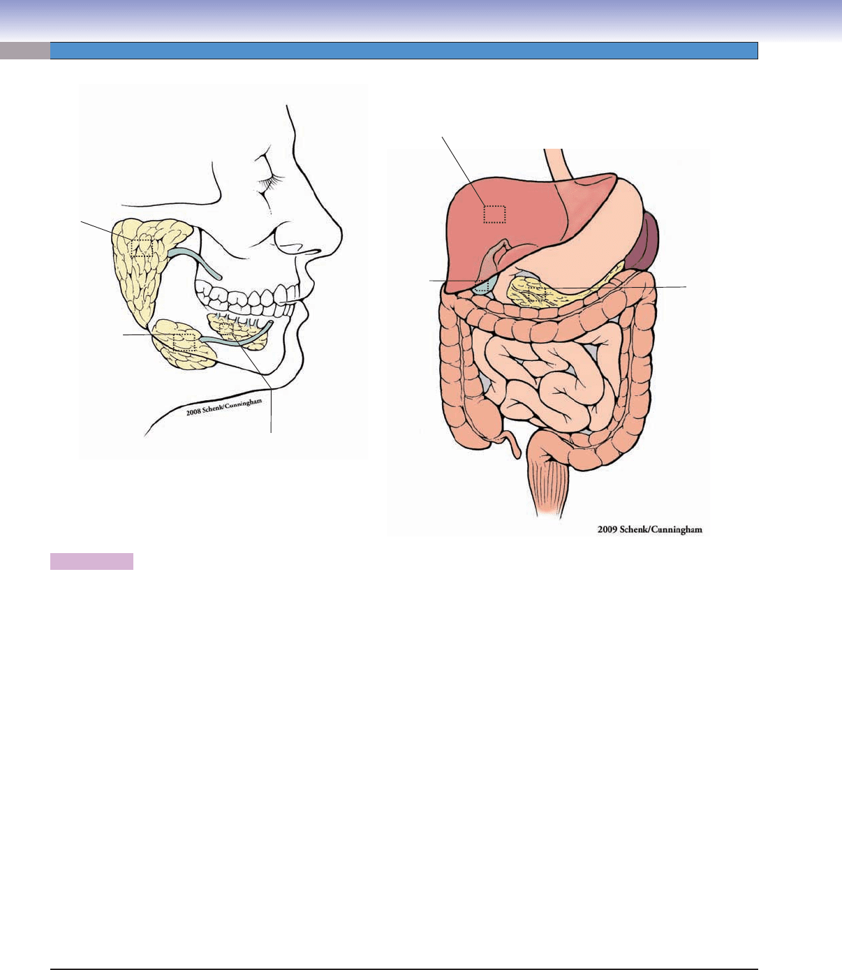
Figure 16-2. Orientation of detailed digestive glands and associated organs illustrations.
Fig. 16-8A,B
Fig. 16- 5A,B
Fig. 16- 6A,B
Fig. 16- 7A,B
Fig. 16-3B
Fig. 16-4A
Fig. 16-9B
Fig. 16-10A,B
Fig. 16-15A,B
Fig. 16-12A,B
Fig. 16-13A,B
Fig. 16-14A,B
CUI_Chap16.indd 308 6/2/2010 6:42:19 PM
CHAPTER 16
■
Digestive Glands and Associated Organs
309
Salivary Glands
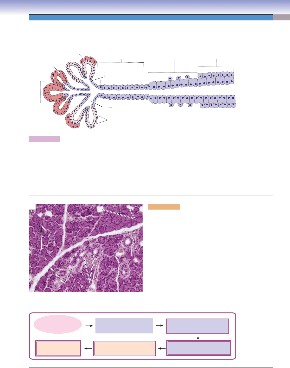
Figure 16-3A. General structure of the major salivary glands.
The parotid, submandibular, and sublingual glands are structurally very similar to one another, although they produce various
secretions. Each unit of the salivary glands can be divided into a secretory portion and a duct system. The secretory portion contains
serous cells, mucous cells, or a mixture of both. These secretory cells are arranged into acini resembling grapes on a stem (interca-
lated duct). Several serous cells form a cap, called a serous demilune, on the outer aspect of mucous cells; this arrangement can be
found in the mixed glands. A capsule (dense connective tissue layer) surrounds an entire gland. Connective tissue septa penetrate the
gland and subdivide it into lobes and lobules. The duct system includes intralobular ducts (located within the lobules), interlobular
ducts (outside or between the lobules), lobar ducts, and a main duct. The intralobular ducts include intercalated ducts and striated
ducts. The main duct empties the secretory products (saliva) into the oral cavity.
Figure 16-3B. Parotid gland. H&E, 130 skin, palm.
The parotid glands are paired glands and are the largest of the major
salivary glands. They are located anterior to and below the lower
half of the ear, and superior, posterior, and deep to the ramus of
the mandible. The main duct of each parotid gland passes through
the cheek and opens into the oral cavity near the second upper
molar tooth (see Fig. 16-1). The parotid glands are composed of
serous acini and ducts. They are classifi ed as compound (branched)
acinar glands based on their duct shape and secretory units (see Fig.
3-27A). This photomicrograph shows striated ducts (intralobular
ducts) located in the lobules. The striated ducts are lined by taller
cuboidal (or columnar) cells with centrally located nuclei (see Fig.
16-7A). Connective tissue septa divide the gland into small lobules,
and there are some adipose cells distributed in among the serous
acini. Each acinus is formed by several serous cells with basally
positioned dark nuclei (Fig. 16-4A).
Salivary Gland Duct System
D. Cui
Lobar duct
(a few branches)
Interlobular duct
(multiple branches)
Intralobular duct
(multiple branches)
Secretory portion
Duct system
Serous
cells
Serous
acinus
Serous
demilune
Intercalated
ducts
Lumen of the
Intercalated ducts
Striated
ducts
Mucous
cells
A
Striated
Striated
ducts
ducts
Striated
ducts
Striated
Striated
duct
duct
Striated
duct
Septa
Septa
Septa
Adipose
Adipose
cells
cells
Adipose
cells
Serous acini
Serous acini
Serous acini
B
D. Cui
(Serous, mucous, or mixed cells)
(Simple low cuboidal epithelium)
Small intralobular duct
(Intercalated duct)
Interlobular duct
Main duct
(Stratified columnar epithelium)
(striated duct)
CUI_Chap16.indd 309 6/2/2010 6:42:20 PM
310
UNIT 3
■
Organ Systems
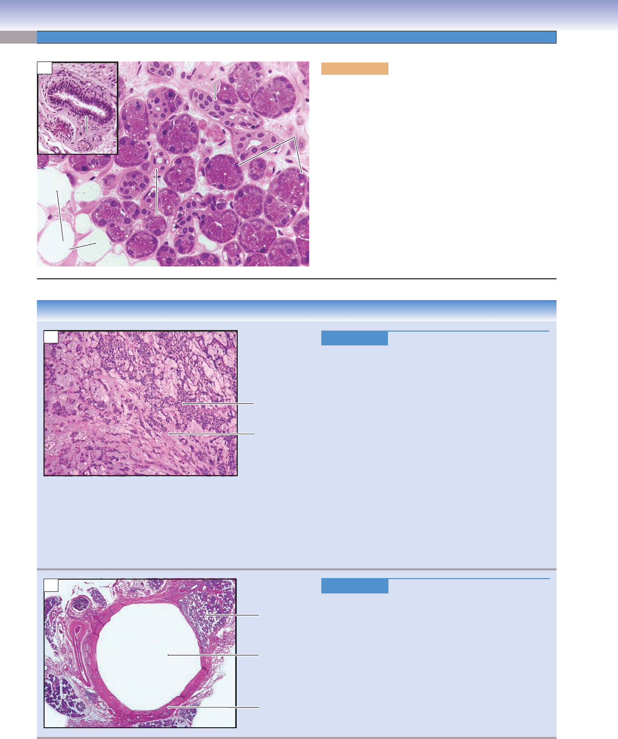
Figure 16-4A. Parotid glands. H&E, 295; inset 97
Parotid glands contain only serous secretory cells. There
are some adipose cells scattered throughout the gland. All
three types of salivary glands have similar duct systems. The
intercalated ducts are shown here; they have a small lumen
and are lined by lower cuboidal cells with basally located
nuclei. The inset shows a large interlobular duct that is
lined by stratifi ed columnar cells and is surrounded by a
large amount of connective tissue. The diameter of the ducts
gradually increases from the intercalated ducts to the large
interlobular ducts, and their lining cells increase in height
and in number of layers (see Fig. 16-3A). The main duct
(duct of Stenson) of the parotid gland traverses the buccina-
tor muscles and opens opposite the secondary upper molars
(see Fig. 16-1). All three types of salivary glands receive para-
sympathetic innervation. The parotid gland is innervated by
the glossopharyngeal nerve (cranial nerve [CN] IX).
Adipose
Adipose
cells
cells
Adipose
cells
Interlobular
Interlobular
duct
duct
Interlobular
duct
Intercalated
Intercalated
duct
duct
Intercalated
duct
Serous
Serous
cells
cells
Serous
cells
Intercalated
Intercalated
duct
duct
Intercalated
duct
A
CLINICAL CORRELATIONS
Figure 16-4B.
Pleomorphic Adenoma. H&E, 55
Pleomorphic adenoma, also called benign mixed tumor,
is the most common benign salivary gland tumor
. The
majority, approximately 80%, occur in the parotid gland,
most of which are in the superfi cial lobe, and are benign
pleomorphic adenomas. The tumor may also involve
submandibular, sublingual, and minor salivary glands. It
is characterized by a slow-growing, mobile, and painless
parotid mass. Most patients are not aware of the tumor
for years. The mass itself is typically well demarcated, but
may be nodular in appearance. Histologically, the neo-
plasm is composed of epithelial and myoepithelial cells in
a chondromyxoid background. Fine needle biopsy is use-
ful in the diagnosis of pleomorphic adenoma. After sur-
gery, pleomorphic adenomas may recur. Rapid growth
of a mass in the same area after surgery may signify
malignant transformation of the residual adenoma called
carcinoma expleomorphic adenoma. This photomicro-
graph shows nests of myoepithelial cells in a myxoid
background.
Figure 16-4C.
Parotid Cyst. H&E, 11
A parotid cyst is a fl uid-fi
lled closed cavity occurring
within the parenchyma of the parotid gland and is usu-
ally caused by trauma, infections, salivary gland stones,
or tumors. Physical exam usually reveals a painless
lump or swelling. As the cyst enlarges, it may interfere
with chewing, swallowing, and speaking. Additionally,
parotid cysts may become infected. Histologically, the
cyst is lined by an epithelium, and the cavity is fi lled
with fl uid or mucus. The surrounding stroma shows
dense fi brosis and may be infi ltrated by aggregates of
lymphocytes, as shown in this slide. If necessary, surgical
removal of the cyst is recommended.
Myoepithelial
cells
Myxoid
background
B
Parotid
gland
Cyst
Stroma
C
CUI_Chap16.indd 310 6/2/2010 6:42:23 PM
CHAPTER 16
■
Digestive Glands and Associated Organs
311
Septum
Septum
Septum
Serous
Serous
demilunes
demilunes
Serous
demilunes
Septum
Septum
Septum
Serous
Serous
acini
acini
Serous
acini
Serous
Serous
demilune
demilun
e
Serous
demilune
Striated
Striated
duct
duct
Striated
duct
Mucous
Mucous
cells
cells
Mucous
cells
A
Figure 16-5A. Submandibular glands. H&E, 136; inset 408
Submandibular glands are also paired glands. They are smaller than
the parotid glands, but larger than the sublingual glands. Each sub-
mandibular gland is divided into superfi cial (large) and deep (small)
lobes and is located under the fl oor of the oral cavity adjacent to
the mandible (see Fig. 16-1). The main duct of the submandibular
gland drains saliva into the oral cavity at the sublingual caruncles on
both sides of the frenulum linguae. The submandibular glands are
mixed glands that contain predominantly serous cells but also some
mucous cells. They are classifi ed as compound (branched) tubulo-
acinar glands (see Fig. 16-3A; see also Chapter 3, “Epithelium and
Glands,” Fig. 3-28A). Serous cells are arranged into many acini, and
mucous cells are arranged as acini or tubular structures, which may
have caps of serous cells (serous demilunes). Submandibular glands
are innervated by parasympathetic nerve fi bers from branches of
the facial nerve (CN VII).
Myoepithelial
cell
Serous acinar
cell
Secretory
granules
Nucleus of the
Nucleus of the
acinar cell
acinar cell
Nucleus of the
acinar cell
Lumen
Lumen
Lumen
RER
RER
RER
B
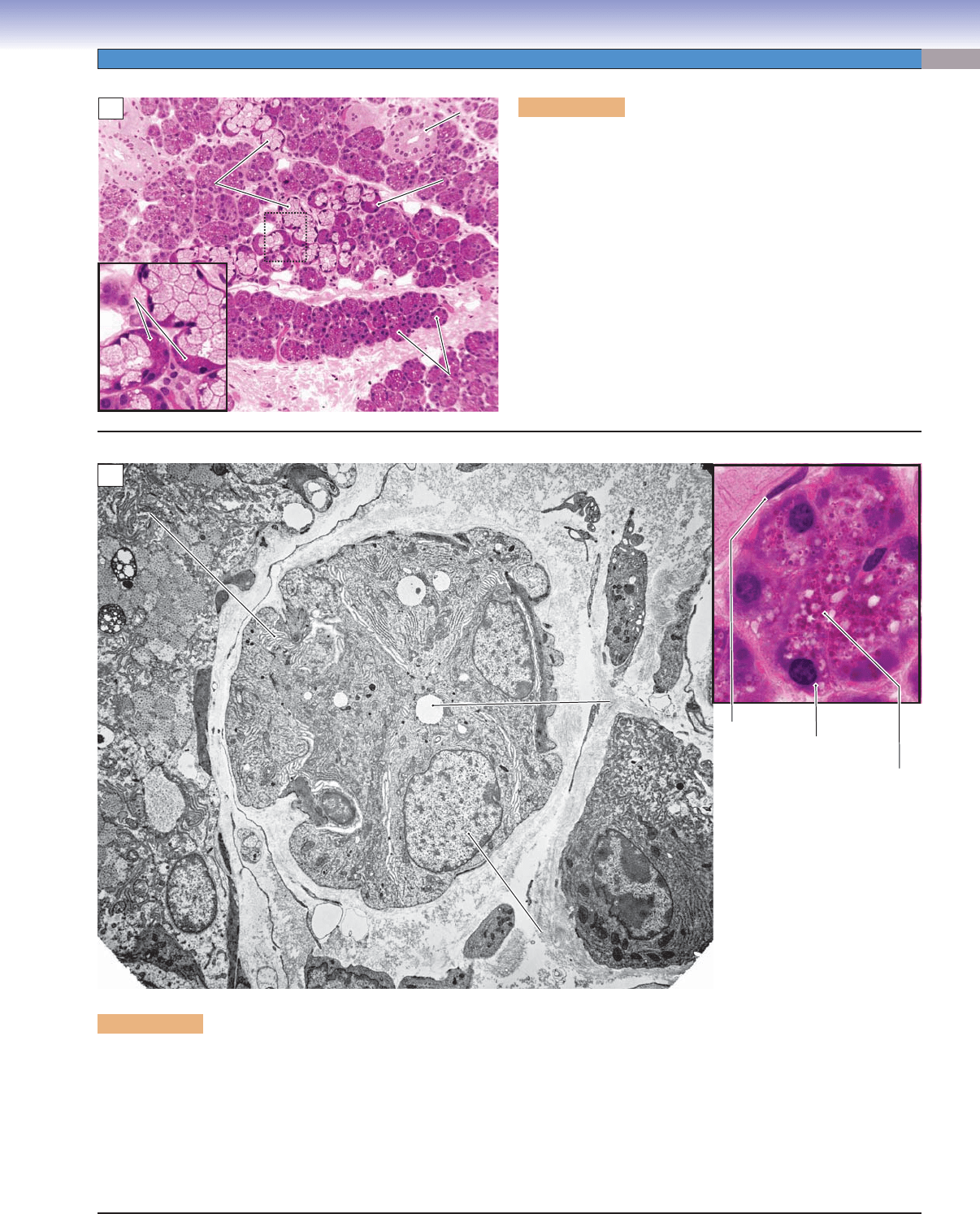
Figure 16-5B. Serous acinus, submandibular gland. EM, 3,937; inset (color), H&E, 1,079
Several serous acinar cells arranged in an acinus and sharing a common lumen are shown. The serous lumens are smaller than those
of a mucous acinus (see Fig. 16-6A). Each secretory cell has a relatively large nucleus and numerous rough endoplasmic reticulum
(RER) cisterns in the basal portion of the cytoplasm. These features indicate their active protein synthesis. Secretory granules are
usually located on the apical region of the cells (color inset). Secretory granules are not seen in this electron micrograph because they
have already been discharged from these particular cells. The inset shows an acinus with secretory cells having round nuclei located
basally and many secretory granules in the apical portions of the secretory cells. A myoepithelial cell with a fl at nucleus is present
here, located beneath the serous cell. It is a contractile cell and contains smooth muscle myosin. The myoepithelial cells contract and
move the secretory products toward the intercalated duct.
CUI_Chap16.indd 311 6/2/2010 6:42:28 PM
312
UNIT 3
■
Organ Systems
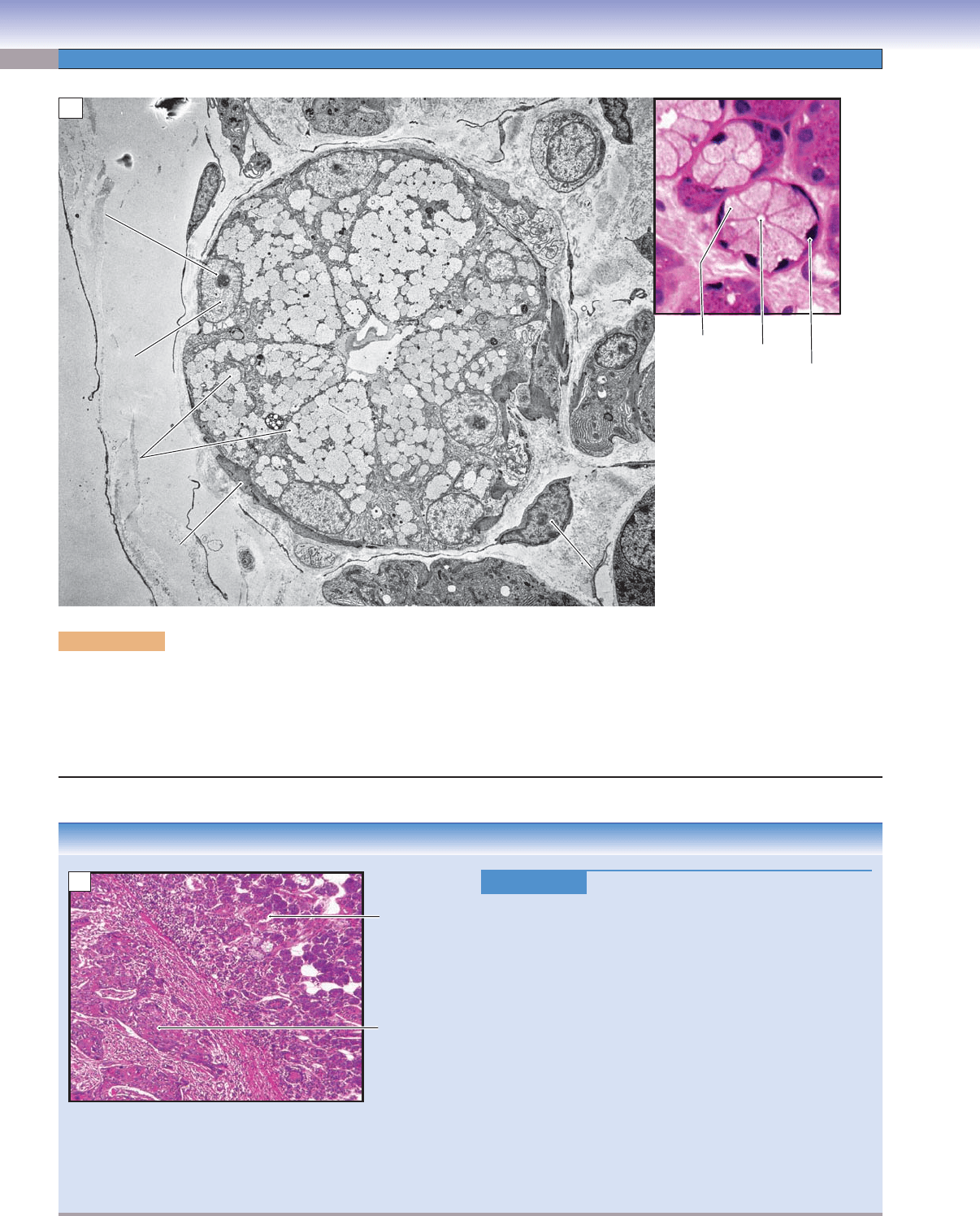
Figure 16-6A. Mucous acinus, submandibular gland. EM, 6,818; inset (color) H&E, 598
This is an example of a mucous acinus, showing numerous large secretory granules in the apical region of the cells. These granules
contain mucins, which are synthesized within the cells. The nuclei of the mucous cells are fl attened and lie against the basement
membrane. Mucous cells have less RER than serous cells. Myoepithelial cells share the basement membrane with mucous cells.
Myoepithelial cells have long cellular processes that can contract, promoting mucous cells to discharge their secretory products into
the lumen of the intercalated ducts.
Nucleolus
Nucleolus
Secretory
Secretory
granules
granules
Secretory
granules
Nucleus of mucous
Nucleus of mucous
acinar cell
acinar cell
Nucleus of mucous
acinar cell
Process of
Process of
myoepithelial cell
myoepithelial cell
Process of
myoepithelial cell
Lumen
Lumen
Lumen
Nucleus of the
Nucleus of the
myoepithelial cell
myoepithelial cell
Nucleus of the
myoepithelial cell
Mucous
acinar cell
Lumen
Lumen
Lumen
Nucleus of
mucous
acinar cell
A
Nucleolus
CLINICAL CORRELATION
Squamous cell
carcinoma
Submandibula
r
gland
B
Figure 16-6B.
Squamous Cell Carcinoma of the Tongue.
H&E, 83
Squamous cell carcinoma of the tongue is a malignant oral
neoplasm representing the most common intraoral cancer.
Squamous cell carcinoma is usually asymptomatic in the early
stages, but local pain, diffi culty swallowing, and dysphagia
are common in the late stages. Clinically, the neoplasm may
appear as a red or white nonhealing ulcer or exophytic mass,
most commonly found on the lateral aspect of the tongue.
This image shows a squamous cell carcinoma on the fl oor
of the mouth invading the submandibular glands. The major
risk factors for oral and tongue squamous cell carcinoma
include smoking and alcohol consumption, chronic irrita-
tion, and chewing tobacco. Pathologically, the cancer cells
show large, irregular and darkly stained nuclei. This cancer
tends to metastasize though the lymphatic system. Surgical
removal of the primary growth and related lymphatic nodes,
chemotherapy, and radiation therapy are treatment options.
CUI_Chap16.indd 312 6/2/2010 6:42:31 PM
CHAPTER 16
■
Digestive Glands and Associated Organs
313
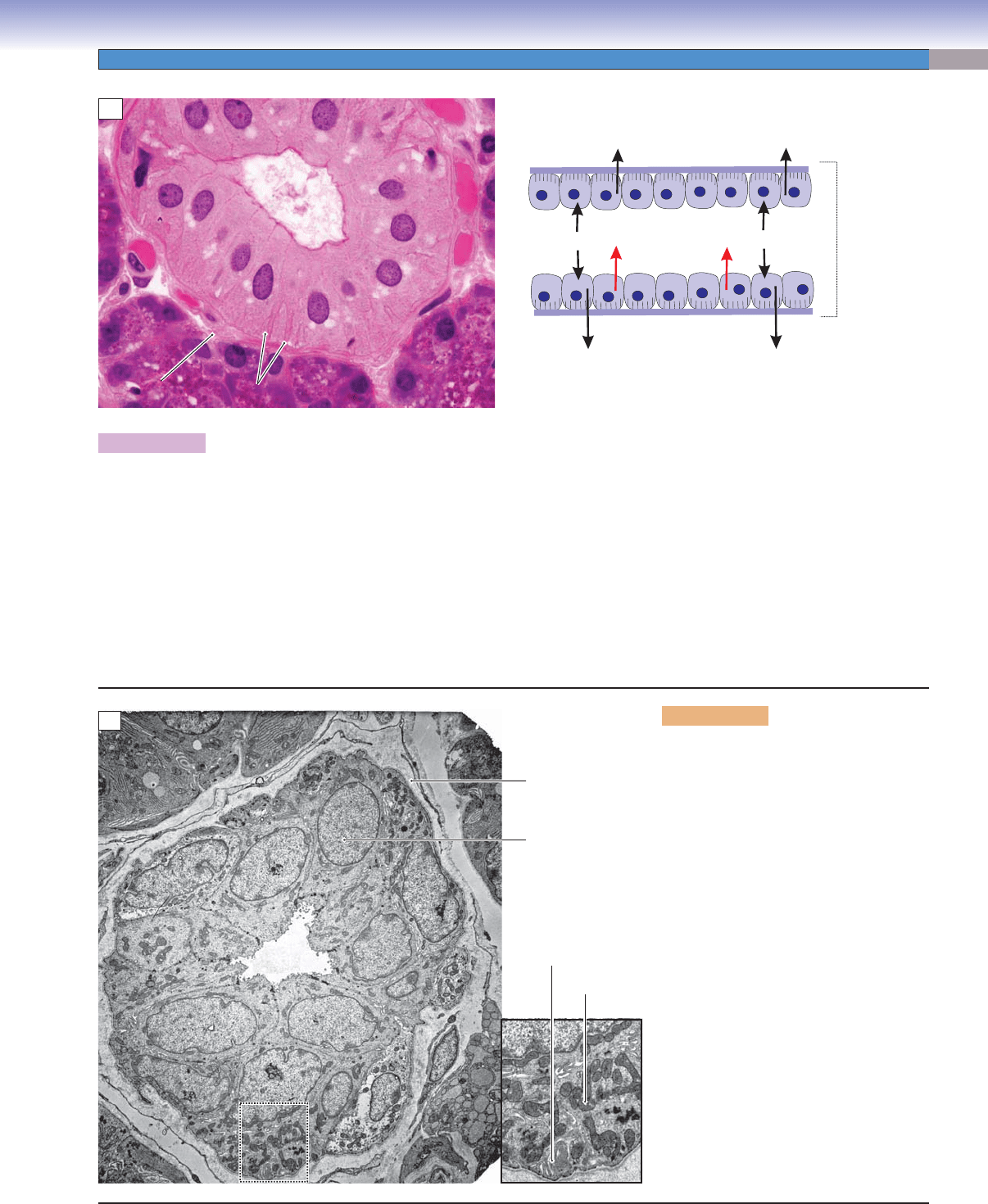
Figure 16-7A. Striated duct, submandibular gland. H&E, 845
Striated ducts are lined by simple cuboidal or columnar cells with centrally positioned nuclei. Their cytoplasm shows striations in
the basolateral region, hence the name. The striations are due to deep indentations in the plasma membrane (basolateral folds),
which increase the surface area occupied by pumps involved in ion and fl uid transport. The folds contain many mitochondria that
provide adenosine triphosphate for active transport. The striated ducts collect primary saliva (produced by acinar cells) from the
intercalated ducts. The main function of the striated ducts is modulation of the pH and ion composition of the saliva. The striated
ducts remove Na
+
and Cl
−
ions in the primary saliva from the lumen and transport these ions to the interstitial tissues. Striated ducts
also pump K
+
and HCO
3
−
into the saliva fl uid. This results in a hypotonic alkaline saliva (secondary saliva), which moves from the
striated ducts to the interlobular ducts, then to the main duct, and eventually enters the oral cavity. Saliva plays important roles in
moistening and cleansing the oral cavity, helping to repair oral tissues, infl uencing the pH (buffering) in the oral cavity, stimulating
the taste buds, helping to digest food, helping in the mineralization and hardening of the enamel of posterupted teeth, and destroying
bacteria by antimicrobial action.
D. Cui
Striations
Striations
Striations
Interstitial
Interstitial
tissue
tissue
Interstitial
tissue
Serous
Serous
acinus
acinus
Serous
acinus
Lumen
Lumen
Lumen
Na
+
Cl
_
Na
+
K
+
Cl
_
HCO
3
_
Na
+
Cl
_
Interstitial
tissue
Striated
duct
A
Basal
membrane
enfolding
Mitochondrion
Nucleus
Basement
membrane
Lumen
B
Figure 16-7B. Intralobular duct, sub-
mandibular gland. EM, 3,382; inset
6,779
The duct shown here appears to be at the
transition between a striated intralobular
duct and an interlobular duct. Some of
the lining cells, such as the one shown in
the inset, have features of a striated duct
in that there are extensive basolateral
folds containing numerous mitochondria.
These are the same features that account
for the eosinophilic striations seen in these
cells in H&E-stained sections for light
microscopy.
CUI_Chap16.indd 313 6/2/2010 6:42:34 PM
314
UNIT 3
■
Organ Systems
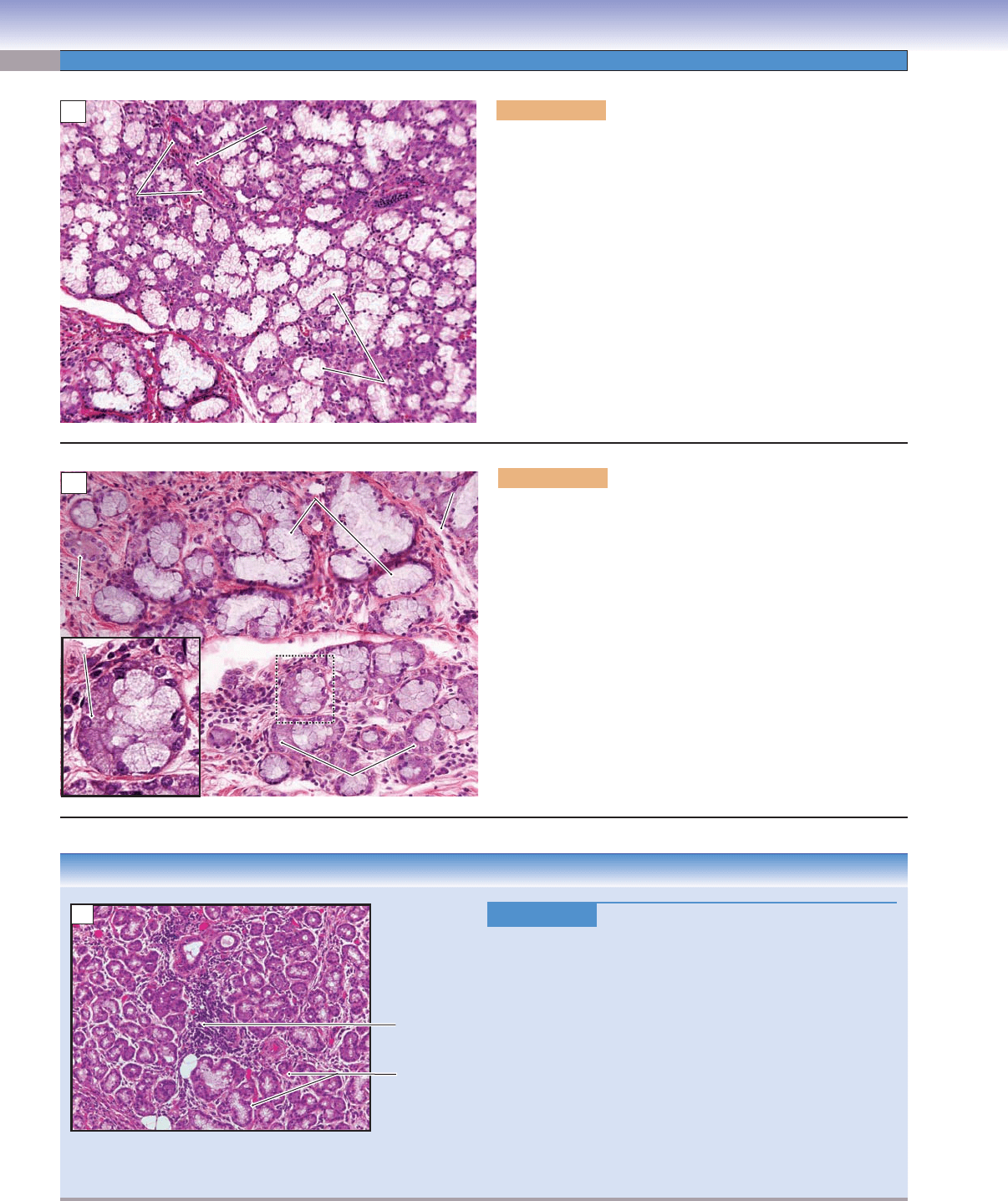
Figure 16-8A. Sublingual glands. H&E, 123
Sublingual glands are the smallest of the three major salivary
glands. They are paired glands and are located deep in the fl oor
of the oral cavity, anterior to the submandibular glands. They
are covered by the oral mucosa, but have no capsule (dense con-
nective tissue) enclosing them as the other two major salivary
glands do. Sublingual glands have about 8 to 20 small ducts,
which open into the crest of the sublingual fold on the fl oor
of the oral cavity. They are mixed glands, and contain serous
and mucous secretory cells, but predominantly mucous cells. As
in submandibular glands, serous demilunes are present; sublin-
gual glands are also classifi ed as compound (branched) tubu-
loacinar glands (see Figs. 16-3A and 3-28A). Two intralobular
ducts located inside connective tissue septa are labeled here.
Sublingual glands are innervated by parasympathetic nerve
fi bers from branches of the facial nerve (CN VII).
Mucous
Mucous
acini
acini
Mucous
acini
Interlobular
Interlobular
ducts
ducts
Interlobular
ducts
Septum
Septum
Septum
A
Mucous cell
Mucous cell
Mucous cell
Serous demilune
Serous demilune
Serous demilune
Capillary
Capillary
Capillary
Mucous acini
Mucous acini
Mucous acini
Intralobular
Intralobular
duct
duct
Intralobular
duct
Serous demilune
Serous demilune
Serous demilune
B
Figure 16-8B. Sublingual gland. H&E, 179; inset 408
Mucous cells make up the majority of the cells in the sublingual
glands and are distributed throughout the gland. Some mucous
cells may be capped with serous cells (serous demilunes); very
occasionally complete serous acini may be present. Mucous cells
stain lighter than serous cells and they are often arranged in an
elongated tubular structure (mucous acinus) with a fl attened or
round lumen. These mucous cells have dark nuclei located at the
basal end of the cells. Nuclei of the mucous cells are smaller and
fl atter than in serous cells. The large intralobular ducts in the
sublingual glands are short, and the striations of the ducts are
not particularly obvious. The small intralobular ducts (interca-
lated ducts) are similar to those of the other two major salivary
glands; they receive secretions directly from secretory cells.
CLINICAL CORRELATION
Lymphocyte
s
Sublingual
gland tissue
C
Figure 16-8C.
Sialadenitis. H&E, 109
Sialadenitis is the infl
ammation of salivary tissues or salivary
glands caused by injuries, viral or bacterial infection, auto-
immune disease, or stones within the salivary gland ducts.
Ductal obstruction due to stones (sialolithiasis) may lead to
painful gland enlargement and abscess, most often because of
bacteria such as Staphylococcus aureus. Histologic fi ndings in
acute sialadenitis would show infi ltration of glandular paren-
chyma by abundant neutrophils. The most common cause of
viral sialadenitis is mumps, often affecting the parotid glands.
Sjögren syndrome is an autoimmune disease characterized by
periductal and periacinar lymphocytic infi ltrates, formation of
lymphatic nodules, periductal fi brosis, and destruction of the
glands. This image shows chronic sialadenitis with a lympho-
cytic infi ltrate within the glandular parenchyma.
CUI_Chap16.indd 314 6/2/2010 6:42:37 PM