Gladstone Geoffrey J., Black Evan H., Myint Sh. Oculoplastic Surgery Atlas
Подождите немного. Документ загружается.


Contraindications 87
Procedure 87
Complications 90
10 Lip Augmentation 91
Etiology 91
Clinical Evaluation 92
Medical Management 92
Surgical Management 93
11 Soft Tissue Augmentation 95
César A. Sierra, MD
Collagen 96
Hyaluronic Acid 99
Techniques in Soft Tissue Enhancement 100
Postoperative Treatment 101
Index 103
Current #1 Head xiii


1
C
OSMETIC FACIAL
ANATOMY
• John G. Rose Jr., MD
• Mark J. Lucarelli, MD
• Bradley N. Lemke, MD
Department of Ophthalmology and Visual Sciences,
University of Wisconsin–Madison, Madison, Wisconsin
P
roper diagnosis and management of facial cosmetic issues hinge
on a thorough understanding of the location of critical struc-
tures and the anatomic relation between them. Accurate intra-
operative identification of anatomy is fundamental when performing
facial cosmetic surgery and for preventing complications.
FOREHEAD AND EYEBROW
As a major determinant of facial expression and an important source
of support for the eyelids, the eyebrows should be included in any
evaluation of eyelid dysfunction. Eyebrow position strongly influences
eyelid position and architecture; and many cases of upper eyelid pto-
sis and apparent dermatochalasis are, in fact, a consequence of eye-
brow ptosis. Similarly, frontalis muscle recruitment can mask sig-
nificant blepharoptosis. In these situations, addressing only the lids
may lead to an inadequate or undesirable surgical result.
1

The ideal contour of the eyebrows (Fig. 1-1) varies according to
age and gender. The female medial brow generally begins superior,
or slightly superonasal, to the medial canthus; and the lateral brow
ends superotemporal to the lateral canthus, at the end of a line ex-
tending from the most lateral extent of the ala of the nose through
the lateral canthus.
1
The medial and lateral ends of the brow are typ-
ically at the same vertical level, although the lateral brow may be
slightly higher. The apex should lie above the region between the lat-
eral limbus and the lateral canthus.
2
The male eyebrow generally
rides lower and flatter than that of the female eyebrow.
3
Eyebrow contour and position are influenced by five principal fore-
head muscles: frontalis, orbicularis, corrugator, procerus, and de-
pressor supercilii. Contraction of the frontalis elevates the eyebrows,
and contraction of the orbicularis depresses them. The corrugator de-
presses the medial eyebrows toward the midline and forms the ver-
tical furrows in the glabella. The procerus depresses the glabella and
forms horizontal wrinkles across the dorsum of the nose. The de-
pressor supercilii also depresses the eyebrows medially, contributing
to the formation of oblique glabellar wrinkles. Cook et al.
4
demon-
strated that the depressor supercilii originates in either one or two
heads that separate the angular vessels. The frontalis lies approxi-
mately 3 mm deep to the skin, and the eyebrow depressors lie ap-
proximately 5 mm deep to the skin.
5
Underneath the eyebrow lies the eyebrow fat pad or retroorbicu-
laris oculi fat (ROOF), which supports the eyebrow over the supra-
orbital ridge. Dense, fibrous attachments anchor the ROOF to the
supraorbital ridge. Because the ridge underlies only the medial one-
third to one-half of the eyebrow, the lateral eyebrow lacks the same
degree of underlying support. This has been proposed as an expla-
nation for the fact that the lateral eyebrow often droops more than
the medial eyebrow with age.
6
2 Cosmetic Facial Anatomy

EYELIDS
Topography
Eyelid topography (Fig. 1-1) is influenced by age, race, ethnicity,
and surrounding facial anatomy, particularly that of the eyebrow. In
most individuals, the lateral canthus sits 2 mm higher than the me-
dial canthus, with slightly more elevation in individuals of Asian de-
scent. The adult interpalpebral distance measures 28–30 mm hori-
zontally and 9–12 mm at its greatest vertical extent centrally. The
Eyelids 3
FIGURE 1-1. Topographic eyelid and eyebrow anatomy in the adult female.
The eyebrow is gently arched, with the highest point above the temporal
limbus. The highest point to the upper eyelid is slightly nasal to the center
of the pupil; the lower eyelid margin lies at the inferior limbus.

upper eyelid margin rests approximately 1–2 mm below the superior
limbus. The lower eyelid margin rests at or slightly above the infe-
rior limbus. Laxity of the canthal ligaments not only causes poor ap-
position of the eyelids to the globe, it also changes the contour of the
interpalpebral fissure. The upper eyelid is gently curved, with the
highest point nasal to the center of the pupil.
7,8
The upper eyelid crease is an important surgical landmark, as it
is often an incision site. The crease is formed by the superficial in-
sertions of the levator aponeurosis
9
and should generally be re-formed
if these attachments are disturbed.
10
It rides parallel to the lid mar-
gin and lies 8–11 mm above the eyelid margin in women and 7–8 mm
in men.
8
In people of European ancestry, the septum-levator inser-
tion occurs 2–5 mm superior to the upper edge of the tarsus.
11
In
Asians, the orbital septum inserts lower on the levator aponeurosis,
below the superior tarsal border,
11,12
yielding a low or generally less
well defined lid crease.
13
This is an important point to keep in mind
when operating on Asian eyelids.
The lower eyelid crease is less prominent. It begins medially 4–5
mm below the lower eyelid margin. It slopes inferiorly as it proceeds
laterally. It is formed by fibers that extend anteriorly from the cap-
sulopalpebral fascia into the subcutaneous tissues.
14
Skin and Margin
The eyelid skin is the thinnest in the body, mainly owing to its
attenuated dermis. Eyelid incisions therefore heal rapidly. The thin-
ness of the skin also helps keep scarring to a minimum. As it crosses
over the orbital rim, the eyelid skin abruptly thickens.
The surface of the eyelid margin contains numerous important
anatomic landmarks (Fig. 1-2) for eyelid surgery. The upper eyelid
margin has approximately 100 eyelashes, and the lower has about
50. Several sebaceous Zeiss glands empty into each lash follicle, and
Moll sweat glands are located between the follicles. Posterior to the
lash line on the eyelid margin is the easily noticeable line of meibo-
mian glands, which emanate from the edge of the tarsus. Between
the lash line and the meibomian line lies a faint gray line, which is
more pronounced in young individuals. This represents the edge of
the muscle of Riolan, a striated muscle in the same plane as, but dis-
tinct from, the orbicularis oculi.
15
The gray line serves as an impor-
tant surgical landmark, separating the eyelid vertically into the an-
terior lamella (skin and orbicularis) and the posterior lamella (tarsus,
retractors, and conjunctiva).
16
4 Cosmetic Facial Anatomy
Connective Tissue
The orbital septum (Fig. 1-2) is the boundary between the eyelids
and the orbit. It is commonly encountered during eyelid surgery and
is easily identified by tugging inferiorly on it to confirm its strong at-
tachment to the orbital rim. The orbital septum is a multilamellar
layer of dense connective tissue that lines the orbit and terminates
by fusing at the periosteum of the orbital rim. This termination forms
the arcus marginalis.
11
Laterally, the septum inserts anteriorly onto
the lateral canthal ligament and posteriorly on Whitnall’s tubercle
on the lateral orbital rim. Medially, the septum splits and inserts in
both the posterior and anterior lacrimal crest. Multiple fibrous at-
tachments emanate from the orbital septum, anchoring it anteriorly
to the orbicularis muscle.
17
The preaponeurotic fat lies immediately
posterior to the orbital septum. In the lower eyelid, the orbital sep-
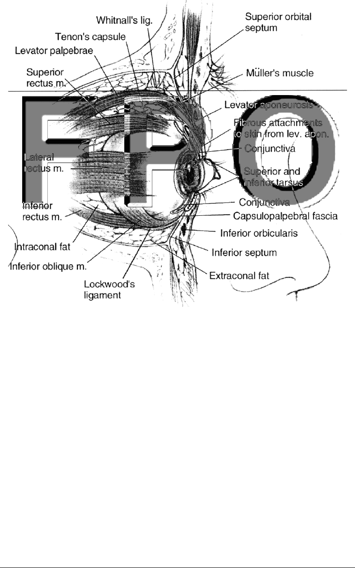
Eyelids 5
FIGURE 1-2. Parasagittal section of the orbit, showing eyelid structures.

tum fuses with the capsulopalpebral fascia 5 mm inferior to the lower
border of the tarsus.
14
In addition, in many Asians a subcutaneous
fat pad exists anterior to the septum.
18
The strength of the orbital septum varies among individuals and
with age. With time, the septum attenuates, resulting in anterior pro-
lapse of orbital fat.
8,19
The tarsal plates (Fig. 1-2) provide rigidity to the eyelids. They
are composed of dense, fibrous connective tissue. The upper tarsus
measures 10–12 mm vertically, and the lower measures 3–5 mm.
20
The tarsal borders adjacent to the lid margin are straight, whereas
the opposite edges have a convex curvature. The posterior edge of the
tarsus is firmly attached to the palpebral conjunctiva, which extends
onto the eyelid margin and terminates at the gray line.
Within the tarsus lie branched, acinar, sebaceous glands with long
central ducts. Known as the meibomian glands, they open at the eye-
lid margin just posterior to the gray line and secrete the oily layer of
the tear film. There are about 25 in the upper eyelid and 20 in the
lower.
11
Inflammation of these glands, known as meibomitis, may,
over a long term, result in distichiasis,
21
or abnormal hair follicles
that, unlike the normal eyelashes, curve inward toward the globe,
causing discomfort and possibly corneal abrasion. A common treat-
ment for distichiasis, electrohyfrecation, may cause focal necrosis of
the tarsus, resulting in notching at the eyelid margin.
8
Similarly, ex-
cessive cryotherapy for distichiasis can cause a wider-than-planned
area of lash loss and scarring.
Emanating from the medial and lateral borders of the tarsi and
anchoring them to the orbital rim are the canthal ligaments. They
are formed by a confluence of the upper and lower crura, the exten-
sions of the margins of the upper and lower tarsi, respectively. They
support not only the tarsi but also the orbicularis. The medial can-
thal ligament splits into three arms: anterior, posterior, and supe-
rior. The anterior arm attaches to the maxillary bone, anterior to the
lacrimal crest; the posterior arm attaches to the posterior lacrimal
crest
22,23
; and the superior arm inserts onto the orbital process of the
frontal bone.
24
The lateral canthal ligament inserts 3–4 mm inside
the lateral orbital rim at Whitnall’s tubercle, on the zygomatic bone.
25, 26
During lower eyelid-tightening procedures, which usually involve sur-
gical manipulation of the lateral aspect of the lower tarsus and the
lateral canthal ligament, the posterior direction and periosteal in-
sertion of the lateral canthal ligament must be reestablished. Laxity
of the canthal ligaments can cause ectropion, as well as cosmetically
apparent shortening of the horizontal palpebral fissure.
27
6 Cosmetic Facial Anatomy

An important support for the upper eyelid is Whitnall’s ligament.
Its role has been debated; it may serve as a fulcrum-like check liga-
ment for the levator or as a swinging suspender, providing vertical
support for the upper eyelid.
17,28,29
Despite this debate, it is under-
stood that Whitnall’s ligament suspends the lacrimal gland, superior
oblique ligament, levator muscle (with the primary support for the
levator coming from the globe), and Tenon’s capsule. Whitnall’s liga-
ment is a transverse fibrous condensation that inserts medially in-
side the superomedial orbital rim on the frontal bone at the trochlea
and laterally inside the superolateral orbital rim, near the frontozy-
gomatic suture, where it fuses with fibers of the lacrimal gland cap-
sule. It encircles the levator complex
30
at the level of the junction of
the levator muscle and the fibrous levator aponeurosis. The aponeu-
rosis extends another 14–20 mm inferior to Whitnall’s ligament to in-
sert on the lower third of the anterior face of the upper tarsus. De-
hiscence of the levator aponeurosis is responsible for most cases of
involutional ptosis.
Musculature
The orbicularis oculi muscle (Fig. 1-2) surrounds the anterior or-
bit and can be divided into three components: pretarsal, preseptal,
and orbital.
31
The pretarsal orbicularis originates from the anterior
and posterior arms of the medial canthal ligament, and it is firmly
adherent to the anterior face of the tarsi. Medially, the pretarsal or-
bicularis divides into a superficial head, which surrounds the canali-
culi, and a deep head, which inserts on the posterior lacrimal crest
and lacrimal fascia. These insertions allow the pretarsal orbicularis
to play an important role in the lacrimal pump mechanism. The pre-
septal orbicularis originates from the upper and lower margins of the
medial canthal ligament and inserts lateral to the orbital rim on the
zygoma. It overlies the orbital septum and orbital rim; it is separated
from the septum by a fibrofatty layer, the postorbicularis fascia.
8
This
layer is an important surgical dissection plane. The orbital orbicu-
laris originates from the maxillary and frontal bones, as well as from
the medial canthal ligament; it overrides the orbital rims and inserts
at the same location as the preseptal orbicularis. The latter two por-
tions of the orbicularis are responsible for forced eyelid closure.
Two important components of the orbicularis are the muscle of
Riolan and Horner’s muscle. The muscle of Riolan is a small segment
of the orbicularis that is separated from the pretarsal orbicularis by
the eyelash follicles.
15
It corresponds to the gray line seen at the eye-
Eyelids 7

lid margin.
16
The deep pretarsal head of the orbicularis is known as
Horner’s muscle. Contraction of this muscle pulls the eyelids medi-
ally and posteriorly. In so doing, Horner’s muscle compresses the
canaliculi and lacrimal ampullae, pushing tears toward the lacrimal
sac and, at the same time, creating negative pressure within the
lacrimal sac.
32
This mechanism, known as the lacrimal pump,
33
can
therefore be compromised by weakening or laxity of the eyelids, re-
sulting in epiphora.
34
The main retractor of the upper eyelid is the levator palpebrae su-
perioris (Fig. 1-2). It originates at the superior edge of the annulus of
Zinn in the orbital apex and courses anteriorly through the superior
orbit, along the superior aspect of the superior rectus muscle. As it ap-
proaches the upper eyelid, the levator is encircled by Whitnall’s liga-
ment.
30
At this point, the levator muscle transitions into the fibrous
levator aponeurosis, which courses inferiorly for another 14–20 mm,
where the posterior one-third of the lamellae attach to the inferior
third of the anterior surface of the tarsus. Also at the level of Whit-
nall’s ligament, the levator sends off lateral and medial horns. The lat-
eral horn attaches to the zygomatic bone. The medial horn fuses with
the posterior arm of the medial canthal ligament and inserts on the
posterior lacrimal crest. The lateral and medial horns help ensure that
the upper eyelid maintains a curvature that keeps it apposed to the
globe during opening.
7
The anterior two-thirds of the lamellae of the
levator aponeurosis sends fibers anteriorly through the septum and or-
bicularis to the skin; these insertions form the upper eyelid crease.
9
Aging affects both the levator muscle and the aponeurosis. Age-
related thinning and dehiscence of the aponeurosis from the tarsus
is a common cause of involutional ptosis.
35,36
In addition, the muscle
belly can become infiltrated with fat and connective tissue.
8
Underlying the levator aponeurosis and attached to it via loose
connective tissue is Müller’s muscle, which is sympathetically inner-
vated and composed of smooth muscle fibers. It originates from the
undersurface of the levator and courses inferiorly for approximately
15 mm to insert with elastic fibers onto the superior edge of the tar-
sus in the upper eyelid. A lateral extension of Müller’s muscle divides
the lacrimal gland into its two lobes.
37
It is generally accepted that
Müller’s muscle is a secondary transmitter of lift to the upper eyelid,
as evidenced by the 2- to 3-mm ptosis seen either in sympathetic den-
ervation syndromes, such as Horner’s syndrome, or in the normal
fatigue-related decrease in sympathetic tone. One group has sug-
gested that Müller’s muscle may serve as a primary transmitter of
levator muscle tone to the tarsal plate.
38
8 Cosmetic Facial Anatomy