Gladstone Geoffrey J., Black Evan H., Myint Sh. Oculoplastic Surgery Atlas
Подождите немного. Документ загружается.


A No. 15 blade is used to make an incision down to the perios-
teum in the central incision. Blunt dissection in the paramedian in-
cision is preferred to prevent bleeding from tributary vessels. The
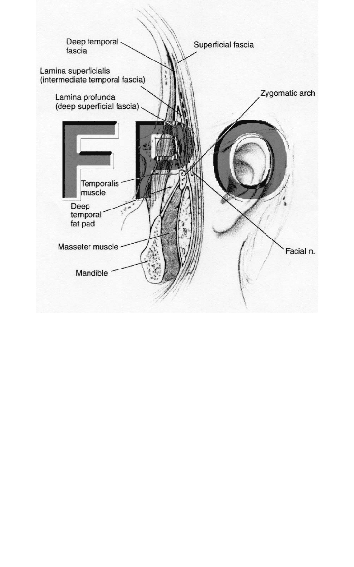
temporal incisions require more meticulous dissection. This incision
is made through the dermis; with blunt dissection, then, spreading
the tissue exposes the temporal parietal fascia, which moves with the
skin. Further dissection exposes the deep temporalis fascia, which
does not move and is superficial to the temporalis muscle. This fas-
cia is a shiny, white, glistening tissue. The plane just above this white
tissue at the superficial layer of the deep temporalis fascia is the
plane of dissection (Fig. 2-2). The temporal branch of the facial nerve
lies within the temporal parietal fascia and is superior to the surgi-
cal plane, out of harm’s way. If desired, a 4 mm drill can be used for
fixation at the posteriormost portions of the two paramedian or lat-
eral incisions. This is done early in the procedure before the perios-
teum is released.
30 Endoscopic Foreheadplasty
Surgical Management 31
FIGURE 2-2. Anatomy in the temporal area. The dotted line is the appro-
priate plane of dissection.

The endoscope is now introduced through the temporal incision
above the deep temporalis fascia. A blunt elevator is used in a di-
rection toward the ear, remaining 1 cm posterior to the facial nerve
marking, and the tissue is carefully elevated. The temporal and cen-
tral pockets are connected blindly, laterally to medially, detaching
the temporal crescent ligament, which is the fusion of the galea
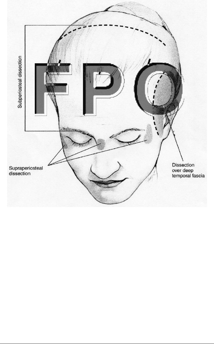
aponeurotica to the crest. Most surgeons prefer to dissect in the sub-
periosteal plane, especially if the forehead is high (Fig. 2-3). The sub-
galeal plane of dissection results in increasing the distance from the
brow to the hairline. The dissection posteriorly is done behind the in-
cision line. Some surgeons choose to extend this farther back toward
the vertex. Continuing in the temporal pocket, dissection is carried
out down toward the lateral canthal angle with direct endoscopic vi-
sualization. It is important to stay on the deep temporalis fascia while
elevating upward gently. At this point the orbicularis-temporal liga-
ment is identified, which is a tough ligament joining the lateral or-
bicularis to the deep temporalis fascia. Carefully dissecting beyond
this point discloses the zygomaticotemporal (sentinal) vein. It is typ-
ically 5 mm temporal to the zygomaticofrontal suture line and is an
important landmark. It is here that branches of the facial nerve are
located, so the dissection should be minimal.
The lateral canthal ligament can be detached with endoscopic vi-
sualization. The arcus marginalis is then released along the supra-
orbital rim, extending medially toward the supraorbital nerve by in-
cising the periosteum. Once the supraorbital nerve is located, a
supraperiosteal pocket is formed above the bridge of the nose to ad-
dress the depressor muscles (procerus, corrugator, depressor super-
cilii, orbicularis). Using blunt dissection, the tissues are moved side
to side to separate the muscles for better visualization. At this point
the tough corrugators can be seen with their insertion and origin on
both sides. The corrugators can be avulsed, rather than cut, to pre-
vent injury to the supratrochlear nerve. This can be accomplished
with endoscopic scissors or laser. Branches of the supratrochlear
nerve are sometimes seen within the corrugator and should be
avoided. The procerus can be addressed in a similar fashion, with
avulsion. The depressor supercilii should also be avulsed and not cut
because of vessels within it. The orbicularis muscle can be visualized
within this area and can be cut vertically. To eliminate the glabellar
lines, vertical incisions can be made in the periosteum centrally, up
to the dermis. At this point, some surgeons choose to perform multi-
ple vertical interfascicular neurotomies to the superficial branches of
32 Endoscopic Foreheadplasty
Fixation 33
FIGURE 2-3. Areas of subperiosteal and supraperiosteal dissection.
the facial nerve along the superior brow lateral to the supraorbital
nerve. This practice theoretically prevents regeneration of the de-
pressor muscles after the surgery, thereby decreasing the chance of
recurrent postoperative brow ptosis.
FIXATION
Once the periosteum has been adequately released, attention is given
to fixation. There are many methods available to fixate the scalp. The
key to the endoscopic forehead procedure is not as much the fixation

as it is the release of the periosteum and the muscles. Anchor or screw
techniques for fixation include use of an internal screw or plate, Mitek
anchor, external screw, and k-wire. Other techniques include galea-
frontalis-occipitalis release, use of lateral suspension sutures or bol-
ster fixation sutures, anterior scalp port excision, galea-frontalis ad-
vancement, creation of a cortical tunnel, and use of tissue adhesives.
Regardless of the technique, it must achieve simplicity, reproducibil-
ity, safety, and long-term results. If the fixation is under tension,
brow ptosis occurs. We prefer the Mitek anchor screw or the cortical
bone tunnel technique for fixation.
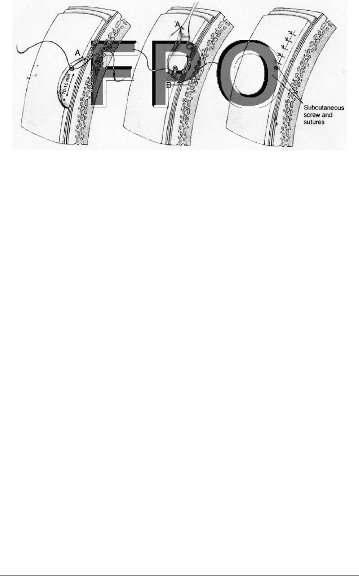
With the Mitek screw, a 4 mm drill bit is used in the outer cal-
varium approximately 1.0–1.5 cm from the anteriormost portions of
the lateral or paramedian incisions. Once drilled, a 2-0 suture and
needle attached to the screw is passed from anterior to posterior
through galea-periosteum at the anterior portion of the lateral or
paramedial incision. This is a double-armed suture; therefore both
are passed anteriorly, elevating the scalp once secured (Fig 2-4). The
temporal region is closed with 3-0 or 4-0 nylon suture from the tem-
poral parietal fascia to the deep temporalis fascia. The remainder of
the skin is closed with surgical staples.
When using the cortical bone tunnel, the instrument is placed in
the lateral or paramedial incision. Sometimes it is necessary to en-
large the incision to accommodate the instrument. A 4 mm drill bit
is used to drill on each side of the instrument, which allows two
45-degree holes that meet in the outer calvarium. A 3-0 or 4-0 nylon
or Ethibond suture is pulled through the hole with a hook and su-
tured in the fashion described earlier.
34 Endoscopic Foreheadplasty
POSTOPERATIVE CARE
After the hair is washed with shampoo, ABD pads and Kerlex dress-
ings are placed around the forehead for 48 hours. Complete wound
healing usually occurs within 42–60 days. Postoperative antibiotics
are not routinely prescribed. A prednisone taper is given, and the pa-
tient is seen in 2 days to remove the dressing. The staples are re-
moved within 7–10 days. Patients usually can return to work within
3–5 days. They are instructed preoperatively that scalp numbness
may persist for 3 months, and “hair shock” can result in some alope-
cia around the incision sites also lasting 3 months.
A worrisome complication is injury to the facial nerve, but it can
usually be avoided by carefully dissecting in the correct plane.
Hematomas and infections are rare. This procedure can be performed
on bald patients with minimal scarring. Endoscopic foreheadplasty is
an accurate procedure that has a higher patient acceptance rate than
the traditional coronal lift. It is a nice addition to the surgeon’s ar-
mamentarium of browlift procedures.
Postoperative Care 35
FIGURE 2-4. Fixation using the Mitek anchor.

3
D
IRECT EYEBROW LIFT
• César A. Sierra, MD
Department of Ophthalmology
Yale University School of Medicine
New Haven, Connecticut
B
efore moving to more complex approaches such as the endo-
scopic eyebrow lift, the surgeon must master the fundamental
concepts of preoperative evaluation, indications for surgery,
surgical anatomy, and the basic direct browlift procedure.
The direct eyebrow lift is generally performed with local anes-
thesia and requires excising the tissues adjacent to the superior line
of the brow follicles. The advantages of a direct approach include less
operating time, less anesthesia, and the ability to address the whole
eyebrow or a specific segment of it by manipulating the shape of the
excised tissue. In general, the closer the surgery is performed to the
eyebrow the more effective it is. The direct browplasty is therefore
an excellent option in cases of severe brow ptosis or in patients with
a receding hairline or baldness where incisions used for other ap-
proaches would leave noticeable scars. The degree of elevation and
the long-term results are excellent when compared with other ap-
proaches. The major negative aspect is the inferior cosmetic outcome
with a supraciliary scar that is sometimes more visible than expected,
depending on the healing ability of the patient.
37

ETIOLOGY
As the aging process continues, the gravitational syndrome occurs at
different rates in different people, resulting in sagging or involutional
ptosis of the eyebrow with redundancy of skin. Downward displace-
ment of the tissues also takes place as a result of facial overactivity
patterns such as squinting and frowning. In these cases the overac-
tive depressor muscles determine the most affected area of the brow.
Paralysis of the facial nerve [cranial nerve (CN) VII] or its temporal
branch also leads to moderate to severe eyebrow ptosis with blunt-
ing of the ipsilateral forehead rhytids due to loss of tone of the
frontalis muscle.
PREOPERATIVE EVALUATION AND
INCISION MARKINGS
The position of the eyebrows is extremely important when evaluat-
ing a patient who complains of superior visual blockage or drooping
eyelids. Commonly, patients who present with upper eyelid ptosis or
dermatochalasis also have eyebrow ptosis. This sagging of the eye-
brow can add fullness and weight that, when combined with an ab-
normal levator muscle or excessive eyelid skin, can result in more
hooding and loss of superior visual field. Recognition is crucial to pre-
vent inadequate surgical management and failure to help the patient.
Gender differences exist, with eyebrows being higher, more
arched, and slightly raised laterally in women. Male eyebrows are
usually described as T-shaped: They are less inclined and lower, with
the inferior border at the level of the superior orbital rim. This base-
line difference could be the reason that brow ptosis is more prevalent
in men than in women.
The preoperative evaluation is always performed with the patient
sitting upright with relaxed frontalis muscle action. A 1:1 lift
desired/skin excised ratio is used. The eyebrow is manually elevated
to an appropriate position. This amount of lift is measured using a
ruler in the lateral, central, and medial aspects of the eyebrow. A
fusiform skin incision pattern is usually obtained where the upper
line of eyebrow hair follicles dictates the inferior marking, and the
superior marking is given by the measurements previously taken for
each portion of the eyebrow. As a general rule, the inferior incision
line should include a small number of the superiormost brow hairs
38 Direct Eyebrow Lift

to conceal the scar. It is also recommended that the supraorbital notch
be marked to maintain awareness of where the supraorbital neu-
rovascular bundle exits and becomes more superficial.
SURGICAL MANAGEMENT
After the skin markings are outlined with a surgical marking pen,
the area is adequately infiltrated with local anesthetic. The incision
(Fig. 3-1) is started at the inferior marking where the superiormost
brow follicles follow a cephalad orientation. The incision must be
made with the blade beveled parallel to these follicles to prevent ex-
Surgical Management 39
FIGURE 3-1. Suprabrow incision after marking the incision site.

cessive damage, loss of eyebrow hair, and a conspicuous scar (Fig.
3-2). The incision on the superior marking follows, with the blade an-
gled in the same direction to provide adequate apposition and ever-
sion of the wound edges. Care should be taken at all times to incise
superficially in the area of the supraorbital neurovascular bundle to
avoid inadvertent injury. Deeper sharp dissection and excision of the
skin and subcutaneous tissue are done laterally, where there is no
risk of damage to the supraorbital neurovascular bundle (Fig. 3-3).
Careful superficial dissection in the medial aspect of the eyebrow and
hemostasis follows.
The wound is first reapproximated with buried subcutaneous 5-0
Vicryl. Each suture must be perfectly aligned in the plane horizontal
to the wound edges. It is also crucial that the bites taken with this
suture maintain exactly the same depth in the vertical aspect of both
sides of the wound to provide precise apposition of the edges, thereby
decreasing scar formation.
The tendency of the scar as it heals is to draw the skin toward
the vertical axis, depressing it. Therefore the epidermis should be
closed with a 6-0 nonabsorbable suture in a vertical or horizontal mat-
tress fashion to achieve an adequate amount of eversion of the skin
edges to allow the expected flattening.
For cases of severe facial nerve (CN VII) paralysis, it is recom-
mended that dissection be carried deeper through the frontalis mus-
cle and nonabsorbable 5-0 or absorbable 4-0 sutures be carefully
passed though the muscle and periosteum in a buried fashion to
achieve better fixation of the ptotic brow. For milder cases of brow
ptosis, this technique would create a more depressed scar and di-
minish the action of the frontalis muscle. The asymmetry created by
unilateral blunting of the deep forehead rhytides in these cases can
be corrected if the incision is created in the mid forehead instead of
the superior edge of the eyebrow.
40 Direct Eyebrow Lift