Gladstone Geoffrey J., Black Evan H., Myint Sh. Oculoplastic Surgery Atlas
Подождите немного. Документ загружается.


Less well defined than their counterparts that elevate the upper
eyelid, the lower eyelid retractors—capsulopalpebral fascia and in-
ferior tarsal muscle—are palpebral extensions of the inferior rectus
muscle. The inferior rectus muscle, through these lower eyelid re-
tractors, is responsible for the full extent of depression of the lower
eyelid during downgaze.
8
A fibrous extension of the inferior rectus
muscle, the capsulopalpebral head of the inferior rectus wraps around
the inferior oblique muscle, at which point the capsulopalpebral head
splits into inferior and superior divisions. The inferior division, which
is the capsulopalpebral fascia, then rejoins the superior division, the
inferior tarsal muscle,
14
which, like Müller’s muscle, is composed of
smooth muscle fibers. These two layers are not generally distinct dur-
ing surgical dissection.
The lower eyelid retractors have three insertions. Posteriorly, the
retractors insert on Tenon’s fascia. Centrally, the inferior tarsal mus-
cle fibers terminate a few millimeters inferior to the tarsus,
14
and a
fibrous continuation attaches to the inferior border of the tarsus. An-
teriorly, the capsulopalpebral fascia fuses with the orbital septum
4 mm inferior to the tarsus. Fibers continue through the septum and
attach to the subcutaneous tissue, forming the lower eyelid crease.
7
Eyelid Fat Pads
The eyelid fat pads (Fig. 1-2) play an important role in the ap-
pearance and contour of the eyelids. In the youthful face, this anterior
orbital fat imparts a fullness and smoothness to the upper and lower
eyelids. With age, atrophy of eyelid fat can cause the eyelids to sink
posteriorly, resulting in involutional enophthalmos and a lid crease
displaced away from the lid margin.
27
In addition, weakening of the
orbital septum can allow anterior prolapse of the anterior orbital fat,
resulting in puffy-appearing eyelids, known as steatoblepharon.
19
The upper eyelid contains two fat pads, separated by the trochlea
and superior oblique tendon, which are located posterior to the or-
bital septum and immediately anterior to the levator muscle and
aponeurosis. This anatomic relation is a reliable guide for the eyelid
surgeon who wishes to combine levator aponeurosis repair with bleph-
aroplasty, fat pad excision, or both. This region of the upper eyelid is
divided into three fibrous compartments. The medial and central com-
partments contain the medial and preaponeurotic fat pads,
39
and the
lateral compartment contains the lacrimal gland. Care must be taken
not to confuse the lacrimal gland with eyelid fat in the upper eyelid.
The lacrimal gland, which sits lateral to the two upper eyelid fat pads,
Eyelids 9

appears pink and firm, in contrast to the glistening, yellow, loose-ap-
pearing fat in the central preaponeurotic pad and the whiter, more
fibrous nasal fat pad.
The lower eyelid contains three fat pads that are enclosed in three
fibrous compartments: medial, central, and lateral. The inferior
oblique muscle courses between the medial and central compartments
in the lower eyelid; and the central and lateral fat pads are separated
by a fibrous acurate extension passing from the inferior oblique to
the inferior lateral orbital wall. Because the eyelid fat pads are an-
terior projections of orbital fat, care must be taken intraoperatively
not to cause excessive traction, as orbital hemorrhage may result dur-
ing the intra- or postoperative period.
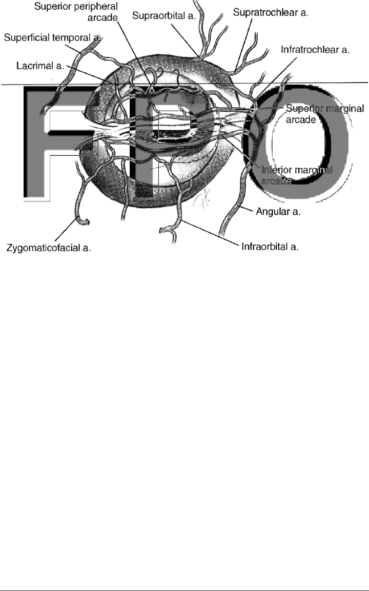
Eyelid Vasculature
Eyelid blood supply (Fig. 1-3) arises from both the external and
internal carotid arteries. The external carotid artery gives rise to the
facial artery, the superficial temporal artery, and the infraorbital ar-
tery. As it courses across the face diagonally toward the nasolabial
folds, the facial artery becomes the angular artery, which lies directly
underneath the orbicularis and feeds the vascular arcades of the eye-
lids at the medial canthus. The internal carotid artery gives rise to
the ophthalmic artery, which in turn terminates as the lacrimal,
frontal, supraorbital, supratrochlear, and nasal arteries. Anastomoses
between the angular, lacrimal, and supratrochlear arteries form the
superior marginal arcade and superior peripheral arcade in the up-
per eyelid. The angular artery anastomoses in the lower eyelid with
the infraorbital and zygomaticofacial arteries to form the inferior
marginal arcade. A poorly developed inferior peripheral arcade is
present in some individuals.
7,8
In the upper and lower eyelids, the marginal arcades lie just an-
terior to the tarsi, 2–4 mm from the eyelid margin. Also in the upper
eyelid, the superior peripheral arcade lies just anterior to Müller’s
muscle, superior to the tarsus. This arcade not only serves the upper
part of the upper eyelid it supplies the superior conjunctival fornix
and communicates with the anterior ciliary vessels near the limbus.
Dissection in the plane of Müller’s muscle can cause hemorrhage from
this arcade.
8
The facial vein is the principal venous drainage source for the eye-
lids. It courses superficial and lateral to the facial artery. It begins
near the medial canthus as the angular vein and anastomoses with
the superior ophthalmic vein via the supraorbital vein.
10 Cosmetic Facial Anatomy
Lymphatic drainage of the eyelids has been somewhat elusive, but
a study by Cook et al.
40,41
demonstrated in a primate model that the
entire upper eyelid drains to the parotid lymph nodes, with additional
drainage from the medial upper eyelid to the submandibular lymph
nodes. The medial canthus and lateral lower eyelid drain to the
parotid lymph nodes. The central and medial lower eyelid drain to
the submandibular lymph nodes.
Innervation
The eyelids are innervated by the facial nerve [cranial nerve (CN)
VII], oculomotor nerve (CN III), trigeminal nerve (CN V), and sym-
pathetic fibers from the superior cervical ganglion. Sensory innerva-
tion to the upper eyelid is provided by the ophthalmic division of
the trigeminal nerve (CN V
1
), which has three branches—lacrimal,
frontal, nasociliary—all of which enter the orbit via the superior or-
bital fissure. The lacrimal nerve supplies the lacrimal gland con-
junctiva and lateral upper eyelid, and it sends off a branch that anas-
tomoses with the zygomaticotemporal nerve. The frontal nerve
Eyelids 11
FIGURE 1-3. Arterial blood supply to the eyelids.

courses anteriorly between the periorbita and levator, dividing into
the supraorbital and supratrochlear nerves. The supratrochlear nerve
innervates the medial upper eyelid and forehead, and the two divi-
sions of the supraorbital nerve innervate most of the remainder of
the forehead. A superficial division passes anterior to the frontalis
muscle to innervate the forehead skin, and a deep division passes lat-
erally anterior to the pericranium and supplies the frontoparietal
scalp.
42
The nasociliary branch gives rise to the posterior and ante-
rior ethmoidal nerves, two or three long ciliary nerves to the globe,
a sensory root to the ciliary ganglion, and the infratrochlear nerve.
8
Sensory innervation to the lower eyelid is provided by the maxil-
lary branch of the trigeminal nerve (CN V
2
). The zygomatic branch
from V
3
divides into the zygomaticofacial and zygomaticotemporal
nerves. The zygomaticofacial nerve courses along the inferolateral or-
bit, passes through the zygomaticofacial foramen, and supplies the
skin of the cheek. The zygomaticotemporal nerve exits the orbit into
the temporal fossa, innervating the lateral forehead. The infraorbital
nerve, a continuation of V
2
, exits via the infraorbital foramen, yield-
ing several terminal branches—inferior palpebral, lateral nasal, and
superior labial nerves—which supply the skin and conjunctiva of the
lower eyelid, the skin and septum of the nose, and the skin and mu-
cosa of the upper lip, respectively.
8
Motor innervation to the levator muscle comes from the superior
division of the oculomotor nerve (CN III). This division courses within
the muscle cone of the orbit, entering the superior rectus from its in-
ferior aspect, 15 mm from the orbital apex. At this point, it also sends
off terminal fibers, which pass around or through the medial aspect
of the superior rectus to innervate the levator.
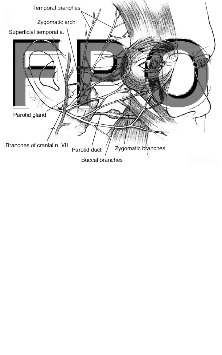
The frontalis and orbicularis muscles are innervated by divisions
of the facial nerve (Fig. 1-4). After originating at its nucleus in the
pons, the facial nerve leaves the facial canal via the stylomastoid fora-
men. It then passes through the parotid gland and gives rise to sev-
eral divisions: temporal, zygomatic, buccal, mandibular, and cervical
nerves. The temporal branch innervates the frontalis muscle and is
one of the most commonly injured nerves during forehead and tem-
poral surgical dissection. The temporal, zygomatic, and buccal divi-
sions all contribute innervation to the orbicularis oculi, with signifi-
cant overlap of the regions innervated by each nerve.
12 Cosmetic Facial Anatomy
MID-FACE AND LOWER FACE
Osteology
The topography of the mid-face is determined to a significant ex-
tent by the bony anatomy. The facial bones are demarcated from the
bones of the cranium at the level of the orbits. The superior extent
of the mid-face is the zygomaticofrontal suture, and the teeth form
the inferior border. The posterior border is defined by the sphe-
noethmoid junction and the pterygoid plates.
Most of the mid-facial bones extend from the borders of the orbit.
The zygoma, which forms the lateral facial buttress, and the greater
wing of the sphenoid together form the lateral orbital wall. The me-
dial orbital wall includes the ethmoid, lacrimal, sphenoid, and max-
illary bones. Associated with them are the bony nasal turbinates. The
maxilla extends from the inferior portion of the medial wall to the or-
bital floor and extends inferiorly to form the anterior bony surface of
the mid-face until giving rise to the upper teeth. The contour of the
maxilla has recently been shown to undergo characteristic changes
Mid-Face and Lower Face 13
FIGURE 1-4. Anatomy of the facial nerve (cranial nerve VII).

with age.
43
The palatine bone, at its superior extent, forms the pos-
terior orbital floor and extends inferiorly into the posterior mid-face.
The mandible provides skeletal support to the lower face. It forms
a synovial joint with the skull at the condyles; the muscles of masti-
cation stabilize this joint.
A series of studies by Pessa et al. indicated that the facial bones
remodel throughout adulthood and are partly responsible for the ag-
ing changes seen in the mid-face. Specifically, the orbital rim appears
to move posteriorly with respect to the plane of the cornea,
44
and
the maxillary arch and the orbital aperture curvature increase with
age.
43,45
Skin and Subcutaneous Tissues
The facial skin and subcutaneous tissues vary in thickness, tex-
ture, color, and mobility, dividing the face into the aesthetic units
discussed in this chapter. The skin consists of three layers: epider-
mis, dermis, and subcutaneous tissue. The epidermis consists of ker-
atinized, stratified, squamous epithelium. The underlying dermis is
divided into a superficial papillary dermis comprised of randomly ori-
ented collagen fibrils and a deeper reticular dermis, which is vascu-
larized and has collagen fibrils oriented parallel to the epidermal
surface.
46
The underlying subcutaneous fibrofatty layer varies
in thickness among individuals and facial aesthetic units, with the
cheeks, temples, and neck being the thickest.
Age-related changes in the skin are usually seen in conjunction
with photodamage. They include loss of elasticity, atrophy of subcu-
taneous fat, and pigmentary changes.
47
Connective Tissue
The predominant connective tissue layer in the midface is the su-
perficial musculoaponeurotic system (SMAS) (Fig. 1-5). First de-
scribed more than 25 years ago,
48
the SMAS has been suggested to
be a transmitter and distributor of facial muscular contractions to
the skin and a key structure in the development of mid-face ptosis.
Further anatomic study has characterized the SMAS in the periocu-
lar and mid-face regions.
49,50
In recent years, the SMAS has been
identified as an important structure in facial rhytidectomy, and the
SMAS-invested muscles are recognized as targets for facial soft-
tissue augmentation.
5
14 Cosmetic Facial Anatomy
Structurally, the SMAS is a fibromuscular plane that divides the
parotid and cheek fat into two layers.
48
It invests the zygomaticus
major, zygomaticus minor, and levator labii superioris
5
and lies 11–13
mm deep to the skin at mid-cheek.
51
It is continuous with the frontalis
in the upper face, the platysma in the lower face,
52–57
and the ante-
rior and posterior orbicularis fascia
50
in the orbital region. The ma-
jor vessels and nerves, including the motor branches of the facial
nerve, lie deep to the SMAS and send perforating branches anteri-
orly through it.
The following soft tissue attachments support the SMAS: the
parotid fascia, the masseteric fascia via masseteric cutaneous liga-
ments,
49
the platysma via platysma auricular ligaments and anterior
Mid-Face and Lower Face 15
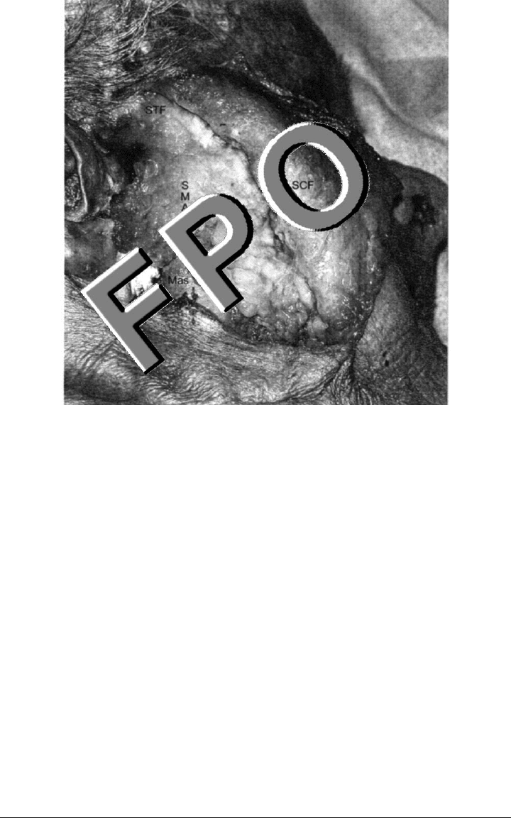
FIGURE 1-5. Human cadaver dissection of the mid-face. Skin and subcuta-
neous fat (SCF), also known as the malar fat pad in this region, are re-
flected anteriorly via a preauricular incision, revealing the superficial mus-
culoaponeurotic system (SMAS), which is continuous with the superficial
temporalis fascia (STF) and lies superficial to the masseter muscle (Mas).

platysma cutaneous ligaments,
58
and the zygomatic major and minor
muscles via their bony attachments. Bony attachments are at the zy-
gomatic arch
48,55
and the mandible,
58
as well as at the inferior or-
bital rim via the orbitomalar ligament,
50
which in turn sends fibers
anteriorly to the skin to form the nasojugal fold.
The SMAS is subject to age-related changes, which are in large
part responsible for mid-face ptosis. Lucarelli et al.
49
demonstrated
age-related attenuation of the orbitomalar, masseteric cutaneous, and
zygomatic ligaments, which support the SMAS and associated malar
and buccal fat pads. Cutaneous projections of the orbitomalar liga-
ment help form the nasojugal and malar skinfolds; increased traction
on the ligament by a descended SMAS may be partly responsible for
the accentuation of the nasojugal and malar skin folds that appear
with age.
50
Involutional descent of the malar fat pad, which attaches
to the superficial surface of the SMAS, results in increased promi-
nence of the nasojugal fold.
59–61
A recent outcomes study by Hamra
62
indicated that the malar fat pad may continue to descend more rap-
idly than the SMAS following facelift surgery.
Musculature
The muscles of facial expression are flat muscles that have high vari-
ability from one individual to another. Freilinger et al.
63
reported in
1987 the three-dimensional arrangement of these muscles in the mid-
face and lower face, dividing the muscles into four layers. The first
and most superficial layer includes the orbicularis oculi, zygomaticus
minor, and depressor anguli oris. The second layer includes the zy-
gomaticus major, levator labii superioris nasae et alaque, platysma,
risorius, and depressor labii inferioris. Progressing deeper, the third
layer includes the orbicularis oris and levator labii superioris. The
fourth and deepest layer includes the mentalis, levator anguli oris,
and buccinator. The facial nerve branches travel between the third
and fourth layers, innervating the first three layers from below and
the fourth layer from above. Subsequent study has shown the zygo-
maticus major, zygomaticus minor, and levator labii superioris all to
be invested in the SMAS,
51
illustrating an evolving understanding of
the muscular anatomy of the mid-face.
The muscles of mastication include the masseter and temporalis,
with two associated muscles: the buccinator and orbicularis oris. The
orbicularis oris acts as a sphincter at the mouth, and the buccinator
provides medially directed tension on the cheeks, keeping food in the
center of the mouth. The masseter originates at the zygomatic arch
16 Cosmetic Facial Anatomy

and inserts in the mandible. The temporalis inserts at the temporalis
fossa and at the medial mandibular ramus and coronoid process. The
temporalis is covered by a tough fascia, the deep temporalis fascia.
Superior to the zygomatic arch, the superficial temporalis fascia
arises and is separated from the deep temporalis fascia by the su-
perficial temporal fat pad. The superficial temporalis fascia is con-
tinuous with the SMAS. Densely adherent to the deep aspect of the
superficial temporal fascia is the temporal branch of the facial nerve;
dissection in this region must therefore be deep to the superficial tem-
poralis fascia, in the plane of the deep temporalis fascia, to avoid in-
juring the nerve.
Mid-face Fat Pads
Apart from the suborbicularis oculi fat pad (SOOF) described
above, the principal fat pads of the mid-face are the malar and buc-
cal fat pads. The malar fat pad comprises the subcutaneous fat in the
cheek and is continuous with both the jowl fat underneath the jaw-
line
60
and the SOOF.
49
The buccal fat pad rests deeper in the face,
bounded medially by the buccal mucosa, with buccal, temporal, and
pterygoid extensions.
64,65
This sub-SMAS fat in the malar region has
been demonstrated to be continuous with the ROOF fat.
50
Although frequently postulated as a rationale for facial soft tis-
sue augmentation,
66–70
few studies since the initial work of Gonzalez-
Ulloa and Flores
71
have demonstrated actual volume loss of facial fat
and muscles with age.
Facial Vasculature
The facial vasculature arises from the internal and external
carotid arteries. The first branch of the internal carotid artery is the
ophthalmic artery, which supplies the eyelids, forehead, and dorsum
of the nose. The forehead is supplied by the supraorbital and supra-
trochlear arteries. The eyelids are vascularized by the infraorbital,
palpebral, and marginal arteries. The nose is supplied by the ante-
rior and posterior ethmoid arteries.
The external carotid artery branches into the facial, internal max-
illary, and superficial temporal arteries. The facial artery supplies
the lips via the superior and inferior labial arteries, as well as the
lateral nose and nasal dorsum, with anastomoses to the anterior and
posterior ethmoidal arteries. Because of these anastomoses, high-
pressure injection of steroids or soft-tissue fillers can result in retro-
Mid-Face and Lower Face 17

grade flow, embolization of the ophthalmic artery or central nervous
system vasculature, and ultimately blindness
72–74
or stroke.
75,76
The
internal maxillary artery gives off the infraorbital artery, which en-
ters the orbit at the infraorbital fissure and courses along the infra-
orbital groove, exiting the orbit at the infraorbital foramen to supply
the lower eyelid. Care must be taken not to damage the infraorbital
artery and associated nerve during surgical dissection along the or-
bital floor and inferior orbital rim. The superficial temporal artery
branches from the external carotid artery in the parotid gland. At the
level of the zygomatic arch, it gives off the transverse facial artery to
supply the lateral canthal area. Superior to the zygomatic arch, the
superficial temporal artery travels within the plane of the SMAS and
gives off the middle temporal artery, which supplies the superficial
temporal fat pad and temporalis muscle. Terminal branches of the
superficial temporal artery supply the parietal area and forehead,
with anastomoses to the supraorbital and supratrochlear arteries.
Facial Innervation
Motor innervation to the mid-face and lower face is via the facial
nerve (CN VII). After exiting the stylomastoid foramen, it enters the
parotid gland, where it divides into its main branches: temporal,
zygomatic, buccal, mandibular, and cervical. The two most subject to
surgical injury are the temporal and mandibular nerves.
The temporal nerve exits the parotid gland at its superior border
and travels superiorly on the underside of the SMAS to reach the un-
derside of the superficial temporalis fascia superior to the zygomatic
arch.
63
Dissection in this region must therefore be deep to the SMAS
and superficial temporalis fascia to avoid damage to the temporal
nerve.
The mandibular nerve, also at risk for surgical injury, passes su-
perior and parallel to the inferior border of the mandible, deep to the
platysma. As it courses medially, it becomes more superficial to
innervate the depressor labii superioris and depressor anguli oris
posteriorly.
77
Although the mandibular nerve is protected during sub-
SMAS dissection laterally, it is subject to injury from medial dissec-
tion along the mandible.
Sensory innervation to the face is via the three divisions of the
trigeminal nerve (CN V): ophthalmic nerve, maxillary nerve, and sen-
sory mandibular branch. The ophthalmic nerve serves the forehead,
upper eyelids, scalp, and dorsum of the nose. Careful attention dur-
ing dissection in the regions of the supraorbital and supratrochlear
18 Cosmetic Facial Anatomy