Higgins M.D. Quantitative textural measurement in igneous and metamorphic petrology
Подождите немного. Документ загружается.

a solid and can be determined from examination of a section. Finally, I will
briefly describe some of the more general methods used to summarise the
distribution of textural parameters, whether determined by 2-D or 3-D analy-
tical methods.
2.1.2 Grain size limits
The size limits and resolution of different analytical methods must be respected
(Figure 2.1). The resolution of many analytical methods is clear – for two-
dimensional images it is commonly the size of the pixels (picture elements –
dots or groups of different coloured dots on the screen). However, other
factors may reduce the resolution, such as the lenses or physical limits. The
minimum size of grain or crystal that can be measured accurately must be
established from this value. For size measurements a typical limit might be ten
times the resolution: for example, each measured crystal must have a length
of at least 10 pixels. If a method uses discrete intervals, then there must also
be sufficient grains or crystals in an interval such that the value is statistically
meaningful. For example, if a size interval contains only one crystal then
that value has very little meaning. These factors generally limit the maximum
size of grains or crystals that can be quantified to about one tenth of the size of
the image.
2.1.3 Edge effects
Almost all analytical methods, both 3-D and 2-D, must deal with the problem
of edge effects of samples. Boundaries of a sample commonly intersect some
crystals, which must be dealt with in a consistent manner. If the sample
contains many crystals then edge effects can be ignored. For smaller samples
two simple solutions are possible. The area or volume of measurement need
not have a simple shape and can be a complex envelope that only includes
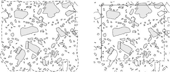
completelyvisiblecrystals(Figure2.2a).Theareaorvolumeofmeasurement
can be a simple rectangle or parallelepiped that is drawn inside the actual limits
ofthesamplesothatpartiallyvisiblecrystalsareexcluded(Figure2.2b).Then
only those crystals that touch two sides of the square, or three edges of the
parallelepiped, are included.
2.1.4 Textural development sequences
When a rock is sampled all that can be generally observed is the final product –
the path taken to achieve that product is not always clear, but may sometimes
2.1 Introduction 9
be revealed by carefully examining other, less developed samples, or by locat-
ing early textures frozen in by other processes. Such sequences of textural
development can help enormously in understanding petrogenetic processes.
For instance a series of lavas or samples taken from a lava lake may show how
magmas crystallise (e.g. Cashman & Marsh, 1988). First-formed plutonic
textures may be seen in oikocrysts and early metamorphic textures may be
preserved in porphyroblasts (e.g. Higgins, 1998). Hence, the methods
described below should be applied not only to the average rock, but also to
special sectors of a sample that can show evidence of earlier textures.
2.2 Complete three-dimensional analytical methods
Three-dimensional analytical methods that conserve the size, shape, orienta-
tion and position of the crystals will be discussed first. Some of these methods
conserve the sample (e.g. X-ray tomography) whereas others are destructive
(e.g. serial sectioning).
2.2.1 Serial sectioning
The complete texture of a sample can be established from serial sectioning
(Bryon et al., 1995). Here, a surface or thin section is cut and recorded as a
photograph or digital image. The surface is then ground away and a new
(a) (b)
Figure 2.2 Two solutions to the problem of edge effects in textural measure-
ments, shown here for a section. (a) Crystals that are not completely visible in
the field of view (open outlines) are excluded and an irregular envelope
passing midway between the crystal edges (dotted line) encloses the crystals
to be measured (grey outlines). The area of the envelope is the area measured.
(b) A rectangle is drawn around all crystals that are completely visible.
Crystals are counted that fall within the rectangle or touch two of the sides.
Those that touch the other two sides (dashed) are excluded as well as those
completely outside the rectangle.
10 General analytical methods
surface or thin section made, parallel to the original section, and the process
repeated. Clearly, the sample is either destroyed or reduced to a series of
thin sections. The resolution of the method is limited by the spacing of the
sections and the resolution of each image, which should ideally be equal
(Marschallinger, 1998b, Marschallinger, 1998a). The images can be processed
as for surface methods to separate out the different minerals (see Sections 2.6.2
and 2.6.3). The processed images can then be combined into a data volume (3-D
image) to establish the complete shape of each crystal (Marschallinger, 2001).
Serial sectioning can give excellent results, especially for small numbers of
irregularly shaped objects, but it is very time consuming and its resolution is
limited by the spacing of the sections. For example, 500 successive images must
be obtained and combined to match an image with a resolution of 500 500
pixels. This is rarely done and the vertical resolution is generally much less
than the resolution in the plane of the images. If the sample is ground away
(‘lapped’) to make the separate images then a resolution as small as 40 mm has
been achieved (Marschallinger, 1998a). If thin sections are used then a much
larger spacing is needed, typically several mm. Of course, the vertical resolu-
tion must be balanced by the need and ability to distinguish individual crystals.
Some textural parameters do not need crystals to be separated (e.g. intercept
orientation method) and in some rocks crystals can be readily isolated in plane
surfaces without optical orientation. However, if crystals must be separated
then it is generally easier to do in a thin section than on a flat surface.
Serial sectioning has been used more extensively in biological and palae-
ontological studies where the interest is in small numbers of very irregular
objects – whereas petrology is more unusually concerned with large numbers
of similarly shaped objects, like crystals.
2.2.2 Optical scanning and confocal microscopy
Optical scanning and confocal microscopy are special techniques that can be
used for the measurement of small proportions of grains in transparent mate-
rials. They give a result similar to serial sectioning, but without destroying the
sample. In optical scanning the section is examined at high magnification with
a large aperture. In this situation the depth of field is small and a narrow range
of depths in the section are focused. A photograph is taken and the sample–
objective distance increased. The process is repeated to build up a complete
3-D reconstruction of the section. The matrix of the crystals must be sufficiently
transparent and the crystal number density must be sufficiently low that the
whole crystal can be observed; hence it can only be applied in special circum-
stances, for instance microlites in a glassy volcanic rock (Castro et al., 2003).
2.2 Complete three-di mensional analytical methods 11
A confocal microscope is designed specifically for these types of application
(e.g. Petford et al., 2001, Bozhilov et al., 2003). Instead of shining a light on the
whole section, only a part of the sample is illuminated with a laser beam that is
scanned across the sample. The image is then reconstructed sequentially, as in
a scanning electron microscope (see below). This method has the advantage
that scattering of light from adjacent crystals and matrix into the volume of
interest is much reduced.
It is not always necessary to reconstruct the whole 3-D structure: the length
and other shape parameters can be determined from the vertical and horizon-
tal position of the ends of the crystal. In some cases the method has been
simplified further by choosing crystals that are nearly parallel to the plane of
the section (Castro et al., 2003).
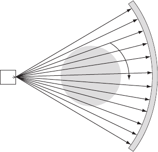
2.2.3 X-ray tomography
Tomography (CAT – computed axial tomography) is the reconstruction of a
section from many separate projections around an object. A series of closely
spaced slices are assembled into a 3-D image. It is commonly applied using
X-rays (Ketcham & Carlson, 2001, Mees et al., 2003), but can also be used with
any radiation that can penetrate the material, such as gamma rays or even light
(Figure 2.3).
X-ray
source
X-ray
detectors
Object
Figure 2.3 X-ray tomography. In most systems used for geological research
the sample is rotated as it is bathed in a flat beam of X-rays. The X-rays that
pass through the sample are detected by a curved bank of detectors. A slice of
the internal structure of the sample can be reconstructed from the quantity of
X-rays recei ved by the detectors. The sample is then moved vertically and
another slice analysed.
12 General analytical methods
Minerals are distinguished in X-ray tomography on the basis of their linear
attenuation coefficient, m. This depends directly on the density of the mineral,
the effective atomic number of the mineral, and the energy of the incoming
X-ray beam. Common minerals have values of m that vary by a factor of three
for 100 keV X-rays, which is much greater than the precision of measurement,
typically 0.1%. However, there is much overlap in the values of m, hence
minerals cannot always be distinguished using this parameter alone. In some
situations the sample can be examined using X-rays of two different energies
(frequencies) and the images combined to separate phases. In general, this
method cannot separate touching crystals of the same mineral. This is because
m does not depend on direction in an anisotropic mineral. This contrasts with
the optical properties of most common anisotropic minerals.
Sample size is limited by the attenuation of X-rays and the physical dimen-
sions of the sample chamber. Many instruments can handle samples up to
40 cm long. Spatial resolution is a function of sample size and the number of
pixels in the image (typically less than 1000 1000 pixels). Resolutions are
commonly 0.1–0.2 mm for centimetric to decimetric size samples. Larger
samples necessarily have a lower resolution.
Recently, synchrotron radiation has been used for micro-tomography (Song
et al., 2001, Cloetens et al., 2002, Ikeda et al., 2004). This has a wide range of
wavelengths and is very intense so that it can be focused in a small volume. It
has also been combined with X-ray fluorescence computed tomography to
give a 3-D compositional map (Lemelle et al., 2004). Although precision is low,
this method shows promise for some materials.
2.2.4 Magnetic resonance imaging
Another 3-D method is magnetic resonance imaging. This is used extensively
in medical applications, but has only recently been applied to textural studies
of rocks. Magnetic resonance generates images that largely reflect the hydro-
gen content of geological materials. So far this method has only been used in a
single textural study, where it was used to help visualise pore distributions in
carbonate rocks (Gingras et al., 2002). Clearly, it may be applied in other
studies of water-bearing rocks, especially if the resolution of the method can be
improved.
2.3 Extraction of grain parameters from data volumes
The digital 3-D analytical methods described above produce a 3-D image
commonly referred to as a data volume, or data brick. It is comprised of
2.3 Extraction of grain parameters 13
voxels, the volumetric equivalent of pixels in images. Two-dimensional image
analysis (see Section 2.6.3) is much more developed than analysis of three-
dimensional images. Many of the 2-D techniques can be extended to 3-D, but
the software for this is still in development.
Extraction of grain and textural parameters from data volumes comprises
three steps: classification, separation and measurement (Ketcham, 2005). The
classification process is commonly the most complex. Many data volumes are
essentially grey-scale images – that is there is only one value for each voxel.
Such images can be segmented by considering a window of acceptable values.
However, mineral phases are not always very regular, and more useful meth-
ods have been developed (Ketcham, 2005). For instance, the ‘seeded threshold’
filter initially accepts voxels within a range of grey-scale values. Each seed
object is then expanded by the addition of connected voxels that have a wider
range of grey-scale values. If the mineral phase is assumed to be spherical then
irregular groups of voxels may be simplified by substituting spheres with
volumes equal to that of the original crystal (Carlson et al., 1995).
Touching crystals or grains are not separated by most 3-D analytical meth-
ods; hence this must be done during data reduction. Voxel groups can be
examined individually and cut apart manually (Ketcham, 2005). An automatic
process was suggested by Proussevitch and Sahagian (2001); interconnected
voxel clusters are ‘peeled’ or eroded until the individual objects are separated,
and finally the crystal centres are defined. The crystals are then rebuilt using an
assumed shape, such as a sphere or equidimensional polyhedron. Another
approach is to use the ‘watershed’ algorithm: the acceptable range of voxels in
a group is reduced until the group separates into distinct objects. The voxel
group is then rebuilt from these centres (Ketcham, 2005).
Measurement of the dimensions of separated groups of voxels is concep-
tually easy, but has not been facilitated by current software. However, new
developments may ease this problem (Ketcham, 2005).
2.4 Destructive partial analytical methods
Some aspects of rock textures can be determined by dismantling the rock and
measuring the dimensions of the separated grains. This avoids problems of
interpreting the grain parameters from sections, but the position and orienta-
tion of the grains are lost. In addition, grains with convoluted shapes may not
be easily separated intact from their matrix and it is difficult to know how to
deal with edge effects. Such analytical methods enables the use of much smaller
samples as many more crystals are encountered in volume compared to those
intersected in a section. However, the minimum crystal size that can be
14 General analytical methods
consistently recognised must be clearly established (and also the maximum size
in rare cases).
2.4.1 Sample disaggregation
Grains can be separated from a rock if it can be readily disaggregated. In some
volcanoes explosive eruption processes may separate crystals mechanically.
However, it is unlikely that this process will be truly unbiased in terms of size
or shape. Other volcanic rocks may be so weak that they can be separated
mechanically with little damage to the crystals (also see sample solution
methods below). Dunbar et al., (1994) did this for bombs from Mt Erebus,
Antarctica, with good results. However, they did have to make a correction for
some broken crystals.
Well-lithified rocks can be disaggregated using more energetic methods. In
electric pulse disintegration a voltage of 100 kV is applied to a rock sample
sitting in a water bath (Rudashevsky et al., 1995). The rock explodes, separat-
ing crystals along grain boundaries. The specialised nature of the equipment
has limited the use of this method so far. Finally, it is possible to dissect a rock
crystal by crystal using a hammer and chisel. Kretz (1993) used this method to
measure the position of each garnet crystal in a schist. He then reconstructed
the whole structure with balls and rods.
Crystals that are already fractured in the rock present special problems. If
one is interested in primary processes then one solution is to examine each
crystal individually and only retain those that are unbroken. This is time
consuming and may bias the sampling of the crystals, but it may enable
some studies that would otherwise be difficult (Gualda et al., 2004). Of course,
if the focus of the study is on the fracturing process then the crystals can be
measured easily.
2.4.2 Sample dissolution
Dissolution is another method for the separation of crystals from a rock. In
carbonate matrix rocks (carbonatites and marbles) some crystals can be sepa-
rated by dissolution of the matrix using HCl or other acids. However, it should
be remembered that some silicates are also slightly soluble in HCl (e.g.
anorthite, olivine) and hence small crystals may be lost.
Similar dissolution methods are commonly used to separate diamonds from
kimberlite or other rocks for exploration purposes. The preferred method is
crushing followed by fusion with alkali flux at 550 8C. Commercially very large
samples are processed – up to 300 kg. Larger crystals may be broken during
2.4 Destructive partial analytical methods 15
crushing that precedes dissolution. In that case the crush size is the maximum
crystal size that can be recognised. In some kimberlites most diamonds are
already broken, probably during emplacement; hence the size measured is that
of the fragments, not the original crystals. The smallest crystals may be lost by
solution or mechanically.
Dissolution methods are also used to extract crystals from silicic volcanic
rocks (Bindeman, 2003). The method works best for light, frothy pumice.
Hydrofluoric (HF), fluorosilicic (H
2
SiF
6
) or fluoroboric (HBF
4
) acid is used
to attack the glass. The acid is applied either until there is complete dissolution
of glass or until the glass is weakened by partial dissolution and the rock can be
easily crushed. Many silicate minerals are also attacked by the acid, but
generally much more slowly than the glass. The surfaces of feldspar crystals
are etched more than quartz, which can be used to distinguish these minerals.
Small crystals may dissolve, stick to the surfaces of the preparation equipment
or be retained on filters. However, good results have been obtained for zircon
and quartz in rhyolitic pumice (Bindeman, 2003).
Mixtures of different minerals extracted by dissolution can be separated by
density using heavy liquids (methylene iodide ¼ diiodomethane, bromoform ¼
tribromomethane, sodium polytungstate) or magnetically using a Frantz#
isodynamic separator (Hutchison, 1974). They can also be hand picked dry,
under alcohol (to reduce reflections) or in immersion oil. In the latter case the
refractive index of the oil can be matched to that of the matrix glass or another
mineral, to make it less visible.
2.5 Surface and section analytical methods
2.5.1 Surface preparation techniques and artefacts
Most quantitative textural studies of rocks start with the preparation of an
artificial flat surface: natural fracture surfaces cannot generally give quantita-
tive results. Commonly, the first step is sawing a rock sample with a diamond-
impregnated circular or wire saw. The rough surface can then be flattened on a
lap (rotating wheel) with abrasive paste. The surface is polished with progres-
sively finer grained abrasives until the necessary degree of flatness has been
achieved. A number of problems and artefacts are commonly encountered:
they should be recognised so that they will not be misinterpreted. This is
especially a problem with automatic analysis systems.
*
Scratches: These are not always easy to remove. They can be a problem if the rock is
comprised of minerals with variable hardness. Surface treatments like etching can
enhance small scratches.
16 General analytical methods
*
Occluded materials: Grains of the grinding material can be pressed into softer
minerals, or can be caught along grain boundaries or cracks in the sample.
*
Pull-outs: Brittle minerals with well-developed cleavages can fracture close to the
surface making small pits.
*
Rounding and surface relief : In polymineralic rocks harder minerals will resist
abrasion and will tend to be higher in the final polished surfac e.
The relief of the surface can be increased by etching. This is commonly used for
Nomarski microscopy (see Section 2.5.3.3) but has also been applied in other
studies. Herwegh (2000) developed a two-stage etching for calcite: the surface
is first immersed in dilute HCl, followed by dilute acetic acid. The surface relief
was then examined with a scanning electron microscope; however, Nomarski
microscopy could also be applied.
2.5.2 Electronic and associated analytical methods
Rock surfaces can be examined using a beam of electrons and the most
common instrument for this is the scanning electron microscope (SEM; Reed,
1996). The sample is placed in a vacuum chamber and a beam of electrons is
scanned across the surface. Interaction of the beam with the material produces
electrons, X-rays and light photons that are detected and measured. The
resolution of the different images is variable, but is theoretically smaller than
for optical measurements as the wavelength of electrons is much smaller than
that of light.
Samples must be flat and highly polished otherwise it is the relief that will be
imaged instead of the composition. Solid samples or polished thin sections can be
used, with the only physical limitation being the size of the sample chamber,
typically less than 10 cm. Samples are commonly coated with carbon or metal to
make them conducting and to reduce charging of the surface (Reed, 1996). In
some instruments the sample chamber is kept at a higher pressure than the
electron gun and hence no coating is necessary (ESEM – environmental scanning
electron microscope). The magnification can be varied enormously, making this
technique very useful over a wide range of sample sizes from 0.1 m m to 1 mm.
The electron microprobe (EMP) is another instrument that uses electrons to
analyse materials. It is mechanically very similar to the SEM, but has been
optimised for different measurements. SEMs are designed for observations at
different magnification scales, but the sample cannot be viewed optically.
EMPs commonly have a magnification fixed to that of the associated optical
system. In addition EMPs are optimised for quantitative chemical analysis.
Recently, there has been a convergence between these two instruments, but
SEMs are still cheaper than EMPs for both purchase and use.
2.5 Surface and section analytical methods 17

2.5.2.1 Backscattered electron images
When a beam of electrons strikes a surface some electrons will pass close to
the nucleus of the atoms and will be scattered by the positive charge of
the protons. If the electron beam is approximately normal to the mineral
surface (‘flat scanning’) then the number of ‘backscattered electrons’ (BSE)
emitted by any part of a rock is proportional to the mean atomic number of the
mineral,
Z:
Z ¼
P
Z
j
N
j
R
j
P
Z
j
R
j
where Z
j
¼ atomic number; N
j
¼ atomic weight and R
j
number of atoms in the
formula of element j. If the beam is strongly inclined to the surface then other
factors come into play (see orientation contrast imaging below).
The spatial resolution of BSE images is limited to about 0.1 mm as the
electrons are produced within a relatively large volume. The atomic number
difference that can be distinguished in a BSE image also decreases with
increasing atomic number (Reed, 1996): At
Z ¼ 10 u (e.g. quartz,
feldspars, Table 2.1) it is 0.1 u and at
Z ¼ 30 u (e.g. Cu-sulphides) it is 0.5 u.
However, the overlap of
Z ranges of potassium feldspar, plagioclase and
quartz is much more of a problem, hence supplemental information, such as
X-ray maps may also be necessary. BSE images are most useful for small
crystals that have significantly different
Z from other minerals and the
groundmass.
Table 2.1 Mean atomic numbers (
Z) for various
minerals. These are derived from actual analyses in
the MinIdent-Win 3 mineral property database
(see Appendix).
Mineral Mean atomic number (
Z)
quartz 10.8
albite 10.8
anorthite 11.9
orthoclase 11.7
fayalite 18.3
forsterite 11.4
orthopyroxene 13.8
18 General analytical methods