Kasper C., van Griensven M., P?rtner R. (Eds.) Bioreactor Systems for Tissue Engineering II: Strategies for the Expansion and Directed Differentiation of Stem Cells
Подождите немного. Документ загружается.

including BMSC [33, 37–47], umbilical cord blood MSC [48], ASC [43, 49], and
stromal cells from dental pulp and trabecular bone [43 ] showing increased clono-
genic efficiency and proliferative capacity compared to standard FCS culture. PL
also stimulates proliferation and collagen produc tion of human tenocytes and
increases the gene expression of matrix-degrading enzymes and angiogenic growth
factors [50]. Furthermore, myelomas, hybridomas, hepatocytes, fibroblasts, and
epithelial cells have already been evaluated using PL with regard to cell growth,
viability, and production efficiency [ 51]. Growth stimulation upon PL treatment
was also demonstrated for primary chondrocytes. However, PL failed to support a
chondrogenic phenotype [52–54] in contrast to BMSC cultures showing increas ed
chondrogenic marker genes in presence of PRP [55]. Primary human skeletal
muscle cells showed decreased differentiation capacity into myotubes and impaired
functionality [56]. Proliferation of primary human osteoblasts was not affected by
addition of PRP to the culture medium [57]. For human dermal and gingival
fibroblasts, contradicting results were obtained for platelet derived products as
culture supplements ranging from growth suppression [58] to growth promotion
[59, 60]. Using ASC, Davenport et al. showed that PL only initially supported cell
proliferation but led to growth arrest shortly after first subcultivation [61]. In
contrast, addition of thrombin activated PRP to the culture medium increased
ASC proliferation and retained their differentiation capacity during long-term
culture [49].
Moreover, platelet derived produc ts and BMSC have already been used clini-
cally both to treat distraction osteogenesis of the lower extremity in patients with
achondroplasia and hypochondroplasia yielding accelerated bone regenera tion [62]
and also to treat successfully a patient with severe radiation burn [63].
However, the influence of platelet derived products for the cultivation of cells
isolated from amniotic membrane has not been addressed before.
1.2.3 Strategies to Circumvent Growth Limitation In Vitro:
Introduction of hTERT
Stem cells needed at therapeutic doses, especially in adults, may require extensive
in vitro expansion. In this regard, one major drawback of thes e cells is their low
proliferative capacity and limited in vitro life span before reaching an irreversible
growth arrest also termed replicative senescence [19]. Additionally, long-term
cultures of human MSC may show altered or reduced responsiveness to differenti-
ation signals [64].
One strategy to circumvent these limitations is the introduction of the catalytic
subunit of human telomerase reverse transcriptase (hTERT) which has been
reported to extend the cellular life span of nume rous cell types including normal
fibroblasts, endothelial or epithelial cells [65–67], in vitro propagated tumor cells
[68–70], and also of stem cells [71, 72]. It has been shown that hTERT immorta-
lized human MSC originating from sources such as bone marrow and adipose tissue
maintain their differentiation potency [72–74]. We report in this section the
6 S. Wolbank et al.
establishment and the characteriza tion of the first hTERT immortalized hAMSC
lines including their immunomodulatory functions, a crucial factor for using these
cell lines in allogeneic cell therapies [75]. Therefore, if cell banking is intended, it is
important to monitor the cells’ ability to alloactivate peripheral blood mononuclear
cells (PBMC) as well as to modulate the proliferation of activated PBMC.
2 Stem Cell Characteristics and Immunomodulatory Potential
of Human Amnion-Derived Stem Cells
2.1 Isolation of Separate Populations of hAEC and hAMSC
Placentae were collected from Cesarean sections after obtaining informed consent
of the mothers according to the approval of the local ethical committee. Amnion
was peeled off the placenta, washed extensively with phosphate buffered saline
(PBS) at 4
C, and dissected in 2–3 cm
2
pieces. Half of these were digested for
3 20 min with 0.05% trypsin/EDTA (PAA, Austria), the other half for 2 h with
1 mg/mL collagenase I (Biochrom, Austria) for isolation of hAEC and hAMSC,
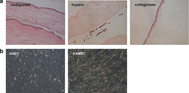
respectively. Hematoxylin/eosin staining of paraffin embedded sections of amnion
demonstrates that digestion of amniotic membrane with trypsi n and collagenase left
an essentially intact mesenchymal and epithelial layer (Fig. 1a).
After addition of ice-cold PBS, cell suspensions were filtere d through a 100-mm
cell strainer, centrifuged, and seeded in culture flasks at a density of 7 10
3
cells/cm
2
for hAMSC and 14 10
3
–21 10
3
cells/cm
2
for hAEC in EGM-2
Fig. 1 Isolation of pure fractions of hAEC and hAMSC. (a) Hematoxylin and eosin stain of fresh
amnion (undigested) and after digestion with trypsin and collagenase, as performed for isolation of
hAEC and hAMSC, respectively. (b) Epithelial and mesenchymal morphology of hAEC and
hAMSC, respectively
Alternative Sources of Adult Stem Cells: Human Amniotic Membrane 7
(Lonza, Belgium). The resulting cultures are composed of pure populations with a
clearly distinguishable epithelial and mesenchymal morphology, respectively
(Fig. 1b).
2.2 Stem Cell Characteristics and Immunomodulation
of hAEC and hAMSC
Both amniotic cell populations are routinely characterized by a common surface
marker expression profile including the presence of CD73, CD90, CD105, and
MHC I, and the concomi tant absence or low levels of CD34, CD45, and MHC II,
analyzed by flow cytometry. Purity of amniotic subpopulations could be determined
by CD49d (a4-integ rin) expression which was 2 2.4% in the hAEC and
96 3.9% in the hAMSC population.
To evaluate the reproducibility of differentiation of hAEC and hAMSC,
osteogenic differentiation was induced 24 h after seeding by changing the medium
to Mesenchymal Stem Cell Osteogenic Stimulatory Kit (O-Kit, Stemcell Tech-
nologies, Canada) and maintaining these cultures for 21 days. Adipogenic dif-
ferentiation was performed according to Portmann-Lanz et al. [23]. Osteogenic
differentiation was demonstrated by spectrophotometric assessment of Alizarin red
(AR). Typically, osteogenic differentiation of both cell types, hAEC (at P1 or P2)
and hAMSC (at P2), was successfully induced in three of four cases. Adipogenic
differentiation was evident for two of four hAMSC isolations, while, in contrast to
published data [22, 23], hAEC did not differentiate along the adipogenic lineage in
our hands (data not shown).
For investigating immunomodulation in vitro, amnion-derived cells were cocul-
tured with PBMC, isolated from whole blood as in mixed lymphocyte reactions
(MLRs). For this, 5 10
4
cells of two different allogeneic PBMC populations
were cocultured in 100 mL PBMC medium/well (RPMI1640, 9% FCS, 2 mM
L-glutamine, 100 U/mL penicillin G, and 0.1 mg/mL streptomycin) in triplicates
in 96-well flat bottom plates. Amnion-derived cells were seeded in the wells and
allowed to adhere before adding PBMC. The stem cells (SC) were added at
SC/PBMC ratios of 1:1 (5 10
4
SC), 1:2, 1:4, 1:8, and 1:16. On day 5, 10 mM
5-bromo-2-deoxyuridine (BrdU) was added and BrdU ELISA (Roche) was per-
formed on day 6 according to the manufacturer’s instructions. Similarly, for
phytohemagglutinin (PHA) activation assay, 5 10
4
PBMC were activated by
5 mg/mL PHA (Sigma) on day 3 of the culture. To examine interaction between
allogeneic SC and unstimulated PBMC, SC were cocultured with unstimul ated
PBMC at 1:1 (5 10
4
SC), 1:2, 1:4, 1:8, and 1:16 ratios in 100 mL. On day 4,
10 mM BrdU was added. The inhibitory effect of SC was calculated as PBMC
proliferation (%) = (E
STIM+SC
E
STIM
) 100. E
STIM+SC
= mean absorption of
stimulated PBMC cocultured with allogeneic SC; E
STIM
= mean absorption of
stimulated PBMC. Data were analyzed by one-way ANOVA and Tukey’s multiple
8 S. Wolbank et al.
comparison test. Data sets of cells at low vs high population doublings (PDs) were
compared by two-tailed Student’s t-test. A p-value less than 0.05 was considered as
significant.
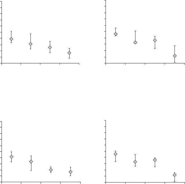
hAMSC and hAEC inhibited proliferation of activated PBMC in a dose-depen-
dent manner as demonstrated by a decrease in proliferation with increasing stem
cell amounts. SC were most effective when added in equal cell numbers compared
to PBMC, significantly reducing PBMC proliferation in MLR experiments to a level
of 34% (range 3–73%) in the case of hAMSC (Fig. 2a) and 23% (range 0–72%) in
the case of hAEC (Fig. 2b). When PBMC were activated by PHA, similar inhibition
was reached, in detail 33% (range 12–66%) for hAMSC (Fig. 3a), and 28% (range
0–60%) for hAEC (Fig. 3b). The lowest SC dose resulting in significant inhibition
of lymphocyte response was 25%, in single cases even 12.5%.
200
a
180
160
140
120
100
80
60
40
20
0
12.5 25
hAMSC added (%)
PBMC proliferation (%)
50 100
200
b
180
160
140
120
100
80
60
40
20
0
12.5 25
hAEC added (%)
PBMC proliferation (%)
50 100
Fig. 2 hAMSC and hAEC inhibit MLR-activated PBMC in a cell dose-dependent manner. PBMC
were cocultured with equal amounts of allogeneic PBMC and different amounts of third party (a)
hAMSC (n ¼ 12), (b) hAEC (n ¼ 9). Median Q1 and Q3 are depicted
200
a
180
160
140
120
100
80
60
40
20
0
12.5 25
hAMSC added (%)
PBMC proliferation (%)
50 100
200
b
180
160
140
120
100
80
60
40
20
0
12.5 25
hAEC added (%)
PBMC proliferation (%)
50 100
Fig. 3 hAMSC and hAEC inhibit PHA-activated lymphocyte proliferation in a cell dose-depen-
dent manner. PBMC were cocultured with (a) hAMSC (n ¼ 12), (b) hAEC (n ¼ 8). Median
Q1 and Q3 are depicted
Alternative Sources of Adult Stem Cells: Human Amniotic Membrane 9
3 Phenotypic Shift and Reduced Osteogenesis During In Vitro
Expansion of Human Amnion Epithelial Cells
For tissue engineering purposes, cells may be applied either directly after isolation
from the tissue or after a period of in vitro expansion to obtain higher cell numbers.
In order to investigate the advantages and drawbacks of these strategies we com-
pared freshly isolated and cultivated hAEC regarding their surface antigen expres-
sion profile and their osteogenic differentiation capacity.
3.1 Shift in Surface Antigen Expression During
Cultivation of hAEC
To investigate the impact of in vitro expansion on the immunophenotype of cells
with potential for regenerative medicine, we carefully characterized the surface
antigen profile of hAEC directly after isolation and during cultivation by flow
cytometry. We focused on hAEC, as recovery of primary hAMSC is usually too
low for thorough analysis. For this purpose, freshly isolated cells and cells during
culture were immunostained for CD14, CD34, CD45, CD13, CD29, CD44, CD49c,
CD49d, CD49e, CD54, CD73, CD90, CD166, Ki67 (BD, Austria), CD105 (Abcam)
and SSEA-4, TRA-1-60, TRA-1-81 (Chemicon), by 7-AAD (BD) for dead cells and
measured by flow cytometry.
Surface antigens were clustered into four groups, according to their expression
patterns (representative histograms are depicted in Fig. 4a, summarized in Fig. 4b).
The first group comprises CD49d (integrin a4; used to differentiate hAEC from
hAMSC) and the hematopoietic markers CD14, CD34, and CD45. These antigens
are hardly detectable on freshly isolated hAEC (preP0) and remain at similar levels
during passaging.
The surface antigens of the second group are uniformly expressed at high levels,
both in primary isolates (preP0) and after further cultivation. This group comprises
the stromal cell markers CD29 (integrin b1), CD49c (integrin a3), CD73 (ecto-5
0
-
nucleotidase), and CD166 (ALCAM), and the embryonic stem cell marker SSEA-4.
Group 3 consists of the stromal cell associated markers CD13 (aminopeptidase
N), CD44 (HCAM), CD49e (integrin a5), CD54 (ICAM-1), CD90 (Thy-1), and
CD105 (endoglin), which are low (medium to undetectable) directly after isolation
(preP0) and are rapidly increased during in vitro cultivation.
Two additional embryonic stem cells markers, TRA-1-60 and TRA-1-81 (group
4), are characterized by medium expression in preP0 cells, which decreases upon
cultivation.
As in vivo, amniotic cells reside within a tissue that remains of approximately
the same size during the last weeks of pregnancy, these cells would probably be in a
quiescent state directly after isolation, but start dividing upon transfer into tissue
culture medium. Therefore, we tested the hypothesis that upregulation of group 3
antigens might be associated with re-entry of the cells into the cell cycle. Only
10 S. Wolbank et al.
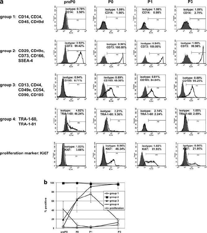
2–6% of freshly isolated hAEC (preP0) were stained for the proliferation marker
Ki67, which is expressed in all phases of the cell cycle but not in G0. Af ter a few
days in cultur e, Ki67 expression increased dram atically (Fig. 4a, b), concomitant
with the observed upregulation of group 3 antigens. However, expression of group 3
antigens is not depend ent on proliferation of hAEC, as expression remained high
Fig. 4 Surface antigen expression of four freshly isolated (preP0) hAEC strains and during
cultivation at various passages (P0–P3) by flow cytometry. (a) Antigens were grouped according
to their expression profile during cultivation (see text) and representative histograms of one
member of each group are shown. Gray peaks represent unspecific isotype controls, solid lines
represent the specific antibodies. (b) Summary: shown are means and corresponding standard
deviations, calculated from the data of all antigens of each group of all four hAEC isolations
(hAEC 87, 88, 90, and 91) at the indicated passages
Alternative Sources of Adult Stem Cells: Human Amniotic Membrane 11
when proliferation slowed down after several passages in vitro, concomitant with a
drastic decrease in Ki67 staining (Fig. 4a, b).
3.2 Osteogenic Differentiation Potential of hAEC Decreases
upon In Vitro Cultivation
We addressed the question as to whether the observed shift in mesenchymal and
embryonic stem cell markers during cultivation of hAEC (Fig. 4) is associated with
alterations of their functional phenotype, i.e., their capacity to differentiate along
the adipogenic and osteogenic lineages. Therefore, adipogenic and osteogenic
conditions (as described in Sect. 2.1) were applied to different hAEC isolations
seeded directly after isolation (P0) and after cultivation (P2 or P3). In addition to
mineralization, quantitative real time PCR was performed analyzing expression of
RUNX2/CBFA1 (core binding factor alpha), alkaline phosphatase (ALPL), bone
gamma-carboxyglutamate protein (BGLAP, osteocalcin), bone morphogenetic pro-
tein receptor 1B (BMPR1B), and bone morphogenetic protein receptor 2 (BMPR2)
using a light cycler TM480 (Roche) and Taqman gene expression assays (Applied
Biosystems). Expression values were normalized to the housekeeping gene hypo-
xanthine-guanine phosphoribosyltransferase (HPRT).
Similar to passaged hAEC, no adipogenic differentiation was observed using
four strains of freshly isolated hAEC (data not shown). In contrast, the same strains
showed predominantly stronger mineralization ability at P0 (in three out of four
cases) when compared to passaged cells (P2–P3). Interestingly, preliminary results
with hAMSC suggest similar mineralization and lipid accumulation after induction
of P0 vs P2 cells (data not shown). We confirmed mineralization by analysis of
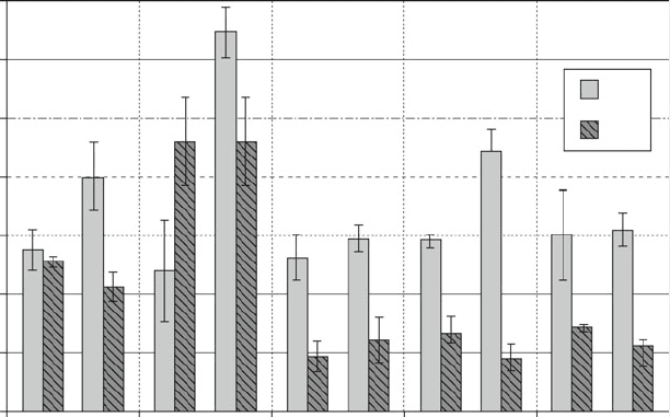
mRNA levels of selected genes involved in osteogenesis. In freshly isolated hAEC
(P0) all osteogenic markers were upregulated upon cultivation in O-Kit for 14 and
21 days (Fig. 5). In cultivated hAEC (P1), RUNX2 and ALPL were also increased
under osteogenic conditions whereas virtually no alteration in transcription of
BGLAP, BMPR1B, and BMPR2 was observed.
4 Platelet Lysate for FCS-Free Expansion and
Cryoconservation of Amnion-Derived Cells
For producing PL, platelet concentrates from 36 healthy donors that could no longer
be used for patients were pooled, frozen at 80
C, and thawed quickly in a water
bath at 37
C, resulting in growth factor release from bursting platelets. Platelet
debris was removed by centrifugation at 2,000g for 10 min while the PL was
filtered using a 0.22-mm filter, aliquoted, and stored until application at 80
C.
For determining growth kinetics, cells were isolated from three donors as
described in Sect. 2.1, and 2.5 10
5
cells were seeded in T-25 flasks and cultured
12 S. Wolbank et al.
for about 100 days in PL expansion medium (DMEM-LG & Ham’s F12, 5% PL,
2mM
L-glutamine, 100 U/mL Penicillin, 0.1 mg/mL Streptomycin (PAA), 2 U/mL
Heparin (Biochrom)) at 37
C, 5% CO
2
, and 95% humidity. PD was calculated at
each subcultivation using the formula: SLN (cells harvested/cells seeded)/LN(2).
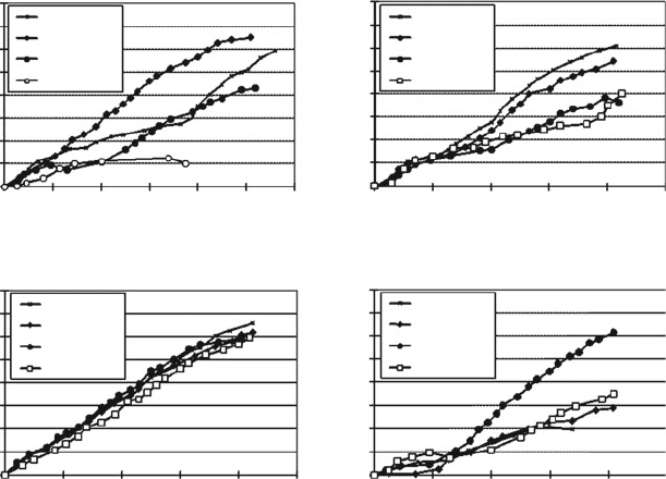
hAMSC from all three donors cultured in PL showed between 21 and 32 PD
without growth arrest whereas control hAMSC cultur ed in EGM-2 (Lonza), a
commercially available medium containing 2% FCS, showed only 5 PD before
proliferation totally ceased (Fig. 6a).
Not only during expansion but also during cryopreservation, substitution of FCS
would be favorable for establishing cell banks. In preliminary experiments, several
media containing PL (5%, 90% PL) were compared to standard FCS media (10%,
90% FCS) as well as a serum-free medium (CryoSFM, PromoCell) for cryopreser-
vation of hAMSC. Then 1 10
6
P1 cells were resuspended in the respective
medium and frozen at a freezing rate of 1
C/min. From these preliminary data,
the best conditions, namely 5% PL medium (5% PL, 10% dimethylsulfoxide
(DMSO), 85% DMEM-LG), 90% FCS medium (90% FCS, 10% DMSO), and
CryoSFM were chosen for further investigation.
After thawing, cell viability was assessed by trypanblue exclusion assay. Addi-
tionally, growth kinetic studies were performed to evaluate characteristics of
hAMSC before and after cryopreservation. Cells cryopreserved in PL showed
7
RUNX2 ALPL BGLAP BMPR1B BMPR2
P 0
P 1
6
5
4
3
2
1
0
d14
fold expression compared to d0
d21 d14 d21 d14 d21 d14 d21 d14 d21
Fig. 5 Osteogenic differentiation of hAEC isolations seeded directly after isolation (P0) and after
cultivation (P1). Expression levels of selected genes implicated in osteogenesis, determined using
quantitative real-time PCR. Expression levels after cultivation in O-Kit for 14 and 21 days (d14
and d21) were normalized to the levels before induction (d0). Shown are means and standard
deviations of three measurements from two individual donors
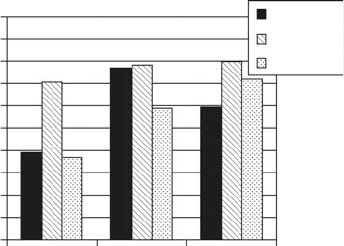
Alternative Sources of Adult Stem Cells: Human Amniotic Membrane 13
lower cell viability after thawing when compared to those stored in FCS medium or
serum-free CryoSFM (Fig. 7). While growth kinetics after thawing seemed to be
unaffected by the cryopreservation medium applied, strong impact of the donors on
PD was evident (Fig. 6b–d). An observed phenomenon was the low attachment
capacity of hAMSC cryopreserved in 5% PL. Hence, coating experiments with
gelatine (1% in PBS) or “Coating matrix Kit” (Invitrogen) were performed but
attachment of cells to the culture vessel surface could not be substantially improved
(data not shown).
5 hTERT Induced Extension of In Vitro Life Span
of Amnion-Derived Stem Cells
For introduction of hTERT a retroviral transfection system was chosen. Therefore,
the cDNA of hTERT (kindly provided by Geron Corp.) was inserted into the
retroviral vector pLXSN (Clontech Laboratories Inc.) and retroviral particles
praecryo
donor 3 - after cryo
donor 1 - after cryo
donor 2 - after cryo
days in culture
PDs
PDs
PDs
PDs
days in culture
days in culture
days in culture
0
0
0
0
0
0
0
0
5
5
5
5
10
10
10
10
15
15
15
15
20
20
20
20
25
25
25
25
30
30
30
30
35
35
35
35
40
40
40
40
20
20
20
20
40
40
40
40
60
60
60
60
80
80
80
80
100
100
100
100
120
5% PL
donor 3 - EGM-2
donor 3 - PL
donor 2 - PL
donor 1 - PL
5% PL
5% PL
5% PL praecryo
5% PL praecryo
5% PL praecryo
cryo-SFM
cryo-SFM
cryo-SFM
90% FCS
90% FCS
90% FCS
ab
dc
Fig. 6 Growth characteristics of hAMSC from three donors before (a) and after cryopreservation
(b–d) cultivated in medium supplemented with either 5% PL or EGM-2 and cryopreserved in FCS,
PL containing media or serum-free Cryomedium (CryoSFM). PDs cumulative population
doublings
14 S. Wolbank et al.
were generated as described previously [70]. Gene transfer was performed at early
PD (<PD8) according to the manufacturer’s instructions (Clontech Laboratories
Inc.). Then 24 h post transduction transfectants were selected using 200 mg/mL
Geneticin Sulfate G418 and arising cell clones were grown as mass cultur e. PD of
transduced cell lines were calculated starting with the first passage post transduc-
tion (PDpT) using the formula stated in Sect. 4. Telomerase activity (TA) was
determined using a modification of the real-time telom eric repeat amplification
protocol (TRAP) assay as described in detail previously [70] and calculated relative
to that of HEK293 cells (positive control). For determination of senescence,
associated b-galactosidase (SA-b-gal) activity cells were fixed with 3% formalde-
hyde and stained as described in detail previously [76]. For characterizing pheno-
type, differentiation potential, and immunom odulatory properties, protocols
according to Sect. 2.2 were performed. For quantitative evaluation, AR was
measured after extraction using 20% methanol/10% acetic acid at 450 nm. For
quantification of intracellular alkaline phosphatase (AP) activity, washed cells were
frozen and thereafter incubated in 0.5% Triton X-100. After incubation with
4-nitrophenolphosphate, samples were measured at 405/620 nm. In addition to
osteogenic marker genes peroxisome proliferator-activated receptor gamma
(PPARg) and leptin (Lep) were evaluated as adipogenic marker genes by quantita-
tive RT-PCR as described in Sect. 3.2.
Immortalized stem cell lines (originating from the mesenchymal layer of the two
amniotic membrane donors hAMSC76 and hAMSC83) were established by over-
expression of hTERT. Human stem cells were isolated from amnion and propagated
in vitro until they reached replicative senescence. Representative growth curves of
hAMSC76 are shown in Fig. 8a. Senescence was evidenced by growth arrest, large
and flat cell morphology (Fig. 8b), and SA-b-gal activity (Fig. 8c). Upon ectopic
expression of hTERT stem cell populations were immortalized (so far expanded
to at least PD60 with no signs of growth retardation; Fig. 9a). Furthermore,
hTERT overexpression maintained many characteristics of the original cellular
phenotype. Figure 9b demonstrates fibroblastoid morphology of transduced cells
(hAMSC76telo-PD78pT, hAMSC83telo-PD43p T) comparable to early passage
100
90
80
70
60
50
40
30
20
10
0
90%FCS
cryomedium
viability (%)
Cryo SFM
hAMSC1
hAMSC2
hAMSC3
5%PL
Fig. 7 Cell viability of
hAMSC from three donors
after cryopreservation in PL,
FCS containing media or
serum-free Cryomedium
(CryoSFM) and thawing as
determined by trypanblue
exclusion assay. Data are
presented in % of viability at
freezing
Alternative Sources of Adult Stem Cells: Human Amniotic Membrane 15