Townsend Courtney M.Jr., Evers B. Mark. Atlas of General Surgical Techniques: Expert Consult
Подождите немного. Документ загружается.


544 Section VII • Pancreas
SELECTED REFERENCES
1. Di Sebastiano P, Di Mola F, Friess H: Management of chronic pancreatitis: Conservative, endoscopic and
surgical. In Blumgart LH (ed): Surgery of the Liver, Biliary Tract and Pancreas, 4th ed. Philadelphia,
Saunders, 2007, pp 728-740.
2. Buchler M, Friess H, Bittner R, et al: Duodenum preserving pancreatic head resection: Long term results.
J Gastrointest Surg 1997;1:13-19.
3. Frey CF, Amikura K: Local resection of the head of the pancreas combined with longitudinal pancreatico-
jejunostomy in the management of patients with chronic pancreatitis. Ann Surg 1994;220:492-507.
545
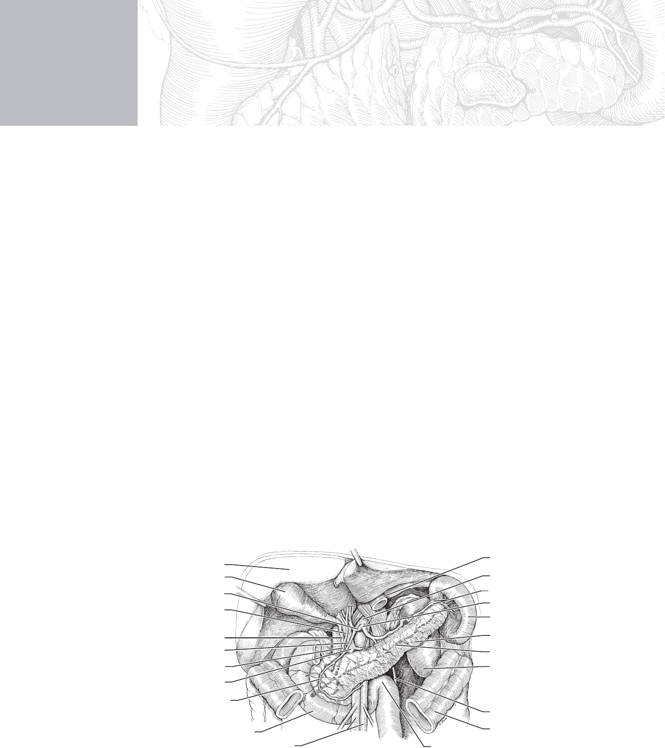
STEP 1: SURGICAL ANATOMY
◆ All pancreatic surgery requires an understanding of the anatomic relationships in the lesser
sac. After either an upper midline or a bilateral subcostal (chevron) incision, one enters the
lesser sac by dissecting along the avascular plane at the points of attachment of the gastro-
colic omentum to the transverse colon (Figure 49-1). The proper plane is between the
anterior and posterior leafl ets. This is my favored point of entry. The alternative entry is by
transversely dividing and ligating the vascular structures embedded in the omentum while
preserving the gastroepiploic vessels located along the greater curvature of the stomach.
◆ Upon entering the lesser sac, one will encounter varying amounts of adhesions between the
posterior wall of the stomach and the anterior surface of the pancreas. These fetal adhesions
do not imply prior infl ammatory events. Considerable dense adhesions may be encountered
in pathologic states.
CHAPTER
49
Pylorus-Saving
Pancreaticoduodenectomy
William H. Nealon
Duodenojejunal flexure
and jejunum
Liver
Duodenum
Superior mesenteric artery and vein
Gallbladder
Cystic duct
Portal vein
Anterior, superior
pancreaticoduodenal artery
Gastroduodenal artery
Common bile duct
Splenic artery and vein
Pancreatic duct
Left kidney
Inferior mesenteric vein
Transverse colon (cut)
Pancreas
Stomach (cut)
Aorta
Celiac trunk
Common hepatic artery
Common hepatic duct
Spleen
FIGURE 49 –1

546 Section VII • Pancreas
◆ The pancreas is essentially encased in a sandwich of major blood vessels. The vena cava and
aorta occupy the posterior surface in the midline. The splenic artery courses along the supe-
rior surface from the aorta toward the tail. The splenic vein occupies the posterior superior
surface of the body and tail of the pancreas. It meets the superior mesenteric vein, which is
oriented vertically in the groove created by the uncinate process in the posterior aspect of the
head of the pancreas and the right lateral and anterior components of the head. The confl u-
ence of these two veins constitutes the portal vein, which traverses this uncinate groove and
emerges to join the bile duct and the hepatic artery in the hepatoduodenal ligament (see
Figures 49-1, 49-3, and 49-4).
◆ The superior mesenteric artery is located in a plane posterior and slightly medial to the supe-
rior mesenteric artery. The common hepatic artery, another branch of the celiac trunk (along
with the splenic artery and left gastric artery), courses along the superior border of the head
of the pancreas to join the hepatoduodenal ligament. Its fi rst branch is the typically minis-
cule right gastric artery. Just distal is the more substantial gastroduodenal artery, which
emerges at a right angle to the hepatic artery from its inferior surface and courses beneath
the pylorus. After sending the right gastroepiploic artery in the plane between the inferior as-
pect of the pylorus and the superior surface of the head of the pancreas, the gastroduodenal
artery pierces the head of the pancreas (see Figures 49-4 and 49-5).
◆ The anterior superior and the posterior superior pancreaticoduodenal arteries also arise from
branches of the gastroduodenal artery. These arteries form an arch medial to the C-loop of the
duodenum, and they collateralize with branches of the anterior and posterior inferior pancreat-
icoduodenal arteries, which are branches of the superior mesenteric artery. Small branches
from these arteries provide blood supply to the duodenum (see Figures 49-3 through 49-6).
◆ Key anatomic features in pancreatic head resections are the network of tributaries projecting
between the superior mesenteric vein/portal vein confl uence and the uncinate process. These
tributaries are located at the right lateral aspect of the veins. These tiny veins exit the pan-
creas at the mid-portion of the groove in which the major veins reside (see Figure 49-12).
◆ Viewed in cross-sectional imaging, the uncinate process forms a C-shaped structure. The
terminal posterior extent of the uncinate process projects in a medial direction as a liga-
mentous structure and contains a variable number of arterial branches from the superior
mesenteric artery, which project at right angles to the major artery and provide blood sup-
ply to the uncinate process. Division of the tiny venous tributaries and the arterial branches
are key steps in respective procedures. This uncinate margin is the most problematic in
managing malignant tumors in the head of the pancreas (see Figure 49-13).
◆ The pancreas is entirely retroperitoneal, and therefore operative procedures will require mo-
bilization of the pancreas from its retroperitoneal position. The plane lateral to the C-loop
of the duodenum is incised in nearly all procedures; this plane is avascular, and its mobili-
zation is termed the Kocher maneuver. This exposes the vena cava and aorta, and it permits
“bimanual palpation” of the head of the pancreas. The dissection may be easily extended to
the fourth portion of the duodenum and the ligament of Treitz (see Figure 49-3).
◆ The inferior border of the body of the pancreas is also avascular, although the inferior mes-
enteric vein may be encountered to the right of the spine (see Figure 49-4).

CHAPTER 49 • Pylorus-Saving Pancreaticoduodenectomy 547
◆ Peritoneum overlies the hepatoduodenal ligament. Dissection reveals the triad in gross ana-
tomic terms, which corresponds to the microscopic portal triad—with portal, hepatic arte-
rial, and biliary structures. The common bile duct is located in an anterior lateral position,
and the hepatic artery is anterior medial. The portal vein is positioned in the posterior
groove created by the apposition of these anterior structures (see Figure 49-5).
◆ Although lymph nodes may be seen at a wide array of locations, there is a constant lymph
node in the groove created by the lateral border of the second portion of the duodenum
and the hepatoduodenal ligament. Dissection of this lymph node is necessary to fully visu-
alize the proximal hepatic artery. Other common sites of lymph nodes are on the lateral
aspect of the mid-portion of the hepatoduodenal ligament, in the fi brovascular bundle sur-
rounding the right gastroepiploic complex, and on the superior border of the confl uence of
the head and body of the pancreas. Beneath this lymph node one fi nds the origins of the
common hepatic artery and the splenic artery.
◆ On the inferior border of the pancreatic head, just where the duodenum dives beneath the
superior mesenteric vein and artery, one may dissect the peritoneum and visualize the supe-
rior mesenteric vein as it passes in a superior direction beneath the head of the pancreas.
◆ The ligament of Treitz is a signifi cant anatomic structure, and it can be accessed by lifting
the transverse colon and omentum in an anterior direction. The ligament can be seen to the
left of the spine.
◆ The main pancreatic duct originates in the tail of the pancreas and traverses the length of
the pancreas to exit in the duodenum through both main ampulla (Vater) and the accessory
ampulla, which is located more proximally in the duodenum. The main pancreatic duct
(Wirsung) and the minor or accessory duct (Santorini) fuse during fetal development at
what is termed the genu or “knee” of the duct.
STEP 2: PREOPERATIVE CONSIDERATIONS
◆ Establishment of the indications for pancreatic resection depends on imaging; pathologic
confi rmation; establishment that curative intent can be applied; and assessment of the
medical status of the patient, including nutritional state.
◆ In the case of cancer, it is not unusual to proceed to resection without pathologic confi rma-
tion. In this case, a very experienced pancreatic surgeon must make that determination
based on strong evidence by imaging that malignant disease exists.
◆ Resectability of pancreatic cancer in the head must be founded on the presence of meta-
static disease (typically in the liver, but potentially many remote sites, as well) and the pres-
ence of what has been termed “locally advanced” disease. In the case of adenocarcinoma of
the head of the pancreas, local invasion and/or encasement of the superior mesenteric artery
or vein are the primary elements that establish this clinical stage. In several large series,
local extension is responsible for establishment of unresectability in half of those deemed
not to be operative candidates.

548 Section VII • Pancreas
◆ Both chronic pancreatitis and cystic neoplasms such as intraductal papillary mucinous neo-
plasm (IPMN) are nonmalignant diagnoses that are treated appropriately with radical resec-
tion. Because pancreatic cancer may masquerade as chronic pancreatitis or coexist with it, a
confi rmation of that distinction is not possible. Suspicions to favor chronic pancreatitis are
the presence of glandular calcifi cation, the presence of a pancreatic mass in the absence of
clinical jaundice, and the chronicity of symptoms. We favor making a strong effort to con-
fi rm the diagnosis of IPMN before proceeding to resection.
◆ Because of the magnitude of the operative procedure, care must be taken to exclude signifi -
cant cardiac, pulmonary, and renal disease and to determine the presence of diabetes melli-
tus. Nutritional needs must also be addressed before surgery.
STEP 3: OPERATIVE STEPS
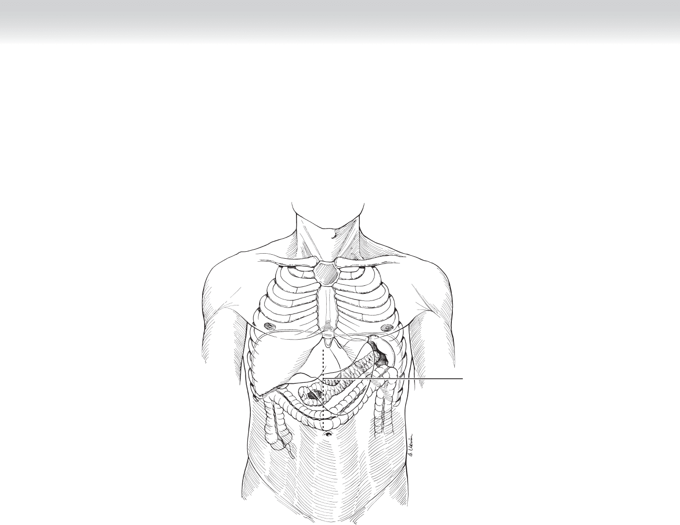
1. INCISION
◆ Midline/xiphoid to some distance below the umbilicus (Figure 49-2)
◆ Self-retaining retractor such as Thompson retractors
CHAPTER 49 • Pylorus-Saving Pancreaticoduodenectomy 549
Midline incision
FIGURE 49 –2

550 Section VII • Pancreas
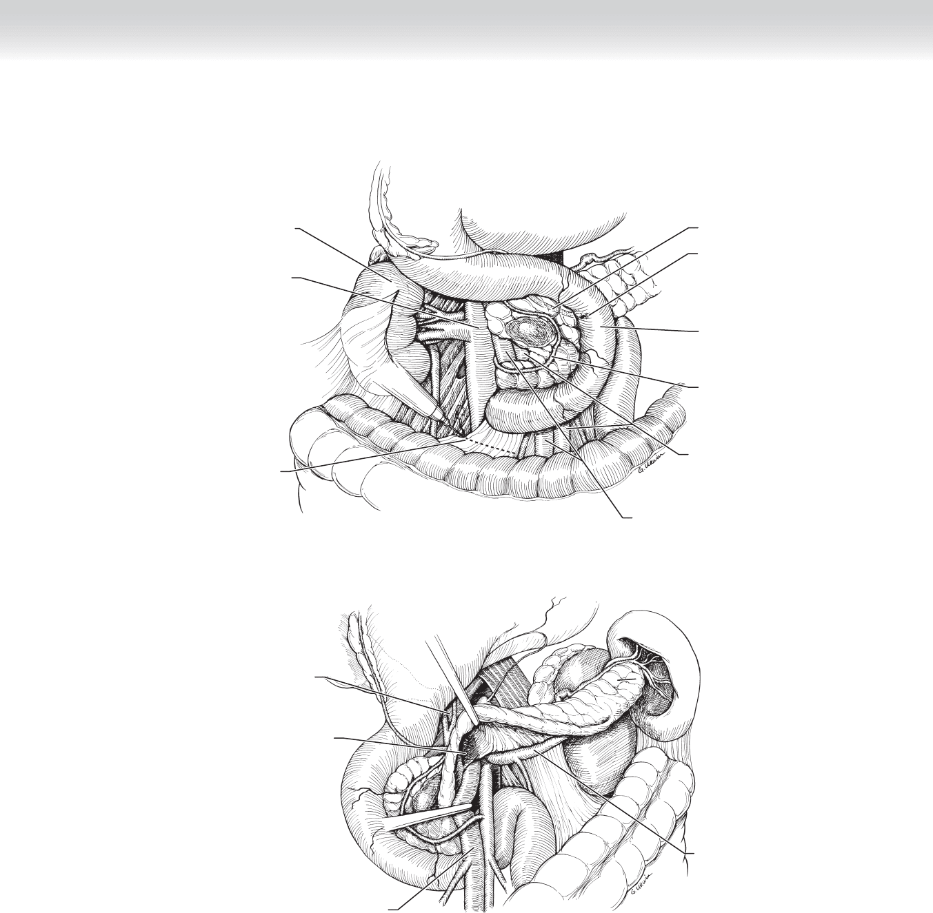
2. DISSECTION
◆ Perform an extended Kocher maneuver, combining mobilization of the duodenum through
to the fourth portion (ligament of Treitz) with mobilization of the hepatic fl exure, which is
refl ected inferiorly (Figure 49-3).
◆ Separate the gastrocolic omentum from its avascular attachment to the transverse colon.
Begin the dissection to the left of the spine, and extend the dissection to the cecum. Care
must be taken to avoid simply creating a window through both leafl ets of the omentum.
Identifi cation of the posterior wall of the stomach confi rms access to the lesser sac.
◆ Mobilize the inferior border of pancreas beginning at the midline and progressing to the
right (Figure 49-4).
◆ The superior mesenteric vein will become apparent to the right of the spine passing over
the third portion of the duodenum on the inferior aspect of the dissection. On the superior
edge of the divided retroperitoneal plane, the superior mesenteric vein courses beneath the
pancreas. Establish this plane posterior to the head of the pancreas and anterior to the
superior mesenteric vein/portal vein confl uence (see Figure 49-4).
CHAPTER 49 • Pylorus-Saving Pancreaticoduodenectomy 551
Posterior inferior
pancreaticoduodenal
artery
Vena cava
Cautery of peritoneum
Superior mesenteric artery
Duodenum
Major duodenal
papilla (of Vater)
Common bile duct
Superior mesenteric vein
Kidney
FIGURE 49 –3
Superior mesenteric vein
Superior mesenteric
vein groove
Splenic vein
Portal vein
FIGURE 49 –4
552 Section VII • Pancreas
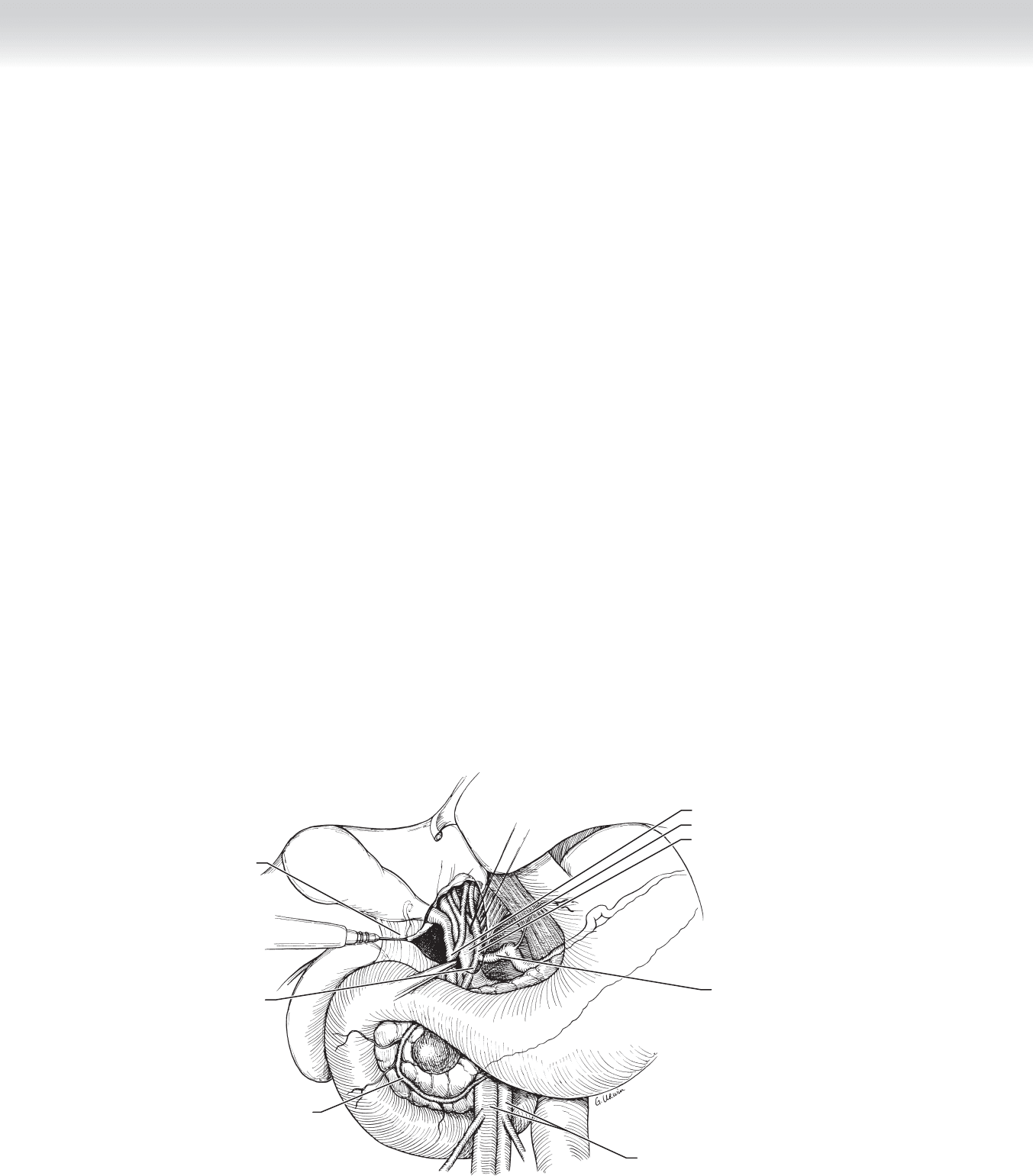
◆ Incise the peritoneum, which envelops the hepatoduodenal ligament in a transverse direc-
tion, beginning in the distal third of this structure. Isolate and encircle, with vessel loops,
the common bile duct and the proper hepatic artery and distract these vessels medially and
laterally to reveal the portal vein (Figure 49-5).
◆ Establish a plane, if possible, extending from the previously dissected inferior plane of dis-
section where the superior mesenteric vein courses beneath the head of the pancreas and
current plane in the hepatoduodenal ligament where the portal vein has been identifi ed.
There is a dense layer of connective tissue overlying the portal vein, and this must be care-
fully incised to reach the proper plane. Carefully dissecting this plane from the superior
aspect permits “meeting in the middle” with the inferior dissection. Pass a
1
⁄2-inch Penrose
drain along this newly established plane and place clamps separately on each end of the
drain (Figure 49-6).
◆ Follow the proper hepatic artery toward the celiac trunk. The proper hepatic artery travels
in a generally transverse direction and takes a right-angle turn directed cephalad. At this
right angle, the gastroduodenal artery continues along the same direction as the proper he-
patic artery. Thus one must identify the common hepatic artery to avoid misidentifying the
proper hepatic artery for the gastroduodenal artery. Dissect the gastroduodenal artery free
and double loop it with a silk suture, but do not tie (Figure 49-7).
◆ At this point, resectability is established and, if determined, then resection is undertaken. In
spite of the fact that 1 or 2 hours of dissection may already have been completed, nothing
has been done to this point in the operation that requires reconstruction.
Dissect hepatoduodenal
ligament
Gastroduodenal artery
Common hepatic artery
Hepatic triad:
Common bile duct
Portal vein
Proper hepatic artery
Anterior pancreaticoduodenal
artery
Superior mesenteric artery and vein
FIGURE 49 –5
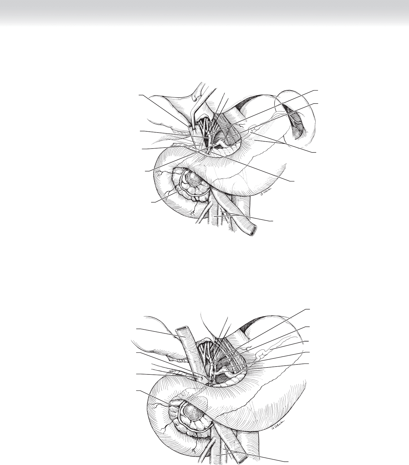
CHAPTER 49 • Pylorus-Saving Pancreaticoduodenectomy 553
Superior mesenteric artery and vein
Proper hepatic artery
Loop around common bile duct
Common hepatic artery
Splenic artery
and vein
Celiac trunk
Portal vein
Penrose drain
Anterior pancreaticoduodenal artery
Gastroduodenal artery
FIGURE 49 –6
Gastroduodenal artery stump
Loop around bile duct
Anterior pancreaticoduodenal
artery
Splenic artery
Superior mesenteric artery
and vein
Portal vein
Proper hepatic artery
Celiac trunk
Common hepatic artery
Penrose drain
FIGURE 49 –7