Yellampalli S. (ed.) Carbon Nanotubes - Synthesis, Characterization, Applications
Подождите немного. Документ загружается.

Aligned Growth of Single-Walled and Double-Walled
Carbon Nanotube Films by Control of Catalyst Preparation
197
buffer layer to prevent the formation of Co silicide during the heating process, just prior to
the introduction of CH
4
gas into the reaction chamber. Co particles were then deposited on
the TiN buffer layer using pulsed arc plasma deposition (Arc plasma gun, ULVAC, Inc.) in
vacuum, at a pressure of 10
-4
Torr at room temperature. The arc plasma gun was operated
intermittently in a pulsed operation. No heat pre-treatment was performed for the catalyst
prior to the CNT growth process. The density of Co nanoparticles on the substrate was
controlled by varying the number of pulses in the range from 50 to 250 shots, corresponding
to the particle number density of 4 × 10
12
to 2 × 10
13
cm
-2
on the surface. A mixture of CH
4
and H
2
was used as the source gas. The flow rates of CH
4
and H
2
were 50 and 70 sccm,
respectively. The microwave power and total pressure were maintained at 900 W and 70
Torr, respectively. CNTs were grown on the Co-catalyzed Si substrates in the presence of a
TiN buffer layer at a substrate temperature of 700 ˚C.
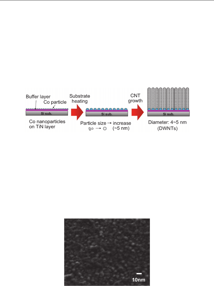
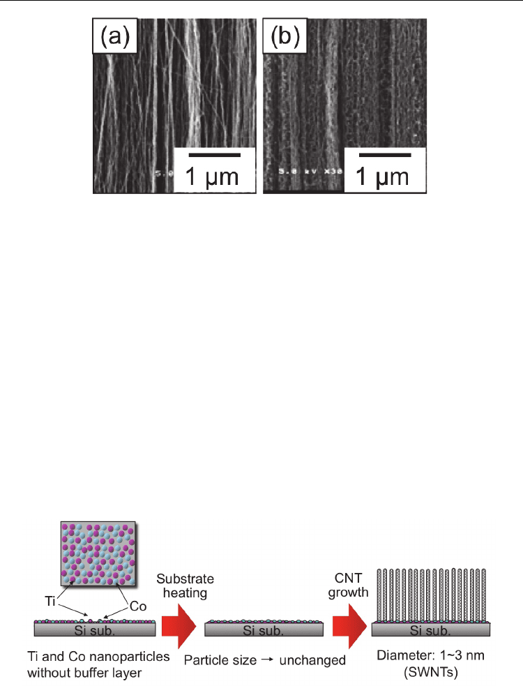
Fig. 7. Schematic of catalyst preparation for growing CNTs with small diameters (DWNTs)
Figure 8 shows a SEM image of Co-catalyzed Si substrate just before introducing CH
4
at
substrate temperature of 700 ˚C. Co particles were prepared using pulsed arc plasma
deposition with 250 pulses, corresponding to the cumulative particle number density of
approximately 2 × 10
13
cm
-2
on the surface. It was found that Co nanoparticles with size of
4–5 nm were formed on the TiN buffer layer. The pulsed arc plasma deposition using the arc
plasma gun yielded Co nanoparticles of about 1–2 nm in size, according to AFM
observations of the Co-catalyzed substrate without the TiN buffer layer as shown in Fig.
3(b). In our system, it takes about 10 min to increase the substrate temperature from room
temperature to 700 ˚C. During this period, the overlapped or closely adjacent particles
would aggregate, presumably resulting in the formation of Co nanoislands of about 3–5 nm
in size, which would be suitable for the nucleation of double-walled CNTs.
Fig. 8. SEM image of Co-catalyzed Si substrate just before introducing CH
4
at substrate
temperature of 700 ˚C. Co particles were prepared using pulsed arc plasma deposition with
250 pulses. The cumulative Co particle number density was 2 × 10
13
cm
-2
on the surface.
Carbon Nanotubes - Synthesis, Characterization, Applications
198
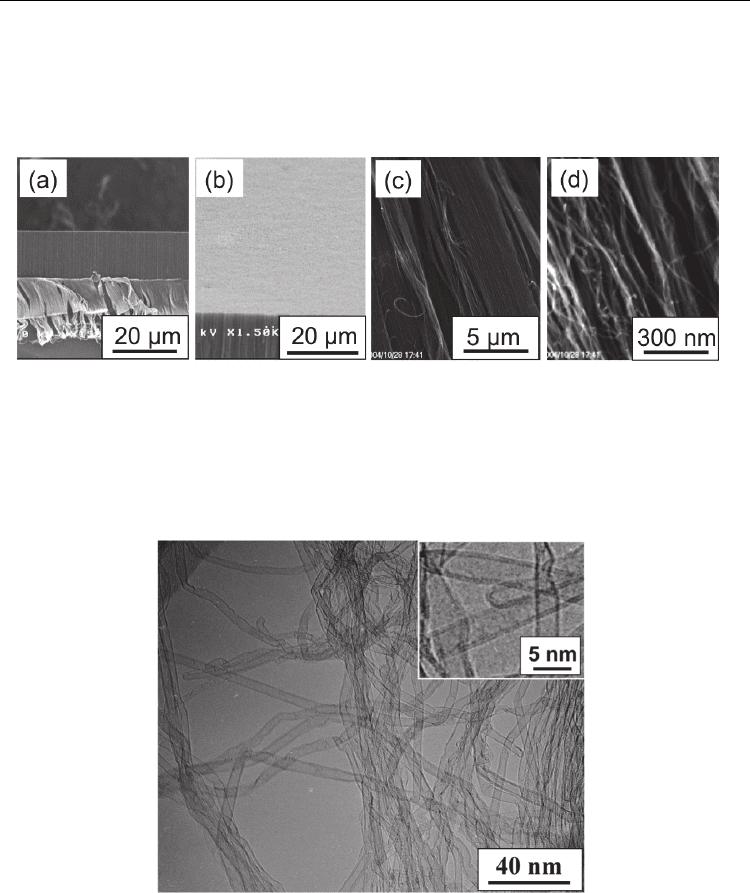
Figures 9(a) and 9(b) show typical cross-sectional and top-view SEM images of the CNT
film, respectively. Figures 9(c) and 9(d) show close-up images of the cleaved CNT film.
From the morphology observed in Fig. 9(d), it can be seen that individual CNT bundles
grew almost vertically via a self-supporting mechanism, due to the extremely high density
of the CNTs.
Fig. 9. SEM micrographs of CNT films grown on a Co-catalyzed Si substrate with a TiN
buffer layer. The Co particles were prepared using pulsed arc plasma deposition with 250
pulses. (a) Cross-sectional SEM image of the dense CNT film. (b) SEM image showing the
surface of the CNT film, (c)(d) Close-up images of cleaved CNT film showing aligned
growth of the nanotubes. (Hiramatsu et al., 2005b) - reproduced with permission from
Institute of Pure and Applied Physics
Fig. 10. TEM image of CNTs scraped from the substrate. The CNTs were grown on the Co-
catalyzed Si substrates at a substrate temperature of 700 ˚C. The inset shows a magnified
TEM image of typical CNTs. (Hiramatsu et al., 2005b) - reproduced with permission from
Institute of Pure and Applied Physics
Figure 10 shows a low-magnification TEM image of typical CNTs. Co nanoarticles were
prepared using pulsed arc plasma deposition with 50 pulses. The TEM specimen in Fig. 10
was scraped away from the substrate and was ultrasonicated in methanol. The CNTs are
Aligned Growth of Single-Walled and Double-Walled
Carbon Nanotube Films by Control of Catalyst Preparation
199
hollow and have a small average diameter of approximately 4.5 nm. Note that no Co
particles were observed at the nanotube tips in the TEM micrographs. As shown in the inset
of Fig. 10, a magnified TEM image reveals that most of CNTs have a double-walled
structure, with a clear inner channel of approximately 4 nm inner diameter. The percentages
of single-, double- and triple-walled CNTs were estimated to be 5%, 80%, 15%, respectively.
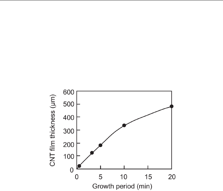
A growth rate curve for the CNT film was obtained by measuring the thickness of the CNT
films for different growth periods, according to the observation of cross-sectional SEM
images of CNT films. Figure 11 shows the average thickness of the aligned CNT film as a
function of the growth period. As shown in Fig. 11, the thickness of the CNT film increased
linearly up to 10 min and the CNTs grew at an extremely high rate of 600 nm/s during the
first 10 min. Dense DWNT films with a thickness over 500 µm were obtained after 20 min.
Fig. 11. Average thickness of aligned CNT films as a function of growth period. Co particles
were prepared using pulsed arc plasma deposition with 250 pulses. The cumulative Co
particle number density was 2 × 10
13
cm
-2
on the surface. (Hiramatsu et al., 2005b) -
reproduced with permission from Institute of Pure and Applied Physics
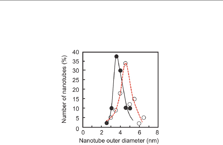
Figure 12 shows distributions of the nanotube outer diameters grown from Co nanoparticles
of different number densities. Co particles were prepared using pulsed arc plasma
deposition with 50 (closed circles) and 250 (open circles) pulses, corresponding to the Co
particle number densities of 4 × 10
12
and 2 × 10
13
cm
-2
on the surface, respectively. In the case
of growth on Co-catalyzed Si substrates with cumulative Co nanoparticle density of 2 × 10
13
cm
-2
(250 pulses), the CNTs had small average diameters of the nanotubes and a narrow
diameter distribution centered around 4.5 nm. On one hand, in the case of the CNTs grown
with Co nanoparticle density of 4 × 10
12
cm
-2
(50 pulses), average outer diameter of CNTs
decreased to 3.5–4 nm. The most significant difference from previous reports is that the
catalyst was prepared originally in the form of nanoparticles in our study. It has been
reported that the nanotube growth rate is inversely proportional to the nanotube diameter
(Bower et al., 2000). Namely, the smaller diameter nanotubes would grow at a faster rate in
terms of height. In this study, the sizes of the Co particles formed by pulsed arc deposition
were relatively small. The resultant Co nanoislands thus clearly play an important role as a
Carbon Nanotubes - Synthesis, Characterization, Applications
200
template for CNT growth, yielding nanotube diameters as small as 4–5 nm in the case of 250
pulses, for example, and consequently, a fast rate of growth could be attained for the CNTs.
The density of nanotubes was roughly estimated to be 10
12
cm
-2
for the CNTs grown on a
Co-catalyzed Si substrate, with a cumulative Co nanoparticle density of 2 × 10
13
cm
-2
(250
pulses).
Fig. 12. Distributions of nanotube outer diameters deduced from TEM observations. Co
particles were prepared using pulsed arc plasma deposition with 50 (closed circles) and 250
(open circles) pulses, corresponding to the cumulative Co particle number density of 4×10
12
and 2×10
13
cm
-2
on the surface, respectively. (Hiramatsu et al., 2005b)
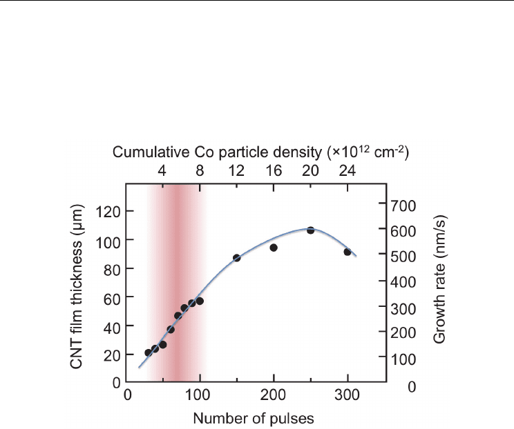
Figure 13 shows a variation of thickness of DWNT films grown for 3 min and corresponding
growth rate as a function of number of pulses of the pulsed arc discharge plasma for the
preparation of Co nanoparticles (or estimated cumulative Co nanoparticle number density
on the TiN buffer layer). As the cumulative Co particle number density on the substrate
increased, the growth rate of the DWNT film increased up to 600 nm/s, as shown in Fig. 13.
Too many Co particles on the substrate lead to the increase of the size of Co nanoislands
formed during the heating process, resulting in the growth of multiwalled (≥ 3 layers) CNTs
with lower growth rate. On the other hand, as the cumulative Co nanoparticle number
density on the substrate decreased, average diameter of grown DWNTs decreased slightly
as can be seen from Fig. 12, and the growth rate of DWNT films decreased. The reduction of
growth rate is attributed to the increase of free space to grow for individual tubes, due to the
decrease in the density of Co nanoislands formed by the aggregation of nanoparticles at the
nucleation stage of growth. In the case of CNT growth without an electrical field, the CNTs
would grow in a curly fashion. If the density of small-sized catalytic islands on the substrate
is low, CNTs would grow in random orientations or in a less-aligned manner, resulting in a
low growth rate of the CNT film. Figure 14(a) shows a cross-sectional SEM image of the
DWNT film grown from Co nanoparticles deposited by 250 shots using pulsed arc plasma
(cumulative Co particle number density of 2 × 10
13
cm
-2
on the surface). These were nearly
optimum conditions for the dense nucleation of DWNTs. As shown in Fig. 14(a), nanotube
bundles grew almost straight up due to the high density of DWNTs, corresponding to the
high growth rate of 600nm/s for a growth time of 5 min. On the other hand, for growth on a
Aligned Growth of Single-Walled and Double-Walled
Carbon Nanotube Films by Control of Catalyst Preparation
201
substrate with low-density Co nanoparticles, the growth rate of the DWNT film decreased.
Figure 14(b) shows a cross-sectional SEM image of DWNT film grown from Co
nanoparticles deposited by 50 shots, corresponding to the Co particle number density of 4 ×
10
12
cm
-2
on the surface. As shown in Fig. 14(b), the DWNTs grew in a curly fashion because
individual nanotubes had more free space to grow, resulting in the formation of wavy tubes
leading to a reduction of the CNT film growth rate (135 nm/s for growth lasting 5 min)
compared to Fig. 14(a).
Fig. 13. Variation of thickness of DWNT films grown for 3 min and corresponding growth
rate as a function of number of pulses of the pulsed arc discharge plasma for the preparation
of Co nanoparticles (or estimated cumulative Co nanoparticle number density on the TiN
buffer layer). Masked area corresponds to the condition where self-assembled cone-shaped
tips composed of CNT bundles were formed; pulsed arc plasma with 50–100 pulses (see
section 4.5).
4.3 SWNT growth from Co and Ti nanoparticles without buffer layer
In the previous section, rapid growth of aligned DWNT films were demonstrated, where
catalytic Co nanoparticles were prepared by pulsed arc deposition on a TiN buffer layer. By
forming metal nanoparticles employing the pulsed arc discharge with a metal electrode, the
density of catalyst nanoparticles with a relatively uniform size can be easily controlled on
the substrate. As shown in Fig. 12, by decreasing the number of arc discharge pulses from
250 down to 50 shots, the average diameter of DWNTs decreased from 4.5 nm down to 3.5
nm. However, aligned SWNTs were not grown even at a low density of Co nanoparticles
less than 50 pulses, probably because closely adjacent or overlapped Co nanoparticles
would easily join together on the TiN surface to increase the size of the nanoislands during
the substrate heating, resulting in the formation of less-aligned, low-density DWNTs or
randomly oriented, sparse SWNTs after all.
Carbon Nanotubes - Synthesis, Characterization, Applications
202
Fig. 14. Cross-sectional SEM images of CNT films grown from Co nanoparticles prepared on
TiN buffer layer using pulsed arc plasma with (a) 250 pulses and (b) 50 pulses. (Hiramatsu
et al., 2005b) - reproduced with permission from Institute of Pure and Applied Physics
In order to grow SWNT film on a Si substrate, a mixture of Co and Ti nanoparticles was
prepared on the Si substrate using pulsed arc plasma deposition with a Co–Ti composite
electrode, without a buffer layer. Figure 15 shows a schematic diagram of catalyst
preparation for growing CNTs with smaller diameters. Ti is highly reactive with Si to form a
silicide, as compared with Co (Murarka, 1983). Accordingly, it is expected that the formation
of a Ti–silicide would precede the formation of a Co–silicide, when Ti is mixed with Co.
Therefore, Ti prevents the formation of Co–silicide to a certain extent in the substrate
heating process, and enables the size of the Co catalyst nanoparticles to be maintained at
approximately 1–2 nm. As a result, the fabrication of films composed of vertically aligned
SWNTs on Si substrates was attained using microwave plasma-enhanced CVD (Hiramatsu
et al., 2007a). Moreover, the controlled preparation of catalyst nanoparticles on the Si
substrate was performed by pulsed arc plasma deposition with the alternate use of Co and
Ti electrodes. This technique has potential for controlling the size of catalyst nanoislands on
the substrate, resulting in the controlled growth of aligned CNTs with single to three walls.
By changing the number of cumulative Co nanoparticles, the fabrications of SWNT and
DWNT films can be controlled (Hiramatsu et al., 2007b).
Fig. 15. Schematic of catalyst preparation for growing CNTs with small diameters (SWNTs)
In the case using sintered Ti–Co composite target electrode, the pulsed arc discharge yields a
mixture of Co and Ti nanoparticles simultaneously. The sintered Ti–Co composite electrode
Aligned Growth of Single-Walled and Double-Walled
Carbon Nanotube Films by Control of Catalyst Preparation
203
is commercially available from ULVAC, Inc., and the Co and Ti contents in the composite
are 55.2 and 44.8 wt.%, respectively. The pulsed arc discharge was operated with sintered
Ti–Co composite electrode at a pressure of 10
-4
Torr at room temperature. Three types of Co-
catalyzed Si substrates (types A, B and C) without buffer layer were prepared by setting the
number of pulsed arc discharges with the Ti–Co composite electrode at 50, 100 and 250
shots, respectively. Ti nanoparticles mixed with the Co nanoparticles prevent the formation
of Co-silicide during the substrate heating process, and enables the size of the Co catalytic
nanoparticles to be maintained at approximately 1–2 nm.
CNT films were grown using microwave plasma-enhanced CVD with a 2.45 GHz, 1.5 kW
microwave generator. A mixture of CH
4
and H
2
was used as the source gas. The flow rates
of CH
4
and H
2
were 50 and 70 sccm, respectively. The microwave power and total pressure
were maintained at 900 W and 70 Torr, respectively. The growth experiments were carried
out for 5 min at a substrate temperature of 700 °C. SEM and TEM were used to evaluate the
morphology of the grown CNTs. Raman spectra for the CNTs were obtained using the 514.5
nm line of Ar laser and the 632.8 nm line of He–Ne laser.
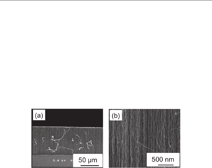
Fig. 16. SEM micrographs of a CNT film grown on a Co-catalyzed Si substrate (type A). The
catalytic nanoparticles were prepared by pulsed arc discharge using a sintered Ti–Co
composite electrode with 50 pulses. (a) Cross-sectional SEM image of the dense CNT film.
(b) Close-up image of the cleaved CNT film showing aligned growth of the nanotubes.
(Hiramatsu et al., 2007a) - reproduced with permission from Elsevier
Figure 16(a) shows a typical cross-sectional SEM image of a cleaved CNT film grown for 5
min on a type A substrate. The catalytic nanoparticles were prepared using pulsed arc
discharges employing the sintered Ti–Co composite electrode with 50 pulses. A dense and
vertically aligned CNT film was observed to grow on the Co-catalyzed Si substrate. Figure
16(b) shows a close-up SEM image of the cleaved CNT film. The morphology in Fig. 16(b)
shows that the CNT bundles were not perfectly straight. In this case, the growth rate of the
CNT film was approximately 190 nm/s.
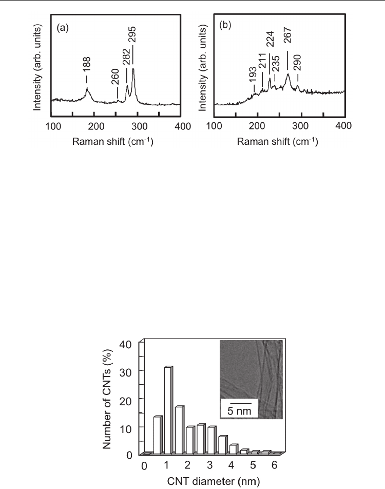
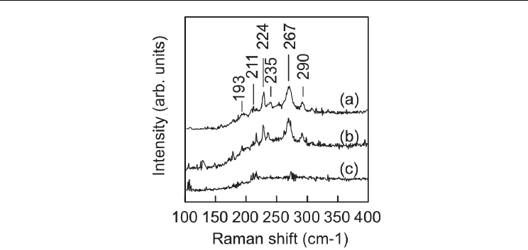
Figure 17(a) shows the Raman spectrum at the low frequency band (100–400 cm
-1
) for the
CNT sample on the type A substrate measured using the 632.8 nm line. The radial breathing
mode (RBM) peaks are clearly observed at 188, 260, 282 and 295 cm
-1
, confirming the
existence of SWNTs. Figure 17(b) shows the Raman spectrum at the low frequency band for
the same CNT sample measured using the 514.5 nm line. In this case, the RBM peaks are
clearly observed at 193, 211, 224, 235, 267 and 290 cm
-1
. The diameter distribution calculated
from the frequency of the RBM peaks for the SWNT bundles is in the range between
0.8 and 1.3 nm.
Carbon Nanotubes - Synthesis, Characterization, Applications
204
Fig. 17. Raman spectra at the low frequency band (100–400 cm
-1
) for the CNT sample grown
on the type ‘A’ substrate. The Raman spectra (a) and (b) were obtained using the 632.8 nm
line of He–Ne laser and the 514.5 nm line of Ar laser, respectively. (Hiramatsu et al., 2007a) -
reproduced with permission from Elsevier
In order to evaluate the morphology and diameter distribution of the CNTs, TEM images
were taken for the same CNT sample used for the measurement of Raman spectra shown in
Figs. 17(a) and 17(b). A distribution histogram of nanotube outer diameters is shown in Fig.
18, and the typical TEM image of the CNTs is shown in the inset in Fig. 18. The TEM
specimen was prepared by scraping from the substrate and ultrasonicating in methanol. The
inset TEM image in Fig. 18 reveals that most CNTs are SWNTs free of metal particles. The
diameters of the SWNTs were measured and the average diameter of the CNTs was
determined to be approximately 1 nm, in agreement with the value calculated from the RBM
peaks of the Raman spectra in Figs. 17(a) and 17(b). The distribution histogram of nanotube
outer diameters shows that the diameters of most CNTs were in the range from 0.5 to 3 nm.
The SWNT and DWNT fractions were estimated to be 80 and 20%, respectively.
Fig. 18. Distribution histogram of nanotube outer diameters deduced from TEM
observations. The CNTs were grown on the type A substrate. The catalytic nanoparticles
were prepared by pulsed arc discharges with the sintered Ti–Co composite electrode with 50
pulses. The inset shows a magnified TEM image of typical CNTs. (Hiramatsu et al., 2007a) -
reproduced with permission from Elsevier
Aligned Growth of Single-Walled and Double-Walled
Carbon Nanotube Films by Control of Catalyst Preparation
205
Fig. 19. Raman spectra at the low frequency band (100–400 cm
-1
), measured using the 514.5
nm line of Ar laser, for CNTs grown on Si substrates with different densities of cumulative
nanoparticles. Raman spectra (a)–(c) were obtained for CNT films grown on Co-catalyzed Si
substrates, types A, B and C, respectively. (Hiramatsu et al., 2007a) - reproduced with
permission from Elsevier
CNTs were grown on Si substrates with different densities of cumulative nanoparticles, and
the Raman spectra for the CNTs were obtained using the 514.5 nm line of an Ar laser. In Fig.
19, the Raman spectra (a)–(c) in the low frequency band were obtained for CNT films grown
on Co-catalyzed Si substrates, types A, B and C, respectively. As mentioned before, in the
Raman spectrum (a) for the CNTs grown on the type A substrate, the RBM peaks are clearly
observed at 188, 217, 260, 279 and 293 cm
-1
, confirming the existence of SWNTs. In the case
of the type B substrate, where the cumulative nanoparticle density was twice that on the
type A substrate, the RBM peaks are also clearly observed in the Raman spectrum (b). The
Raman spectrum (b) is almost identical to the Raman spectrum (a). On the other hand, the
growth rate of the CNT film using the type B substrate was approximately 350 nm/s, which
is almost twice that of the type A substrate. When using a substrate with low-density
catalytic nanoparticles (type A substrate), the CNT bundles had more space to grow and
thereby grew in a curly fashion, as shown in Fig. 16(b). With increasing cumulative density
of catalytic nanoparticles, as in the case using the type B substrate, SWNT film with a high
growth rate was attained due to the dense nucleation of CNTs from the doubled density of
the catalytic nanoparticles as compared to the case using the type A substrate. In contrast,
with further increase in the cumulative catalytic nanoparticle density, the RBM peaks
disappeared, as seen in the Raman spectrum (c) for the CNTs grown on the type C substrate,
resulting in the growth of MWNTs with two walls or more.
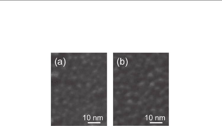
Figures 20(a) and 20(b) show SEM images of the surfaces of the type B and C Co-catalyzed
Si substrates respectively, just before the introduction of CH
4
at a substrate temperature of
700 °C. In Fig. 20(a), nanoparticles with a size of approximately 2 nm were formed on the
type B substrate. On the other hand, nanoparticles with a size of approximately 3–4 nm were
observed on the type C substrate, as seen in the SEM image in Fig. 20(b). Pulsed arc plasma
deposition with the Co electrode yielded Co nanoparticles of about 1–2 nm in size. At an
appropriate density of catalytic nanoparticles, the catalytic nanoparticles deposited by
Carbon Nanotubes - Synthesis, Characterization, Applications
206
pulsed arc discharge play an important role as a template for the growth of SWNTs with
diameters as small as 1–2 nm. On the other hand, in the case of growth on a substrate with
an excess density of cumulative nanoparticles, MWNTs with 2 or 3 walls were primarily
grown. In our system, it takes about 10 min to increase the substrate temperature from room
temperature to 700 °C. During this period, overlapping or closely adjacent particles can
merge, resulting in the formation of catalytic nanoparticles of approximately 3–4 nm in size.
Fig. 20. SEM images of the surface of Co-catalyzed Si substrates: (a) type B and (b) type C,
just before introducing CH
4
at a substrate temperature of 700 °C. (Hiramatsu et al., 2007a) -
reproduced with permission from Elsevier
Further controlled preparation of catalyst nanoparticles on the Si substrate was performed
by pulsed arc plasma deposition with the alternate use of Co and Ti electrodes. This
technique has potential for controlling the size of catalyst nanoparticles on the substrate,
resulting in the controlled growth of aligned CNTs with single to three walls. By changing
the number of cumulative Co nanoparticles, the fabrications of SWNT and DWNT films can
be controlled.
In this case, Co and Ti nanoparticles are deposited alternately on the Si substrate without a
buffer layer, by pulsed arc discharge with the alternate use of Ti and Co electrodes at a
pressure of 10
-4
Torr at room temperature. Ti and Co electrodes were used in turn after
every 10 shots of pulsed discharge. The number of cumulative pulsed discharges with the Ti
electrode was the same as that with the Co electrode for each substrate prepared for the
CNT growth experiment hereafter. The density of Co nanoparticles on the substrate was
controlled by varying the number of pulsed discharges with the Co electrode in the range
from 50 to 250 shots, resulting in cumulative Co particle densities of 4 × 10
12
to 2 × 10
13
cm
-2
.
From the TEM observation, the pulsed arc discharge with the Ti electrode yields Ti
nanoparticles as small as the Co nanoparticles. For Co or Ti film deposition by pulsed arc
discharge, the deposition rate of the Co film using the Co electrode was almost the same as
that of the Ti film using the Ti electrode. Therefore, it would be assumed that the cumulative
densities of Ti and Co nanoparticles are almost the same for each substrate prepared.
Figure 21(a) shows a typical cross-sectional SEM image of a cleaved CNT film grown for 5 min
on a Co-catalyzed Si substrate with a cumulative Co nanoparticle density of 4 × 10
12
cm
-2
. A
dense and vertically aligned CNT film was grown on the Co-catalyzed Si substrate. Figure
21(b) shows a magnified SEM image of the cleaved CNT film. From the morphology shown
in Fig. 21(b), it can be observed that CNT bundles with a diameter of less than 10 nm grew
almost vertically, due to the high density of the CNTs. In this case, the growth rate of the
SWNT film was approximately 200 nm/s. Figure 21(c) is a typical top-view SEM image