Berg J.M., Tymoczko J.L., Stryer L. Biochemistry
Подождите немного. Документ загружается.

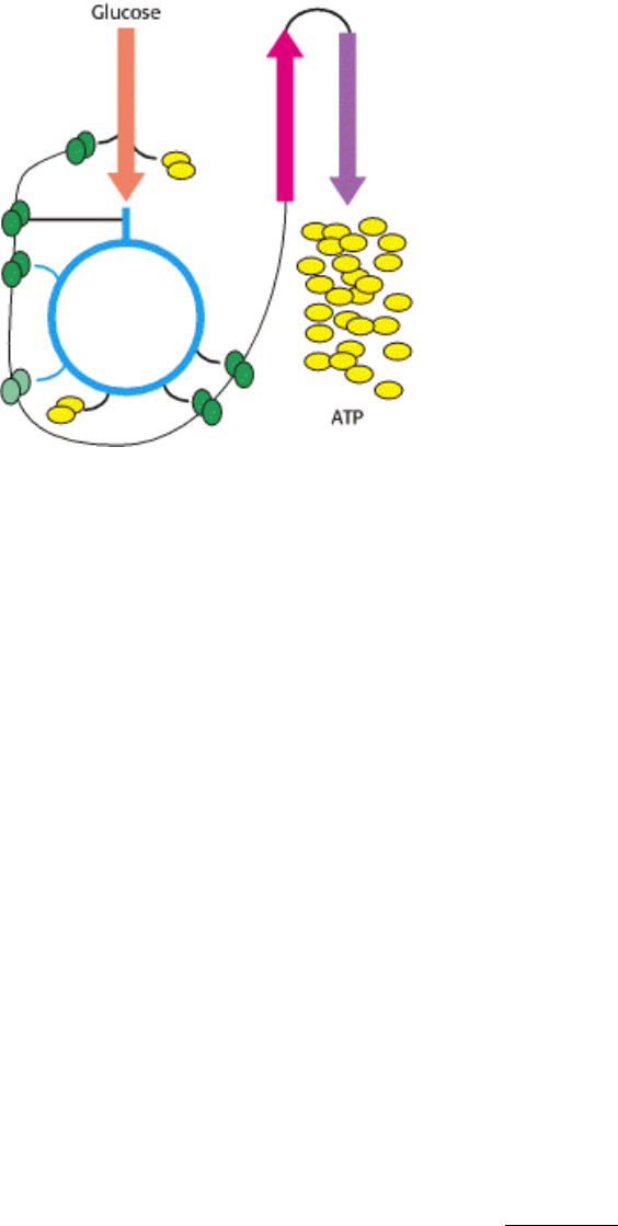
Interplay of metabolic pathways for energy production. At left, the image shows a detail of runners on a Greek
amphora painted in the sixth century, B.C. Athletic feats, and others as seemingly simple as maintenance of blood
glucose levels, require elaborate metabolic integration. The schematic above represents the oxidation of glucose to yield
ATP in a process requiring interplay among glycolysis, the citric acid cycle, and oxidative phosphorylation. These are a
few of the many metabolic pathways that must be coordinated to meet the demands of living. [(Left) Metropolitan
Museum of Art, Rogers Fund, 1914 (14.130.12). Copyright © 1977 by the Metropolitan Museum of Art.]
III. Synthesizing the Molecules of Life 30. The Integration of Metabolism
30.1. Metabolism Consist of Highly Interconnected Pathways
The basic strategy of catabolic metabolism is to form ATP, reducing power, and building blocks for biosyntheses. Let us
briefly review these central themes:
1. ATP is the universal currency of energy. The high phosphoryl transfer potential of ATP enables it to serve as the
energy source in muscle contraction, active transport, signal amplification, and biosyntheses. The hydrolysis of an ATP
molecule changes the equilibrium ratio of products to reactants in a coupled reaction by a factor of about 10
8
. Hence, a
thermodynamically unfavorable reaction sequence can be made highly favorable by coupling it to the hydrolysis of a
sufficient number of ATP molecules.
2. ATP is generated by the oxidation of fuel molecules such as glucose, fatty acids, and amino acids. The common
intermediate in most of these oxidations is acetyl CoA. The carbon atoms of the acetyl unit are completely oxidized to
CO
2
by the citric acid cycle with the concomitant formation of NADH and FADH
2
. These electron carriers then transfer
their highpotential electrons to the respiratory chain. The subsequent flow of electrons to O
2
leads to the pumping of
protons across the inner mitochondrial membrane (Figure 30.1). This proton gradient is then used to synthesize ATP.
Glycolysis also generates ATP, but the amount formed is much smaller than that in oxidative phosphorylation. The
oxidation of glucose to pyruvate yields only 2 molecules of ATP, whereas the complete oxidation of glucose to CO
2
yields 30 molecules of ATP.
3. NADPH is the major electron donor in reductive biosyntheses. In most biosyntheses, the products are more reduced
than the precursors, and so reductive power is needed as well as ATP. The high-potential electrons required to drive
these reactions are usually provided by NADPH. The pentose phosphate pathway supplies much of the required NADPH.
4. Biomolecules are constructed from a small set of building blocks. The highly diverse molecules of life are synthesized
from a much smaller number of precursors. The metabolic pathways that generate ATP and NADPH also provide
building blocks for the biosynthesis of more-complex molecules. For example, acetyl CoA, the common intermediate in
the breakdown of most fuels, supplies a two-carbon unit in a wide variety of biosyntheses, such as those leading to fatty
acids, prostaglandins, and cholesterol. Thus, the central metabolic pathways have anabolic as well as catabolic roles.

5. Biosynthetic and degradative pathways are almost always distinct. For example, the pathway for the synthesis of fatty
acids is different from that of their degradation. This separation enables both biosynthetic and degradative pathways to
be thermodynamically favorable at all times. A biosynthetic pathway is made exergonic by coupling it to the hydrolysis
of a sufficient number of ATP molecules. The separation of biosynthetic and degradative pathways contributes greatly to
the effectiveness of metabolic control.
"To every thing there is a season, and a time to every purpose under
the heaven:
A time to be born, and a time to die; a time to plant, and a time to
pluck up that which is planted;
A time to kill, and a time to heal; a time to break down, and a time to
build up."
E
cclesiastes 3:1-3
30.1.1. Recurring Motifs in Metabolic Regulation
Anabolism and catabolism must be precisely coordinated. Metabolic networks sense and respond to information on the
status of their component pathways. The information is received and metabolism is controlled in several ways:
Pasteur effect-
The inhibition of glycolysis by respiration, discovered by Louis
Pasteur in studying fermentation by yeast. The consumption of
carbohydrate is about sevenfold lower under aerobic conditions than
under anaerobic ones. The inhibition of phosphofructokinase by
citrate and ATP accounts for much of the Pasteur effect.
1. Allosteric interactions. The flow of molecules in most metabolic pathways is determined primarily by the activities of
certain enzymes rather than by the amount of substrate available. Enzymes that catalyze essentially irreversible reactions
are likely control sites, and the first irreversible reaction in a pathway (the committed step) is nearly always tightly
controlled. Enzymes catalyzing committed steps are allosterically regulated, as exemplified by phosphofructokinase in
glycolysis and acetyl CoA carboxylase in fatty acid synthesis. Allosteric interactions enable such enzymes to rapidly
detect diverse signals and to adjust their activity accordingly.
2. Covalent modification. Some regulatory enzymes are controlled by covalent modification in addition to allosteric
interactions. For example, the catalytic activity of glycogen phosphorylase is enhanced by phosphorylation, whereas that
of glycogen synthase is diminished. Specific enzymes catalyze the addition and removal of these modifying groups
(Figure 30.2). Why is covalent modification used in addition to noncovalent allosteric control? The covalent
modification of an essential enzyme in a pathway is often the final step in an amplifying cascade and allows metabolic
pathways to be rapidly switched on or off by very low concentrations of triggering signals. In addition, covalent
modifications usually last longer (from seconds to minutes) than do reversible allosteric interactions (from milliseconds
to seconds).
3. Enzyme levels. The amounts of enzymes, as well as their activities, are controlled. The rates of synthesis and
degradation of many regulatory enzymes are altered by hormones. The basics of this control were considered in Chapter
28; we will return to the topic in Chapter 31.

4. Compartmentation. The metabolic patterns of eukaryotic cells are markedly affected by the presence of compartments
(Figure 30.3). The fates of certain molecules depend on whether they are in the cytosol or in mitochondria, and so their
flow across the inner mitochondrial membrane is often regulated. For example, fatty acids are transported into
mitochondria for degradation only when energy is required, whereas fatty acids in the cytosol are esterified or exported.
5. Metabolic specializations of organs. Regulation in higher eukaryotes is enhanced by the existence of organs with
different metabolic roles. Metabolic specialization is the result of differential gene expression.
30.1.2. Major Metabolic Pathways and Control Sites
Let us now review the roles of the major pathways of metabolism and the principal sites for their control:
1. Glycolysis. This sequence of reactions in the cytosol converts one molecule of glucose into two molecules of pyruvate
with the concomitant generation of two molecules each of ATP and NADH. The NAD
+
consumed in the reaction
catalyzed by glyceraldehyde 3-phosphate dehydrogenase must be regenerated for glycolysis to proceed. Under anaerobic
conditions, as in highly active skeletal muscle, this regeneration is accomplished by the reduction of pyruvate to lactate.
Alternatively, under aerobic conditions, NAD
+
is regenerated by the transfer of electrons from NADH to O
2
through the
electron-transport chain. Glycolysis serves two main purposes: it degrades glucose to generate ATP, and it provides
carbon skeletons for biosyntheses.
Phosphofructokinase, which catalyzes the committed step in glycolysis, is the most important control site. ATP is both a
substrate in the phosphoryl transfer reaction and a regulatory molecule. A high level of ATP inhibits
phosphofructokinase the regulatory sites are distinct from the substratebinding sites and have a lower affinity for the
nucleotide. This inhibitory effect is enhanced by citrate and reversed by AMP (Figure 30.4). Thus, the rate of glycolysis
depends on the need for ATP, as signaled by the ATP/AMP ratio, and on the availability of building blocks, as signaled
by the level of citrate. In liver, the most important regulator of phosphofructokinase activity is fructose 2,6-bisphosphate
(F-2,6-BP). Recall that the level of F-2,6-BP is determined by the activity of the kinase that forms it from fructose 6-
phosphate and of the phosphatase that hydrolyzes the 2-phosphoryl group (Section 16.2.2). When the blood-glucose
level is low, a glucagon-triggered cascade leads to activation of the phosphatase and inhibition of the kinase in the liver.
The level of F-2,6-BP declines and, consequently, so does phosphofructokinase activity. Hence, glycolysis is slowed,
and the spared glucose is released into the blood for use by other tissues.
2. Citric acid cycle and oxidative phosphorylation. The reactions of this common pathway for the oxidation of fuel
molecules carbohydrates, amino acids, and fatty acids take place inside mitochondria. Most fuels enter the cycle as
acetyl CoA. The complete oxidation of an acetyl unit by the citric acid cycle generates one molecule of GTP and four
pairs of electrons in the form of three molecules of NADH and one molecule FADH
2
. These electrons are transferred to
O
2
through the electron-transport chain, which results in the formation of a proton gradient that drives the synthesis of
nine molecules of ATP. The electron donors are oxidized and recycled back to the citric acid cycle only if ADP is
simultaneously phosphorylated to ATP. This tight coupling, called respiratory control, ensures that the rate of the citric
acid cycle matches the need for ATP. An abundance of ATP also diminishes the activities of two enzymes in the cycle
isocitrate dehydrogenase and α-ketoglutarate dehydrogenase. The citric acid cycle has an anabolic role as well. In
concert with pyruvate carboxylase, the citric acid cycle provides intermediates for biosyntheses, such as succinyl CoA
for the formation of porphyrins and citrate for the formation of fatty acids.
3. Pentose phosphate pathway. This series of reactions, which takes place in the cytosol, consists of two stages. The first
stage is the oxidative decarboxylation of glucose 6-phosphate. Its purpose is the production of NADPH for reductive
biosyntheses and the formation of ribose 5-phosphate for the synthesis of nucleotides. Two molecules of NADPH are
generated in the conversion of glucose 6-phosphate into ribose 5- phosphate. The dehydrogenation of glucose 6-
phosphate is the committed step in this pathway. This reaction is controlled by the level of NADP
+
, the electron acceptor

(Figure 30.5).
The second stage of the pentose phosphate pathway is the nonoxidative, reversible metabolism of five-carbon
phosphosugars into phosphorylated three-carbon and six-carbon glycolytic intermediates. Thus, the nonoxidative branch
can either introduce riboses into glycolysis for catabolism or generate riboses from glycolytic intermediates for
biosyntheses.
4. Gluconeogenesis. Glucose can be synthesized by the liver and kidneys from noncarbohydrate precursors such as
lactate, glycerol, and amino acids. The major entry point of this pathway is pyruvate, which is carboxylated to
oxaloacetate in mitochondria. Oxaloacetate is then metabolized in the cytosol to form phosphoenolpyruvate. The other
distinctive means of gluconeogenesis are two hydrolytic steps that bypass the irreversible reactions of glycolysis.
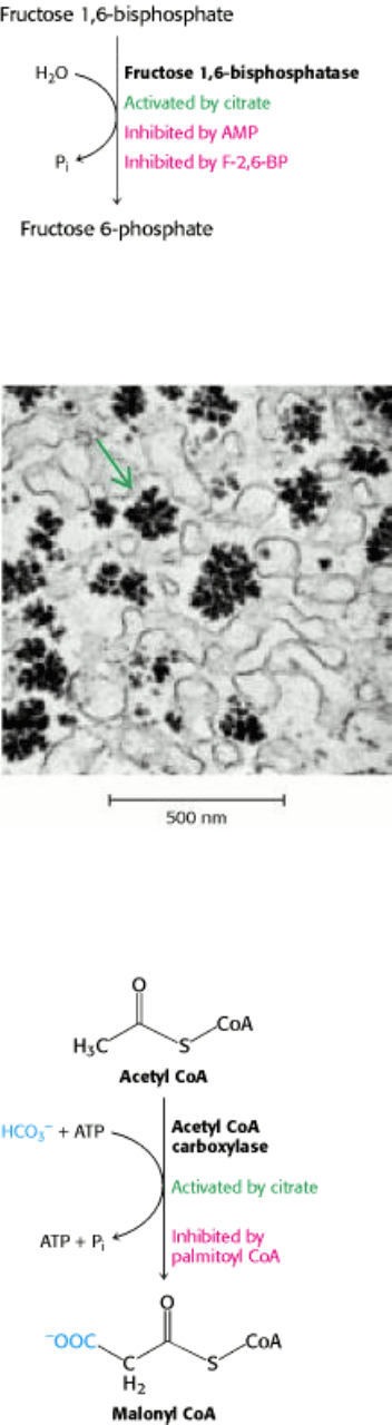
Gluconeogenesis and glycolysis are usually reciprocally regulated so that one pathway is minimally active while the
other is highly active. For example, AMP inhibits and citrate activates fructose 1,6-bisphosphatase, an essential enzyme
in gluconeogenesis, whereas these molecules have opposite effects on phosphofructokinase, the pacemaker of glycolysis
(Figure 30.6). Fructose-2,6-bisphosphate also coordinates these processes by inhibiting fructose 1,6-bisphosphatase.
Hence, when glucose is abundant, the high level of F-2,6-BP inhibits gluconeogenesis and activates glycolysis.
5. Glycogen synthesis and degradation. Glycogen, a readily mobilizable fuel store, is a branched polymer of glucose
residues (Figure 30.7). In glycogen degradation, a phosphorylase catalyzes the cleavage of glycogen by orthophosphate
to yield glucose 1-phosphate, which is rapidly converted into glucose 6-phosphate for further metabolism. In glycogen
synthesis, the activated intermediate is UDP-glucose, which is formed from glucose 1-phosphate and UTP. Glycogen
synthase catalyzes the transfer of glucose from UDP-glucose to the terminal glucose residue of a growing strand.
Glycogen degradation and synthesis are coordinately controlled by a hormonetriggered amplifying cascade so that the
phosphorylase is active when synthase is inactive and vice versa. Phosphorylation and noncovalent allosteric interactions
(Section 21.5) regulate these enzymes.
6. Fatty acid synthesis and degradation. Fatty acids are synthesized in the cytosol by the addition of two-carbon units to
a growing chain on an acyl carrier protein. Malonyl CoA, the activated intermediate, is formed by the carboxylation of
acetyl CoA. Acetyl groups are carried from mitochondria to the cytosol as citrate by the citrate-malate shuttle. In the
cytosol, citrate is cleaved to yield acetyl CoA. In addition to transporting acetyl CoA, citrate in the cytosol stimulates
acetyl CoA carboxylase, the enzyme catalyzing the committed step. When ATP and acetyl CoA are abundant, the level of
citrate increases, which accelerates the rate of fatty acid synthesis (Figure 30.8).
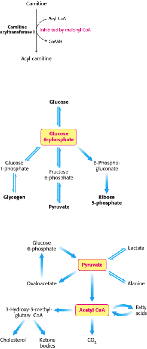
A different pathway in a different compartment degrades fatty acids. Carnitine transports fatty acids into mitochondria,
where they are degraded to acetyl CoA in the mitochondrial matrix by β-oxidation. The acetyl CoA then enters the citric
acid cycle if the supply of oxaloacetate is sufficient. Alternatively, acetyl CoA can give rise to ketone bodies. The
FADH
2
and NADH formed in the β-oxidation pathway transfer their electrons to O
2
through the electron-transport
chain. Like the citric acid cycle, β-oxidation can continue only if NAD
+
and FAD are regenerated. Hence, the rate of
fatty acid degradation also is coupled to the need for ATP. Malonyl CoA, the precursor for fatty acid synthesis, inhibits
fatty acid degradation by inhibiting the formation of acyl carnitine by carnitine acyl transferase 1, thus preventing the
translocation of fatty acids into mitochondria (Figure 30.9).
30.1.3. Key Junctions: Glucose 6-phosphate, Pyruvate, and Acetyl CoA
The factors governing the flow of molecules in metabolism can be further understood by examining three important
molecules: glucose 6-phosphate, pyruvate, and acetyl CoA. Each of these molecules has several contrasting fates:
1. Glucose 6-phosphate. Glucose entering a cell is rapidly phosphorylated to glucose 6-phosphate and is subsequently
stored as glycogen, degraded to pyruvate, or converted into ribose 5-phosphate (Figure 30.10). Glycogen is formed when

glucose 6-phosphate and ATP are abundant. In contrast, glucose 6-phosphate flows into the glycolytic pathway when
ATP or carbon skeletons for biosyntheses are required. Thus, the conversion of glucose 6-phosphate into pyruvate can be
anabolic as well as catabolic. The third major fate of glucose 6-phosphate, to flow through the pentose phosphate
pathway, provides NADPH for reductive biosyntheses and ribose 5-phosphate for the synthesis of nucleotides. Glucose
6-phosphate can be formed by the mobilization of glycogen or it can be synthesized from pyruvate and glucogenic amino
acids by the gluconeogenic pathway.
2. Pyruvate. This three-carbon α-ketoacid is another major metabolic junction (Figure 30.11). Pyruvate is derived
primarily from glucose 6-phosphate, alanine, and lactate. Pyruvate can be reduced to lactate by lactate dehydrogenase to
regenerate NAD
+
. This reaction enables glycolysis to proceed transiently under anaerobic conditions in active tissues
such as contracting muscle. The lactate formed in active tissue is subsequently oxidized back to pyruvate, in other
tissues. The essence of this interconversion buys time and shifts part of the metabolic burden of active muscle to other
tissues. Another readily reversible reaction in the cytosol is the transamination of pyruvate, an α-ketoacid, to alanine, the
corresponding amino acid. Conversely, several amino acids can be converted into pyruvate. Thus, transamination is a
major link between amino acid and carbohydrate metabolism.
A third fate of pyruvate is its carboxylation to oxaloacetate inside mitochondria, the first step in gluconeogenesis. This
reaction and the subsequent conversion of oxaloacetate into phosphoenolpyruvate bypass an irreversible step of
glycolysis and hence enable glucose to be synthesized from pyruvate. The carboxylation of pyruvate is also important for
replenishing intermediates of the citric acid cycle. Acetyl CoA activates pyruvate carboxylase, enhancing the synthesis
of oxaloacetate, when the citric acid cycle is slowed by a paucity of this intermediate.
A fourth fate of pyruvate is its oxidative decarboxylation to acetyl CoA. This irreversible reaction inside mitochondria is
a decisive reaction in metabolism: it commits the carbon atoms of carbohydrates and amino acids to oxidation by the
citric acid cycle or to the synthesis of lipids. The pyruvate dehydrogenase complex, which catalyzes this irreversible
funneling, is stringently regulated by multiple allosteric interactions and covalent modifications. Pyruvate is rapidly
converted into acetyl CoA only if ATP is needed or if two-carbon fragments are required for the synthesis of lipids.
3. Acetyl CoA. The major sources of this activated two-carbon unit are the oxidative decarboxylation of pyruvate and the
β-oxidation of fatty acids (see Figure 30.11). Acetyl CoA is also derived from ketogenic amino acids. The fate of acetyl
CoA, in contrast with that of many molecules in metabolism, is quite restricted. The acetyl unit can be completely
oxidized to CO
2
by the citric acid cycle. Alternatively, 3-hydroxy-3-methylglutaryl CoA can be formed from three
molecules of acetyl CoA. This six-carbon unit is a precursor of cholesterol and of ketone bodies, which are transport
forms of acetyl units released from the liver for use by some peripheral tissues. A third major fate of acetyl CoA is its
export to the cytosol in the form of citrate for the synthesis of fatty acids.
III. Synthesizing the Molecules of Life 30. The Integration of Metabolism 30.1. Metabolism Consist of Highly Interconnected Pathways
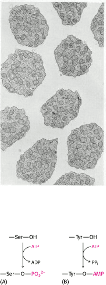
Figure 30.1. Electron Micrograph of Mitochondria. Numerous mitochondria occupy the inner segment of retinal rod
cells. These photoreceptor cells generate large amounts of ATP and are highly dependent on a continuous supply of O
2
.
[Courtesy of Dr. Michael Hogan.]
III. Synthesizing the Molecules of Life 30. The Integration of Metabolism 30.1. Metabolism Consist of Highly Interconnected Pathways
Figure 30.2. Covalent Modifications. Covalent modifications. Examples of reversible covalent modifications of
proteins: (A) phosphorylation, (B) adenylation.
III. Synthesizing the Molecules of Life 30. The Integration of Metabolism 30.1. Metabolism Consist of Highly Interconnected Pathways
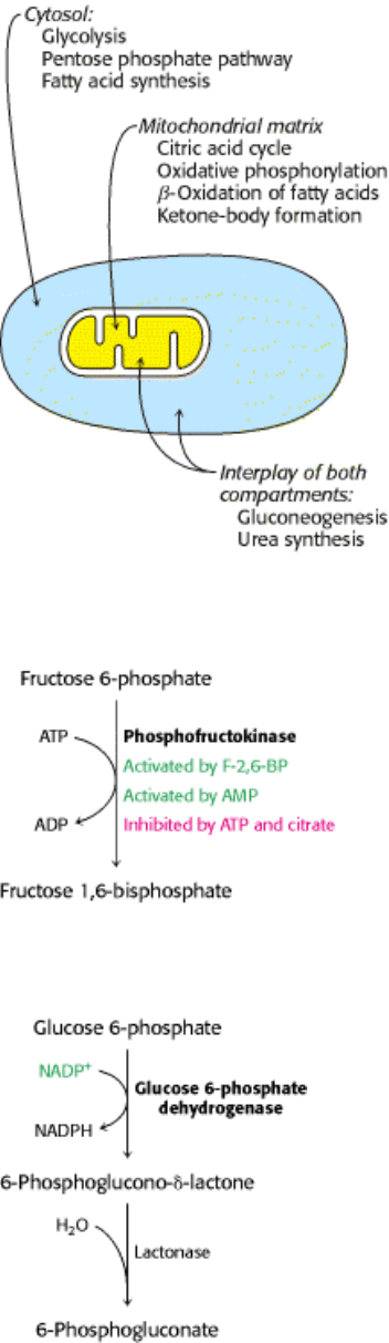
Figure 30.3. Compartmentation of the Major Pathways of Metabolism.
III. Synthesizing the Molecules of Life 30. The Integration of Metabolism 30.1. Metabolism Consist of Highly Interconnected Pathways
Figure 30.4. Regulation of Glycolysis. Phosphofructokinase is the key enzyme in the regulation of glycolysis.
III. Synthesizing the Molecules of Life 30. The Integration of Metabolism 30.1. Metabolism Consist of Highly Interconnected Pathways
Figure 30.5. Regulation of the Pentose Phosphate Pathway. The dehydrogenation of glucose 6-phosphate is the
committed step in the pentose phosphate pathway.
III. Synthesizing the Molecules of Life 30. The Integration of Metabolism 30.1. Metabolism Consist of Highly Interconnected Pathways
Figure 30.6. Regulation of Gluconeogenesis. Fructose 1,6-bisphosphatase is the principal enzyme controlling the rate
of gluconeogenesis.
III. Synthesizing the Molecules of Life 30. The Integration of Metabolism 30.1. Metabolism Consist of Highly Interconnected Pathways
Figure 30.7. Glycogen Granules. The electron micrograph shows part of a liver cell containing glycogen particles.
[Courtesy of Dr. George Palade.]
III. Synthesizing the Molecules of Life 30. The Integration of Metabolism 30.1. Metabolism Consist of Highly Interconnected Pathways
Figure 30.8. Regulation of Fatty Acid Synthesis. Acetyl CoA carboxylase is the key control site in fatty acid synthesis.
III. Synthesizing the Molecules of Life 30. The Integration of Metabolism 30.1. Metabolism Consist of Highly Interconnected Pathways
Figure 30.9. Control of Fatty Acid Degradation. Malonyl CoA inhibits fatty acid degradation by inhibiting the
formation of acyl carnitine.
III. Synthesizing the Molecules of Life 30. The Integration of Metabolism 30.1. Metabolism Consist of Highly Interconnected Pathways
Figure 30.10. Metabolic Fates of Glucose 6-Phosphate.
III. Synthesizing the Molecules of Life 30. The Integration of Metabolism 30.1. Metabolism Consist of Highly Interconnected Pathways
Figure 30.11. Major Metabolic Fates of Pyruvate and Acetyl CoA in Mammals.

III. Synthesizing the Molecules of Life 30. The Integration of Metabolism
30.2. Each Organ Has a Unique Metabolic Profile
The metabolic patterns of the brain, muscle, adipose tissue, kidney, and liver are strikingly different. Let us consider how
these organs differ in their use of fuels to meet their energy needs:
1. Brain. Glucose is virtually the sole fuel for the human brain, except during prolonged starvation. The brain lacks fuel
stores and hence requires a continuous supply of glucose. It consumes about 120 g daily, which corresponds to an energy
input of about 420 kcal (1760 kJ), accounting for some 60% of the utilization of glucose by the whole body in the resting
state. Much of the energy, estimates suggest from 60% to 70%, is used to power transport mechanisms that maintain the
Na
+
-K
+
membrane potential required for the transmission of the nerve impulses. The brain must also synthesize
neurotransmitters and their receptors to propagate nerve impulses. Overall, glucose metabolism remains unchanged
during mental activity, although local increases are detected when a subject performs certain tasks.
Glucose is transported into brain cells by the glucose transporter GLUT3. This transporter has a low value of K
M
for
glucose (1.6 mM), which means that it is saturated under most conditions. Thus, the brain is usually provided with a
constant supply of glucose. Noninvasive
13
C nuclear magnetic resonance measurements have shown that the
concentration of glucose in the brain is about 1 mM when the plasma level is 4.7 mM (84.7 mg/dl), a normal value.
Glycolysis slows down when the glucose level approaches the K
M
value of hexokinase (~50 µM), the enzyme that traps
glucose in the cell (Section 16.1.1). This danger point is reached when the plasma-glucose level drops below about 2.2
mM (39.6 mg/dl) and thus approaches the K
M
value of GLUT3.
Fatty acids do not serve as fuel for the brain, because they are bound to albumin in plasma and so do not traverse the
blood-brain barrier. In starvation, ketone bodies generated by the liver partly replace glucose as fuel for the brain.
2. Muscle. The major fuels for muscle are glucose, fatty acids, and ketone bodies. Muscle differs from the brain in
having a large store of glycogen (1200 kcal, or 5000 kJ). In fact, about three-fourths of all the glycogen in the body is
stored in muscle (Table 30.1). This glycogen is readily converted into glucose 6-phosphate for use within muscle cells.
Muscle, like the brain, lacks glucose 6-phosphatase, and so it does not export glucose. Rather, muscle retains glucose, its
preferred fuel for bursts of activity.
In actively contracting skeletal muscle, the rate of glycolysis far exceeds that of the citric acid cycle, and much of the
pyruvate formed is reduced to lactate, some of which flows to the liver, where it is converted into glucose (Figure 30.12).
These interchanges, known as the Cori cycle (Section 16.4.2), shift part of the metabolic burden of muscle to the liver. In
addition, a large amount of alanine is formed in active muscle by the transamination of pyruvate. Alanine, like lactate,
can be converted into glucose by the liver. Why does the muscle release alanine? Muscle can absorb and transaminate
branched-chain amino acids; however, it cannot form urea. Consequently, the nitrogen is released into the blood as
alanine. The liver absorbs the alanine, removes the nitrogen for disposal as urea, and processes the pyruvate to glucose or
fatty acids. The metabolic pattern of resting muscle is quite different. In resting muscle, fatty acids are the major fuel,
meeting 85% of the energy needs.
Unlike skeletal muscle, heart muscle functions almost exclusively aerobically, as evidenced by the density of
mitochondria in heart muscle. Moreover, the heart has virtually no glycogen reserves. Fatty acids are the heart's main
source of fuel, although ketone bodies as well as lactate can serve as fuel for heart muscle. In fact, heart muscle
consumes acetoacetate in preference to glucose.