Berg J.M., Tymoczko J.L., Stryer L. Biochemistry
Подождите немного. Документ загружается.


The two steps are explained by the reaction of the serine nucleophile with the substrate to form the covalently bound
enzyme-substrate intermediate (Figure 9.5). First, the highly reactive serine 195 hydroxyl group attacks the carbonyl
group of the substrate to form the acyl-enzyme intermediate, releasing the alcohol p-nitrophenol (or an amine if the
substrate is an amide rather than an ester). Second, the acyl-enzyme intermediate is hydrolyzed to release the carboxylic
acid component of the substrate and regenerate the free enzyme. Thus, p-nitrophenolate is produced rapidly on the
addition of the substrate as the acyl-enzyme intermediate is formed, but it takes longer for the enzyme to be "reset" by
the hydrolysis of the acyl-enzyme intermediate.
9.1.3. Serine is Part of a Catalytic Triad That Also Includes Histidine and Aspartic
Acid
Structural Insights, Chymotrypsin: A Serine Protease. Work with
interactive molecular models to learn more about the structural bases of active
site specificity and reactivity, and some of the ways in which active site
residues can be identified.
The determination of the three-dimensional structure of chymotrypsin by David Blow in 1967 was a source of further
insight into its mechanism of action. Overall, chymotrypsin is roughly spherical and comprises three polypeptide chains,
linked by disulfide bonds. It is synthesized as a single polypeptide, termed chymotrypsinogen, which is activated by the
proteolytic cleavage of the polypeptide to yield the three chains. The active site of chymotrypsin, marked by serine 195,
lies in a cleft on the surface of the enzyme (Figure 9.6). The structural analysis revealed the chemical basis of the special
reactivity of serine 195 (Figure 9.7). The side chain of serine 195 is hydrogen bonded to the imidazole ring of histidine
57. The -NH group of this imidazole ring is, in turn, hydrogen bonded to the carboxylate group of aspartate 102. This
constellation of residues is referred to as the catalytic triad. How does this arrangement of residues lead to the high
reactivity of serine 195? The histidine residue serves to position the serine side chain and to polarize its hydroxyl group.
In doing so, the residue acts as a general base catalyst, a hydrogen ion acceptor, because the polarized hydroxyl group of
the serine residue is poised for deprotonation. The withdrawal of the proton from the hydroxyl group generates an
alkoxide ion, which is a much more powerful nucleophile than an alcohol is. The aspartate residue helps orient the
histidine residue and make it a better proton acceptor through electrostatic effects.
These observations suggest a mechanism for peptide hydrolysis (Figure 9.8). After substrate binding (step 1), the
reaction begins with the hydroxyl group of serine 195 making a nucleophilic attack on the carbonyl carbon atom of the
substrate (step 2). The nucleophilic attack changes the geometry around this carbon atom from trigonal planar to
tetrahedral. The inherently unstable tetrahedral intermediate formed bears a formal negative charge on the oxygen atom
derived from the carbonyl group. This charge is stabilized by interactions with NH groups from the protein in a site
termed the oxyanion hole (Figure 9.9). These interactions also help stabilize the transition state that precedes the
formation of the tetrahedral intermediate. This tetrahedral intermediate then collapses to generate the acyl-enzyme (step
3). This step is facilitated by the transfer of a proton from the positively charged histidine residue to the amino group
formed by cleavage of the peptide bond. The amine component is now free to depart from the enzyme (step 4) and is
replaced by a water molecule (step 5). The ester group of the acyl-enzyme is now hydrolyzed by a process that is
essentially a repeat of steps 2 through 4. The water molecule attacks the carbonyl group while a proton is concomitantly
removed by the histidine residue, which now acts as a general acid catalyst, forming a tetrahedral intermediate (step 6).
This structure breaks down to form the carboxylic acid product (step 7). Finally, the release of the carboxylic acid
product (step 8) readies the enzyme for another round of catalysis.
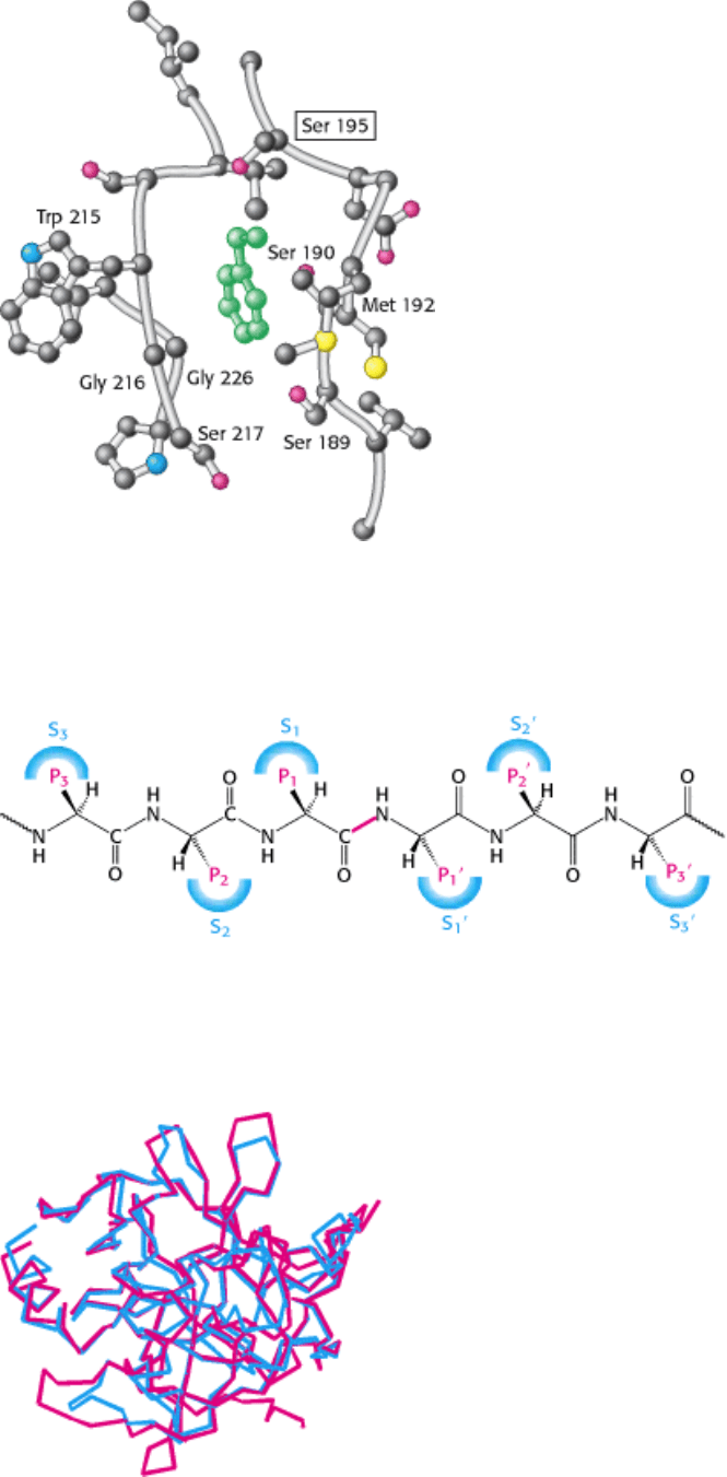
This mechanism accounts for all characteristics of chymotrypsin action except the observed preference for cleaving the
peptide bonds just past residues with large, hydrophobic side chains. Examination of the threedimensional structure of
chymotrypsin with substrate analogs and enzyme inhibitors revealed the presence of a deep, relatively hydrophobic
pocket, called the S
1
pocket, into which the long, uncharged side chains of residues such as phenylalanine and
tryptophan can fit. The binding of an appropriate side chain into this pocket positions the adjacent peptide bond into the
active site for cleavage (Figure 9.10). The specificity of chymotrypsin depends almost entirely on which amino acid is
directly on the amino-terminal side of the peptide bond to be cleaved. Other proteases have more-complex specificity

patterns, as illustrated in Figure 9.11. Such enzymes have additional pockets on their surfaces for the recognition of other
residues in the substrate. Residues on the amino-terminal side of the scissile bond (the bond to be cleaved) are labeled
P
1
, P
2
, P
3
, and so forth, indicating their positions in relation to the scissile bond. Likewise, residues on the carboxyl side
of the scissile bond are labeled P
1
, P
2
, P
3
, and so forth. The corresponding sites on the enzyme are referred to as S
1
,
S
2
or S
1
, S
2
, and so forth.
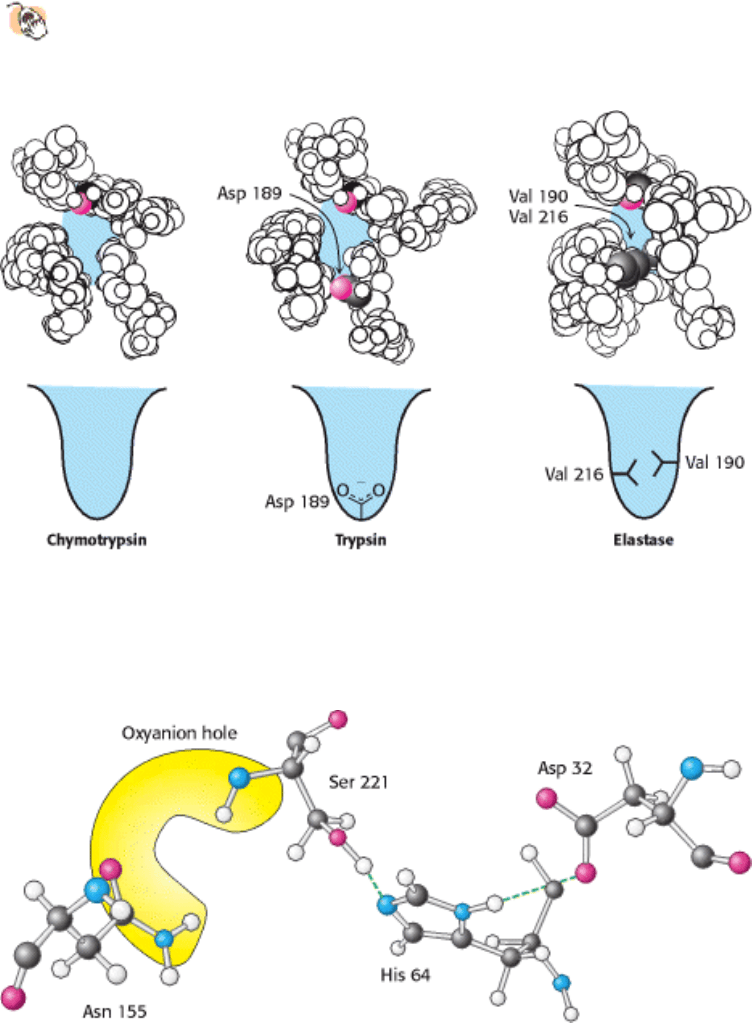
9.1.4. Catalytic Triads Are Found in Other Hydrolytic Enzymes
Many other proteins have subsequently been found to contain catalytic triads similar to that discovered in
chymotrypsin. Some, such as trypsin and elastase, are obvious homologs of chymotrypsin. The sequences of these
proteins are approximately 40% identical with that of chymotrypsin, and their overall structures are nearly the same
(Figure 9.12). These proteins operate by mechanisms identical with that of chymotrypsin. However, they have very
different substrate specificities. Trypsin cleaves at the peptide bond after residues with long, positively charged side
chains namely, arginine and lysine whereas elastase cleaves at the peptide bond after amino acids with small side
chains such as alanine and serine. Comparison of the S
1
pockets of these enzymes reveals the basis of the specificity.
In trypsin, an aspartate residue (Asp 189) is present at the bottom of the S
1
pocket in place of a serine residue in
chymotrypsin. The aspartate residue attracts and stabilizes a positively charged arginine or lysine residue in the substrate.
In elastase, two residues at the top of the pocket in chymotrypsin and trypsin are replaced with valine (Val 190 and Val
216). These residues close off the mouth of the pocket so that only small side chains may enter (Figure 9.13).
Other members of the chymotrypsin family include a collection of proteins that take part in blood clotting, to be
discussed in Chapter 10. In addition, a wide range of proteases found in bacteria and viruses also belong to this clan.
Furthermore, other enzymes that are not homologs of chymotrypsin have been found to contain very similar active sites.
As noted in Chapter 7, the presence of very similar active sites in these different protein families is a consequence of
convergent evolution. Subtilisin, a protease in bacteria such as Bacillus amyloliquefaciens, is a particularly well
characterized example. The active site of this enzyme includes both the catalytic triad and the oxyanion hole. However,
one of the NH groups that forms the oxyanion hole comes from the side chain of an asparagine residue rather than from
the peptide backbone (Figure 9.14). Subtilisin is the founding member of another large family of proteases that includes
representatives from Archaea, Eubacteria, and Eukarya.
Yet another example of the catalytic triad has been found in carboxypeptidase II from wheat. The structure of this
enzyme is not significantly similar to either chymotrypsin or subtilisin (Figure 9.15). This protein is a member of an
intriguing family of homologous proteins that includes esterases such as acetylcholine esterase and certain lipases. These
enzymes all make use of histidine-activated nucleophiles, but the nucleophiles may be cysteine rather than serine.
Finally, other proteases have been discovered that contain an active-site serine or threonine residue that is activated not
by a histidine-aspartate pair but by a primary amino group from the side chain of lysine or by the N-terminal amino
group of the polypeptide chain.
Thus, the catalytic triad in proteases has emerged at least three times in the course of evolution. We can conclude that
this catalytic strategy must be an especially effective approach to the hydrolysis of peptides and related bonds.
9.1.5. The Catalytic Triad Has Been Dissected by Site-Directed Mutagenesis
The techniques of molecular biology discussed in Chapter 6 have permitted detailed examination of the catalytic triad. In
particular, site-directed mutagenesis has been used to test the contribution of individual amino acid residues to the
catalytic power of an enzyme. Subtilisin has been extensively studied by this method. Each of the residues within the
catalytic triad, consisting of aspartic acid 32, histidine 64, and serine 221, has been individually converted into alanine,
and the ability of each mutant enzyme to cleave a model substrate has been examined (Figure 9.16). As expected, the
conversion of active-site serine 221 into alanine dramatically reduced catalytic power; the value of k
cat
fell to less than
one-millionth of its value for the wild-type enzyme. The value of K
M
was essentially unchanged: its increase by no more
than a factor of two indicated that substrate binding is not significantly affected. The mutation of histidine 64 to alanine

had very similar effects. These observations support the notion that the serine-histidine pair act together to generate a
nucleophile of sufficient power to attack the carbonyl group of a peptide bond. The conversion of aspartate 32 into
alanine had a smaller effect, although the value of k
cat
still fell to less than 0.005% of its wild-type value. The
simultaneously conversion of all three catalytic triad residues into alanine was no more deleterious than the conversion
of serine or histidine alone. Despite the reduction in their catalytic power, the mutated enzymes still hydrolyze peptides a
thousand times as rapidly as does buffer at pH 8.6.
Because the oxyanion hole of subtilisin includes a side-chain NH group in addition to backbone NH groups, it is possible
to probe the importance of the oxyanion hole for catalysis by site-directed mutagenesis. The mutation of asparagine 155
to glycine reduced the value of k
cat
to 0.2% of its wild-type value but increased the value of K
M
by only a factor of two.
These observations demonstrate that the NH group of the asparagine residue plays a significant role in stabilizing the
tetrahedral intermediate and the transition state leading to it.
9.1.6. Cysteine, Aspartyl, and Metalloproteases Are Other Major Classes of Peptide-
Cleaving Enzymes
Not all proteases utilize strategies based on activated serine residues.
Classes of proteins have been discovered that employ three alternative approaches to peptide-bond hydrolysis (Figure
9.17). These classes are the (1) cysteine proteases, (2) aspartyl proteases, and (3) metalloproteases. In each case, the
strategy generates a nucleophile that attacks the peptide carbonyl group (Figure 9.18).
The strategy used by the cysteine proteases is most similar to that used by the chymotrypsin family. In these enzymes, a
cysteine residue, activated by a histidine residue, plays the role of the nucleophile that attacks the peptide bond (see
Figure 9.18), in a manner quite analogous to that of the serine residue in serine proteases. An ideal example of these
proteins is papain, an enzyme purified from the fruit of the papaya. Mammalian proteases homologous to papain have
been discovered, most notably the cathepsins, proteins having a role in the immune and other systems. The cysteine-
based active site arose independently at least twice in the course of evolution; the caspases, enzymes that play a major
role in apoptosis (Section 2.4.3), have active sites similar to that of papain, but their overall structures are unrelated.
The second class comprises the aspartyl proteases. The central feature of the active sites is a pair of aspartic acid
residues that act together to allow a water molecule to attack the peptide bond. One aspartic acid residue (in its
deprotonated form) activates the attacking water molecule by poising it for deprotonation, whereas the other aspartic acid
residue (in its protonated form) polarizes the peptide carbonyl, increasing its susceptibility to attack (see Figure 9.18).
Members of this class include renin, an enzyme having a role in the regulation of blood pressure, and the digestive
enzyme pepsin. These proteins possess approximate twofold symmetry, suggesting that the two halves are evolutionarily
related. A likely scenario is that two copies of a gene for the ancestral enzyme fused to form a single gene that encoded a
single-chain enzyme. Each copy of the gene would have contributed an aspartate residue to the active site. The human
immunodeficiency virus (HIV) and other retroviruses contain an unfused dimeric aspartyl protease that is similar to the
fused protein, but the individual chains are not joined to make a single chain (Figure 9.19). This observation is consistent
with the idea that the enzyme may have originally existed as separate subunits.
The metalloproteases constitute the final major class of peptide-cleaving enzymes. The active site of such a protein
contains a bound metal ion, almost always zinc, that activates a water molecule to act as a nucleophile to attack the
peptide carbonyl group. The bacterial enzyme thermolysin and the digestive enzyme carboxypeptidase A are classic
examples of the zinc proteases. Thermolysin, but not carboxypeptidase A, is a member of a large and diverse family of
homologous zinc proteases that includes the matrix metalloproteases, enzymes that catalyze the reactions in tissue
remodeling and degradation.
In each of these three classes of enzymes, the active site includes features that allow for the activation of water or
another nucleophile as well as for the polarization of the peptide carbonyl group and subsequent stabilization of a
tetrahedral intermediate (see Figure 9.18).
9.1.7. Protease Inhibitors Are Important Drugs
Compounds that block or modulate the activities of proteases can have dramatic biological effects. Most natural
protease inhibitors are similar in structure to the peptide substrates of the enzyme that each inhibits (Section
10.5.4). Several important drugs are protease inhibitors. For example, captopril, an inhibitor of the metalloprotease
angiotensin-converting enzyme (ACE), has been used to regulate blood pressure. Crixivan, an inhibitor of the HIV
protease, is used in the treatment of AIDS. This protease cleaves multidomain viral proteins into their active forms;
blocking this process completely prevents the virus from being infectious (see Figure 9.19). To prevent unwanted side
effects, protease inhibitors used as drugs must be specific for one enzyme without inhibiting other proteins within the
body.
Let us examine the interaction of Crixivan with HIV protease in more detail. Crixivan is constructed around an alcohol
that mimics the tetrahedral intermediate; other groups are present to bind into the S
2
, S
1
, S
1
, and S
2
recognition sites
on the enzyme (Figure 9.20). The results of x-ray crystallographic studies revealed the structure of the enzyme-Crixivan
complex, showing that Crixivan adopts a conformation that approximates the twofold symmetry of the enzyme (Figure
9.21). The active site of HIV protease is covered by two apparently flexible flaps that fold down on top of the bound
inhibitor. The hydroxyl group of the central alcohol interacts with two aspartate residues of the active site, one in each
subunit. In addition, two carbonyl groups of the inhibitor are hydrogen bonded to a water molecule (not shown), which,
in turn, is hydrogen bonded to a peptide NH group in each of the flaps. This interaction of the inhibitor with water and
the enzyme is not possible with cellular aspartyl proteases such as renin and thus may contribute to the specificity of
Crixivan and other inhibitors for HIV protease.
I. The Molecular Design of Life 9. Catalytic Strategies 9.1. Proteases: Facilitating a Difficult Reaction
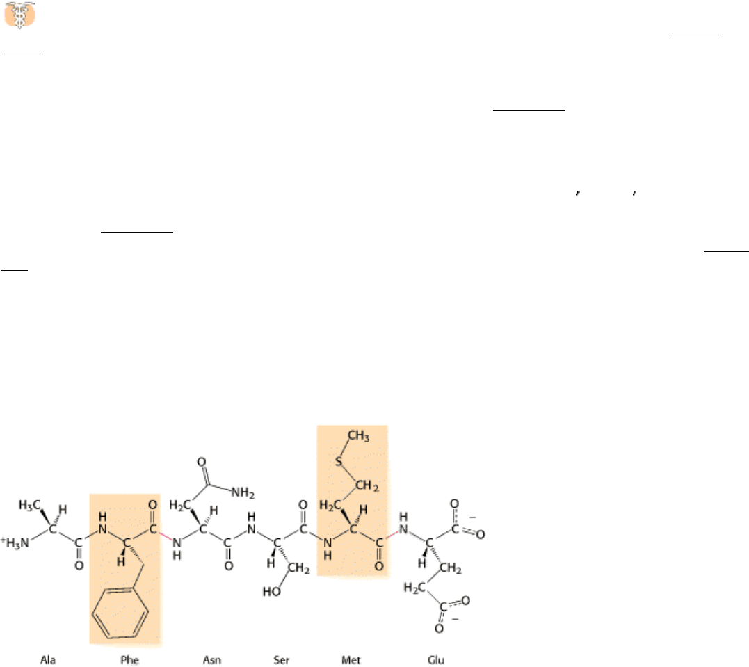
Figure 9.1. Specificity of Chymotrypsin. Chymotrypsin cleaves proteins on the carboxyl side of aromatic or large
hydrophobic amino acids (shaded yellow). The likely bonds cleaved by chymotrypsin are indicated in red.
I. The Molecular Design of Life 9. Catalytic Strategies 9.1. Proteases: Facilitating a Difficult Reaction
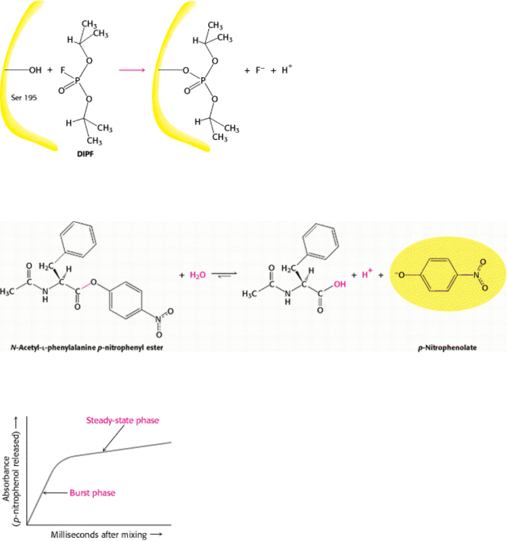
Figure 9.2. An Unusually Reactive Serine in Chymotrypsin. Chymotrypsin is inactivated by treatment with
diisopropylphosphofluoridate (DIPF), which reacts only with serine 195 among 28 possible serine residues.
I. The Molecular Design of Life 9. Catalytic Strategies 9.1. Proteases: Facilitating a Difficult Reaction
Figure 9.3. Chromogenic Substrate. N-Acetyl-
l-phenylalanine p-nitrophenyl ester yields a yellow product, p-
nitrophenolate, on cleavage by chymotrypsin. p-Nitrophenolate forms by deprotonation of p-nitrophenol at pH 7.
I. The Molecular Design of Life 9. Catalytic Strategies 9.1. Proteases: Facilitating a Difficult Reaction
Figure 9.4. Kinetics of Chymotrypsin Catalysis. Two stages are evident in the cleaving of N-acetyl-
l-phenylalanine p-
nitrophenyl ester by chymotrypsin: a rapid burst phase (pre-steady state) and a steady-state phase.
I. The Molecular Design of Life 9. Catalytic Strategies 9.1. Proteases: Facilitating a Difficult Reaction
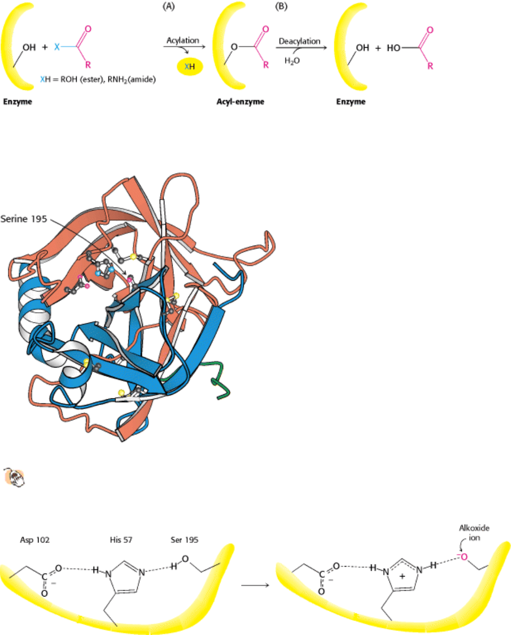
Figure 9.5. Covalent Catalysis. Hydrolysis by chymotrypsin takes place in two stages: (A) acylation to form the acyl-
enzyme intermediate followed by (B) deacylation to regenerate the free enzyme.
I. The Molecular Design of Life 9. Catalytic Strategies 9.1. Proteases: Facilitating a Difficult Reaction
Figure 9.6. Three-Dimensional Structure of Chymotrypsin.
The three chains are shown in ribbon form in orange,
blue, and green. The side chains of the catalytic triad residues, including serine 195, are shown as ball-and-stick
representations, as are two intrastrand and interstrand disulfide bonds.
I. The Molecular Design of Life 9. Catalytic Strategies 9.1. Proteases: Facilitating a Difficult Reaction
Figure 9.7. The Catalytic Triad. The catalytic triad, shown on the left, converts serine 195 into a potent nucleophile, as
illustrated on the right.
I. The Molecular Design of Life 9. Catalytic Strategies 9.1. Proteases: Facilitating a Difficult Reaction
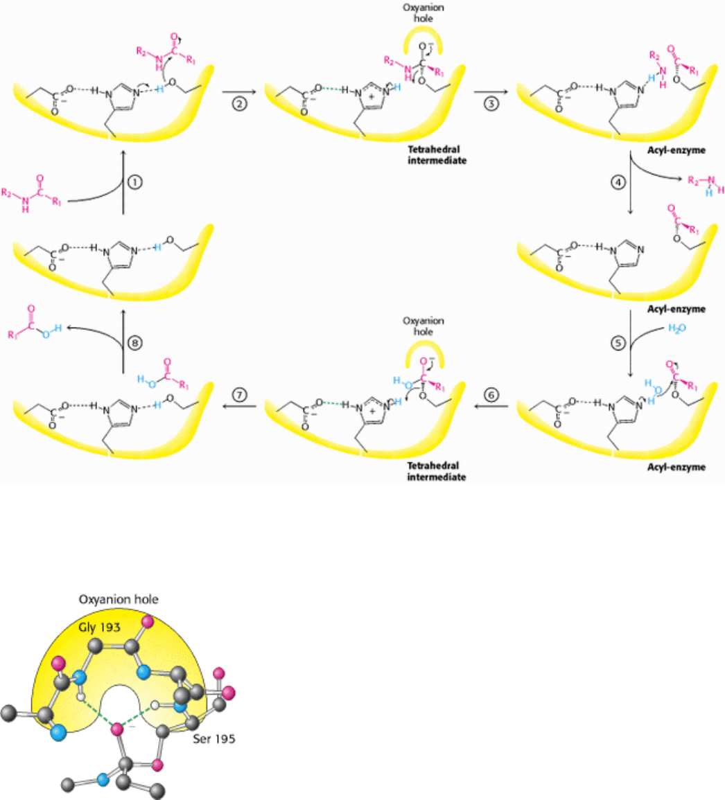
Figure 9.8. Peptide Hydrolysis by Chymotrypsin. The mechanism of peptide hydrolysis illustrates the principles of
covalent and acid-base catalysis. The dashed green lines indicate favorable interactions between the negatively charged
aspartate residue and the positively charged histidine residue, which make the histidine residue a more powerful base.
I. The Molecular Design of Life 9. Catalytic Strategies 9.1. Proteases: Facilitating a Difficult Reaction
Figure 9.9. The Oxyanion Hole. The structure stabilizes the tetrahedral intermediate of the chymotrypsin reaction.
Hydrogen bonds (shown in green) link peptide NH groups and the negatively charged oxygen.
I. The Molecular Design of Life 9. Catalytic Strategies 9.1. Proteases: Facilitating a Difficult Reaction
Figure 9.10. The Hydrophobic Pocket of Chymotrypsin. The hydrophobic pocket of chymotrypsin is responsible for
its substrate specificity. The key amino acids that constitute the binding site are labeled, including the active-site serine
residue (boxed). The position of an aromatic ring bound in the pocket is shown in green.
I. The Molecular Design of Life 9. Catalytic Strategies 9.1. Proteases: Facilitating a Difficult Reaction
Figure 9.11. Specificity Nomenclature for Protease-Substrate Interactions. The potential sites of interaction of the
substrate with the enzyme are designated P (shown in red), and corresponding binding sites on the enzyme are
designated S. The scissile bond (also shown in red) is the reference point.
I. The Molecular Design of Life 9. Catalytic Strategies 9.1. Proteases: Facilitating a Difficult Reaction
Figure 9.12. Structural Similarity of Trypsin and Chymotrypsin. An overlay of the structure of chymotrypsin (red)
on that of trypsin (blue) shows the high degree of similarity. Only α-carbon atom positions are shown. The mean
deviation in position between corresponding α-carbon atoms is 1.7 Å.
I. The Molecular Design of Life 9. Catalytic Strategies 9.1. Proteases: Facilitating a Difficult Reaction
Figure 9.13. The S
1
Pockets of Chymotrypsin, Trypsin, and Elastase. Certain residues play key roles in determining
the specificity of these enzymes. The side chains of these residues, as well as those of the active-site serine residues, are
shown in color.
I. The Molecular Design of Life 9. Catalytic Strategies 9.1. Proteases: Facilitating a Difficult Reaction
Figure 9.14. The Catalytic Triad and Oxyanion Hole of Subtilisin. The peptide bond attacked by nucleophilic serine
221 of the catalytic triad will develop a negative charge, which is stabilized by enzyme NH groups (both in the backbone
and in the side chain of Asn 155) located in the oxyanion hole.
I. The Molecular Design of Life 9. Catalytic Strategies 9.1. Proteases: Facilitating a Difficult Reaction
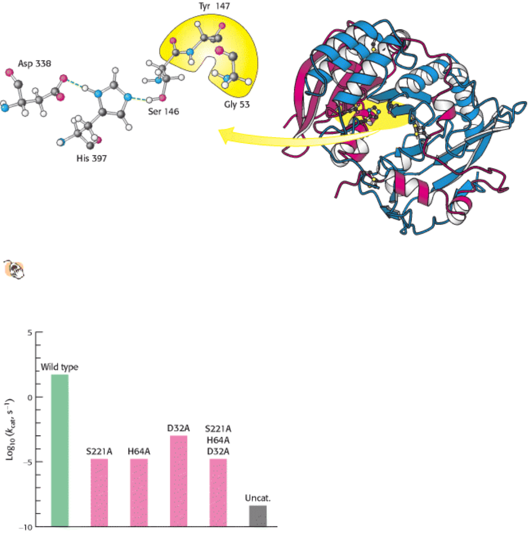
Figure 9.15. Carboxypeptidase II.
The structure of carboxypeptidase II from wheat (right) is illustrated with its two
chains (blue and red). The catalytic triad of carboxypeptidase II (left) is composed of the same amino acids as
those in chymotrypsin, despite the fact that the enzymes display no structural similarity. The residues that form the
oxyanion hole are highlighted in yellow.
I. The Molecular Design of Life 9. Catalytic Strategies 9.1. Proteases: Facilitating a Difficult Reaction
Figure 9.16. Site-Directed Mutagenesis of Subtilisin. Residues of the catalytic triad were mutated to alanine, and the
activity of the mutated enzyme was measured. Mutations in any component of the catalytic triad cause a dramatic loss of
enzyme activity. Note that the activity is displayed on a logarithmic scale. The mutations are identified as follows: the
first letter is the one-letter abbreviation for the amino acid being altered; the number identifies the position of the residue
in the primary structure; and the second letter is the one-letter abbreviation for the amino acid replacing the original one.