Berg J.M., Tymoczko J.L., Stryer L. Biochemistry
Подождите немного. Документ загружается.


The incoming nucleophile attacks the phosphorus atom, and a pentacoordinate transition state is formed. This species has
a trigonal bipyramidal geometry centered at the phosphorus atom, with the incoming nucleophile at one apex of the two
pyramids and the group that is displaced (the leaving group, L) at the other apex. The two mechanisms differ in the
number of times the displacement occurs in the course of the reaction.
In the first type of mechanism, a nucleophile in the enzyme (analogous to serine 195 in chymotrypsin) attacks the
phosphoryl group to form a covalent intermediate. In a second step, this intermediate is hydrolyzed to produce the final
products. Because two displacement reactions take place at the phosphorus atom in the first mechanism, the
stereochemical configuration at the phosphorus atom would be inverted and then inverted again, and the overall
configuration would be retained. In the second type of mechanism, analogous to that used by the aspartyl and
metalloproteases, an activated water molecule attacks the phosphorus atom directly. In this mechanism, a single
displacement reaction takes place at the phosphorus atom. Hence, the stereochemical configuration of the tetrahedral
phosphorus atom is inverted each time a displacement reaction takes place. Monitoring the stereochemical changes of the
phosphorus could be one approach to determining the mechanism of restriction endonuclease action.
A difficulty is that the phosphorus centers in DNA are not chiral, because two of the groups bound to the phosphorus
atom are simple oxygen atoms, identical with each other. This difficulty can be circumvented by preparing DNA
molecules that contain chiral phosphoryl groups, made by replacing one oxygen atom with sulfur (called a
phosphorothioate). Let us consider EcoRV endonuclease. This enzyme cleaves the phosphodiester bond between the T
and the A at the center of the recognition sequence 5
-GATATC-3 . The first step in monitoring the activity of the
enzyme is to synthesize an appropriate substrate for EcoRV containing phosphorothioates at the sites of cleavage (Figure
9.34). The reaction is then performed in water that has been greatly enriched in
18
O to allow the incoming oxygen atom
to be marked. The location of the
18
O label with respect to the sulfur atom indicates whether the reaction proceeds with
inversion or retention of stereochemistry. The analysis revealed that the stereochemical configuration at the phosphorus
atom was inverted only once with cleavage. This result is consistent with a direct attack of water at phosphorus and rules
out the formation of any covalently bound intermediate (Figure 9.35).
9.3.2. Restriction Enzymes Require Magnesium for Catalytic Activity
Restriction endonucleases as well as many other enzymes that act on phosphate-containing substrates require Mg
2+
or
some other similar divalent cation for activity. What is the function of this metal?
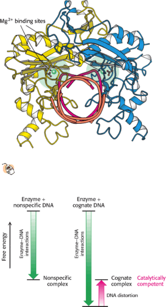
It has been possible to examine the interactions of the magnesium ion when it is bound to the enzyme. Crystals have
been produced of EcoRV endonuclease bound to oligonucleotides that contain the appropriate recognition sequences.
These crystals are grown in the absence of magnesium to prevent cleavage; then, when produced, the crystals are soaked
in solutions containing the metal. No cleavage takes place, allowing the location of the magnesium ion binding sites to
be determined (Figure 9.36). The magnesium ion was found to be bound to six ligands: three are water molecules, two
are carboxylates of the enzyme's aspartate residues, and one is an oxygen atom of the phosphoryl group at the site of
cleavage. The magnesium ion holds a water molecule in a position from which the water molecule can attack the
phosphoryl group and, in conjunction with the aspartate residues, helps polarize the water molecule toward
deprotonation. Because cleavage does not take place within these crystals, the observed structure cannot be the true
catalytic conformation. Additional studies have revealed that a second magnesium ion must be present in an adjacent site
for EcoRV endonuclease to cleave its substrate.
9.3.3. The Complete Catalytic Apparatus Is Assembled Only Within Complexes of
Cognate DNA Molecules, Ensuring Specificity
We now return to the question of specificity, the defining feature of restriction enzymes. The recognition sequences for
most restriction endonucleases are inverted repeats. This arrangement gives the three-dimensional structure of the
recognition site a twofold rotational symmetry (Figure 9.37). The restriction enzymes display a corresponding symmetry
to facilitate recognition: they are dimers whose two subunits are related by twofold rotational symmetry. The matching

symmetry of the recognition sequence and the enzyme has been confirmed by the determination of the structure of the
complex between EcoRV endonuclease and DNA fragments containing its recognition sequence (Figure 9.38). The
enzyme surrounds the DNA in a tight embrace. Examination of this structure reveals features that are highly significant
in determining specificity.
A unique set of interactions occurs between the enzyme and a cognate DNA sequence. Within the 5
-GATATC-3
sequence, the G and A bases at the 5
end of each strand and their Watson-Crick partners directly contact the enzyme by
hydrogen bonding with residues that are located in two loops, one projecting from the surface of each enzyme subunit
(Figure 9.39). The most striking feature of this complex is the distortion of the DNA, which is substantially kinked in the
center (Figure 9.40). The central two TA base pairs in the recognition sequence play a key role in producing the kink.
They do not make contact with the enzyme but appear to be required because of their ease of distortion. 5
-TA-3
sequences are known to be among the most easily deformed base pairs. The distortion of the DNA at this site has severe
effects on the specificity of enzyme action.
Specificity is often determined by an enzyme's binding affinity for substrates. In regard to EcoRV endonuclease,
however, binding studies performed in the absence of magnesium have demonstrated that the enzyme binds to all
sequences, both cognate and noncognate, with approximately equal affinity. However, the structures of complexes
formed with noncognate DNA fragments are strikingly different from those formed with cognate DNA: the noncognate
DNA conformation is not substantially distorted (Figure 9.41). This lack of distortion has important consequences with
regard to catalysis. No phosphate is positioned sufficiently close to the active-site aspartate residues to complete a
magnesium ion binding site (see Figure 9.36). Hence, the nonspecific complexes do not bind the magnesium ion and the
complete catalytic apparatus is never assembled. The distortion of the substrate and the subsequent binding of the
magnesium ion account for the catalytic specificity of more than 1,000,000-fold that is observed for EcoRV
endonuclease despite very little preference at the level of substrate binding.
We can now see the role of binding energy in this strategy for attaining catalytic specificity. In binding to the enzyme,
the DNA is distorted in such a way that additional contacts are made between the enzyme and the substrate, increasing
the binding energy. However, this increase is canceled by the energetic cost of distorting the DNA from its relaxed
conformation (Figure 9.42). Thus, for EcoRV endonuclease, there is little difference in binding affinity for cognate and
nonspecific DNA fragments. However, the distortion in the cognate complex dramatically affects catalysis by
completing the magnesium ion binding site. This example illustrates how enzymes can utilize available binding energy to
deform substrates and poise them for chemical transformation. Interactions that take place within the distorted substrate
complex stabilize the transition state leading to DNA hydrolysis.
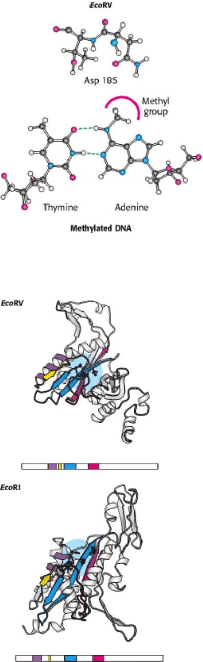
The distortion in the DNA explains how methylation blocks catalysis and protects host-cell DNA. When a methyl group
is added to the amino group of the adenine nucleotide at the 5
end of the recognition sequence, the methyl group's
presence precludes the formation of a hydrogen bond between the amino group and the side-chain carbonyl group of
asparagine 185 (Figure 9.43). This asparagine residue is closely linked to the other amino acids that form specific
contacts with the DNA. The absence of the hydrogen bond disrupts other interactions between the enzyme and the DNA
substrate, and the distortion necessary for cleavage will not take place.
9.3.4. Type II Restriction Enzymes Have a Catalytic Core in Common and Are
Probably Related by Horizontal Gene Transfer
Type II restriction enzymes are prevalent in Archaea and Eubacteria. What can we tell of the evolutionary history
of these enzymes? Comparison of the amino acid sequences of a variety of type II restriction endonucleases did
not reveal significant sequence similarity between most pairs of enzymes. However, a careful examination of three-
dimensional structures, taking into account the location of the active sites, revealed the presence of a core structure
conserved in the different enzymes. This structure includes β strands that contain the aspartate (or, in some cases,
glutamate) residues forming the magnesium ion binding sites (Figure 9.44).
These observations indicate that many type II restriction enzymes are indeed evolutionary related. Analyses of the
sequences in greater detail suggest that bacteria may have obtained genes encoding these enzymes from other species by
horizontal gene transfer, the passing between species of pieces of DNA (such as plasmids) that provide a selective
advantage in a particular environment. For example, EcoRI (from E. coli) and RsrI (from Rhodobacter sphaeroides) are
50% identical in sequence over 266 amino acids, clearly indicative of a close evolutionary relationship. However, these
species of bacteria are not closely related, as is known from sequence comparisons of other genes and other evidence.
Thus, it appears that these species obtained the gene for this restriction endonuclease from a common source more
recently than the time of their evolutionary divergence. Moreover, the gene encoding EcoRI endonuclease uses particular
codons to specify given amino acids that are strikingly different from the codons used by most E. coli genes, which
suggests that the gene did not originate in E. coli. Horizontal gene transfer may be a relatively common event. For
example, genes that inactivate antibiotics are often transferred, leading to the transmission of antibiotic resistance from
one species to another. For restriction-modification systems, protection against viral infections may have favored
horizontal gene transfer.
I. The Molecular Design of Life 9. Catalytic Strategies 9.3. Restriction Enzymes: Performing Highly Specific DNA-Cleavage Reactions
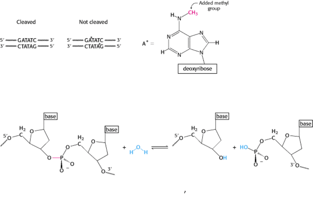
Figure 9.32. Protection by Methylation. The recognition sequence for EcoRV endonuclease (left) and the sites of
methylation (right) in DNA protected from the catalytic action of the enzyme.
I. The Molecular Design of Life 9. Catalytic Strategies 9.3. Restriction Enzymes: Performing Highly Specific DNA-Cleavage Reactions
Figure 9.33. Hydrolysis of a Phosphodiester Bond. All restriction enzymes catalyze the hydrolysis of DNA
phosphodiester bonds, leaving a phosphoryl group attached to the 5
end. The bond that is cleaved is shown in red.
I. The Molecular Design of Life 9. Catalytic Strategies 9.3. Restriction Enzymes: Performing Highly Specific DNA-Cleavage Reactions
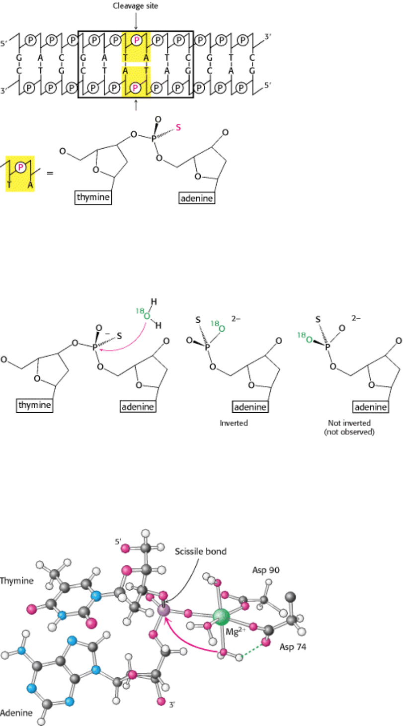
Figure 9.34. Labeling with Phosphorothioates. Phosphorothioates, groups in which one of the nonbridging oxygen
atoms is replaced with a sulfur atom, can be used to label specific sites in the DNA backbone to determine the overall
stereochemical course of a displacement reaction. Here, a phosphorothioate is placed at sites that can be cleaved by
EcoRV endonuclease.
I. The Molecular Design of Life 9. Catalytic Strategies 9.3. Restriction Enzymes: Performing Highly Specific DNA-Cleavage Reactions
Figure 9.35. Stereochemistry of Cleaved DNA. Cleavage of DNA by EcoRV endonuclease results in overall inversion
of the stereochemical configuration at the phosphorus atom, as indicated by the stereochemistry of the phosphorus atom
bound to one bridging oxygen atom, one
16
O, one
18
O, and one sulfur atom. This configuration strongly suggests that the
hydrolysis takes place by the direct attack of water on the phosphorus atom.
I. The Molecular Design of Life 9. Catalytic Strategies 9.3. Restriction Enzymes: Performing Highly Specific DNA-Cleavage Reactions
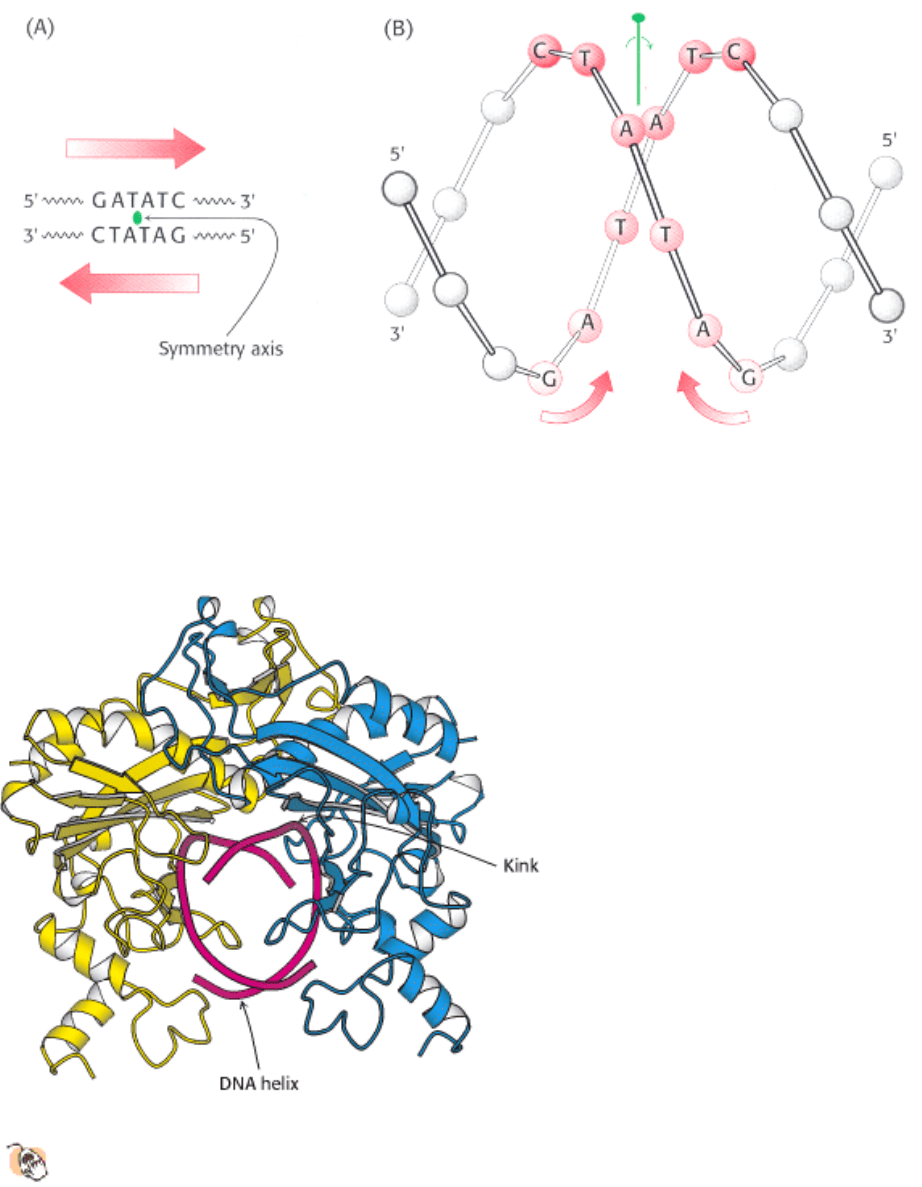
Figure 9.36. Magnesium Ion Binding Site in ECORV Endonuclease. The magnesium ion helps to activate a water
molecule and positions it so that it can attack the phosphate.
I. The Molecular Design of Life 9. Catalytic Strategies 9.3. Restriction Enzymes: Performing Highly Specific DNA-Cleavage Reactions
Figure 9.37. Structure of the Recognition Site of ECORV Endonuclease. (A) The sequence of the recognition site,
which is symmetric around the axis of rotation designated in green. (B) The inverted repeat within the recognition
sequence of EcoRV (and most other restriction endonucleases) endows the DNA site with twofold rotational symmetry.
I. The Molecular Design of Life 9. Catalytic Strategies 9.3. Restriction Enzymes: Performing Highly Specific DNA-Cleavage Reactions
Figure 9.38. Structure of the ECORV - Cognate DNA Complex.
This view of the structure of EcoRV endonuclease
bound to a cognate DNA fragment is down the helical axis of the DNA. The two protein subunits are in yellow
and blue, and the DNA backbone is in red. The twofold axes of the enzyme dimer and the DNA are aligned.
I. The Molecular Design of Life 9. Catalytic Strategies 9.3. Restriction Enzymes: Performing Highly Specific DNA-Cleavage Reactions
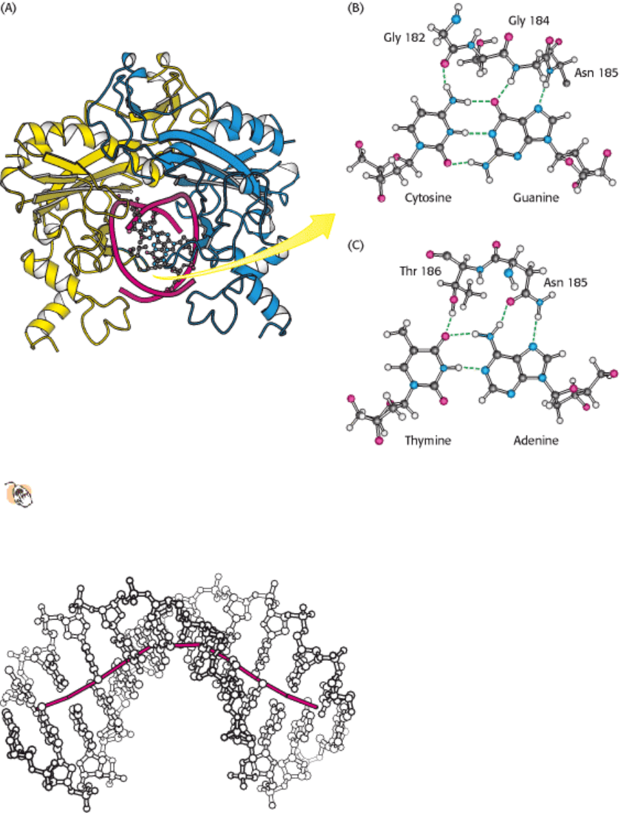
Figure 9.39. Hydrogen Bonding Interactions between ECORV Endonuclease and Its DNA Substrate.
One of the
DNA-binding loops (in green) of EcoRV endonuclease is shown interacting with the base pairs of its cognate
DNA binding site. Key amino acid residues are shown hydrogen bonding with (B) a CG base pair and (C) an AT
base pair.
I. The Molecular Design of Life 9. Catalytic Strategies 9.3. Restriction Enzymes: Performing Highly Specific DNA-Cleavage Reactions
Figure 9.40. Distortion of the Recognition Site. The DNA is represented as a ball-and-stick model. The path of the
DNA helical axis, shown in red, is substantially distorted on binding to the enzyme. For the B form of DNA, the axis is
straight (not shown).
I. The Molecular Design of Life 9. Catalytic Strategies 9.3. Restriction Enzymes: Performing Highly Specific DNA-Cleavage Reactions
Figure 9.41. Nonspecific and Cognate DNA within ECORV Endonuclease.
A comparison of the positions of the
nonspecific (orange) and the cognate DNA (red) within EcoRV reveals that, in the nonspecific complex, the DNA
backbone is too far from the enzyme to complete the magnesium ion binding sites.
I. The Molecular Design of Life 9. Catalytic Strategies 9.3. Restriction Enzymes: Performing Highly Specific DNA-Cleavage Reactions
Figure 9.42. Greater Binding Energy of EcoRV Endonuclease Bound to Cognate Versus Noncognate Dna. The
additional interactions between EcoRV endonuclease and cognate DNA increase the binding energy, which can be used
to drive DNA distortions necessary for forming a catalytically competent complex.
I. The Molecular Design of Life 9. Catalytic Strategies 9.3. Restriction Enzymes: Performing Highly Specific DNA-Cleavage Reactions
Figure 9.43. Methylation of Adenine. The methylation of adenine blocks the formation of hydrogen bonds between
EcoRV endonuclease and cognate DNA molecules and prevents their hydrolysis.
I. The Molecular Design of Life 9. Catalytic Strategies 9.3. Restriction Enzymes: Performing Highly Specific DNA-Cleavage Reactions
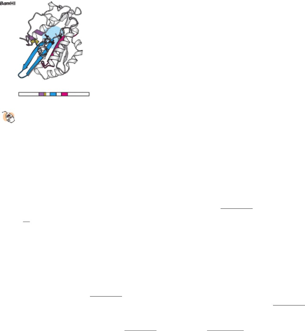
Figure 9.44. A Conserved Structural Core in Type II Restriction Enzymes.
Four conserved structural elements,
including the active-site region (in blue), are highlighted in color in these models of a single monomer from each
dimeric enzyme. The positions of the amino acid sequences that form these elements within each overall sequence
are represented schematically below each structure.
I. The Molecular Design of Life 9. Catalytic Strategies
9.4. Nucleoside Monophosphate Kinases: Catalyzing Phosphoryl Group Exchange
between Nucleotides Without Promoting Hydrolysis
The final enzymes that we shall consider are the nucleoside monophosphate kinases (NMP kinases), typified by
adenylate kinase. These enzymes catalyze the transfer of the terminal phosphoryl group from a nucleoside triphosphate
(NTP), usually ATP, to the phosphoryl group on a nucleoside monophosphate (Figure 9.45). The challenge for NMP
kinases is to promote the transfer of the phosphoryl group from NTP to NMP without promoting the competing
reaction the transfer of a phosphoryl group from NTP to water; that is, NTP hydrolysis. We shall see how the use of
induced fit by these enzymes is used to solve this problem. Moreover, these enzymes employ metal ion catalysis; but, in
this case, the metal forms a complex with the substrate to enhance enzyme-substrate interaction.
9.4.1. NMP Kinases Are a Family of Enzymes Containing P-Loop Structures
X-ray crystallographic methods have yielded the three-dimensional structures of a number of different NMP kinases,
both free and bound to substrates or substrate analogs. Comparison of these structures reveals that these enzymes form a
family of homologous proteins (Figure 9.46). In particular, such comparisons reveal the presence of a conserved NTP-
binding domain. This domain consists of a central β sheet, surrounded on both sides by α helices (Figure 9.47). A
characteristic feature of this domain is a loop between the first β strand and the first helix. This loop, which typically has
an amino acid sequence of the form Gly-X-X-X-X-Gly-Lys, is often referred to as the P-loop because it interacts with
phosphoryl groups on the bound nucleotide (Figure 9.48). As described in Section 9.4.4, similar domains containing P-
loops are present in a wide variety of important nucleotide-binding proteins.
9.4.2. Magnesium (or Manganese) Complexes of Nucleoside Triphosphates Are the
True Substrates for Essentially All NTP-Dependent Enzymes
Kinetic studies of NMP kinases, as well as many other enzymes having ATP or other nucleoside triphosphates as a
substrate, reveal that these enzymes are essentially inactive in the absence of divalent metal ions such as magnesium
(Mg
2+
) or manganese (Mn
2+
), but acquire activity on the addition of these ions. In contrast with the enzymes discussed
so far, the metal is not a component of the active site. Rather, nucleotides such as ATP bind these ions, and it is the metal
ion-nucleotide complex that is the true substrate for the enzymes. The dissociation constant for the ATP-Mg
2+
complex
is approximately 0.1 mM, and thus, given that intracellular Mg
2+
concentrations are typically in the millimolar range,
essentially all nucleoside triphosphates are present as NTP-Mg
2+
complexes.

How does the binding of the magnesium ion to the nucleotide affect catalysis? There are a number of related
consequences, but all serve to enhance the specificity of the enzyme-substrate interactions by enhancing binding energy.
First, the magnesium ion neutralizes some of the negative charges present on the polyphosphate chain, reducing
nonspecific ionic interactions between the enzyme and the polyphosphate group of the nucleotide. Second, the
interactions between the magnesium ion and the oxygen atoms in the phosphoryl group hold the nucleotide in well-
defined conformations that can be specifically bound by the enzyme (Figure 9.49). Magnesium ions are typically
coordinated to six groups in an octahedral arrangement. Typically, two oxygen atoms are directly coordinated to a
magnesium ion, with the remaining coordination positions often occupied by water molecules. Oxygen atoms of the α
and β, β and γ, or α and γ phosphoryl groups may contribute, depending on the particular enzyme. In addition, different
stereoisomers are produced, depending on exactly which oxygen atoms bind to the metal ion. Third, the magnesium ion
provides additional points of interaction between the ATP-Mg
2+
complex and the enzyme, thus increasing the binding
energy. In some cases, such as the DNA polymerases (Section 27.2.2), side chains (often aspartate and glutamate
residues) of the enzyme can bind directly to the magnesium ion. In other cases, the enzyme interacts indirectly with the
magnesium ion through hydrogen bonds to the coordinated water molecules (Figure 9.50). Such interactions have been
observed in adenylate kinases bound to ATP analogs.
9.4.3. ATP Binding Induces Large Conformational Changes
Comparison of the structure of adenylate kinase in the presence and absence of an ATP analog reveals that substrate
binding induces large structural changes in the kinase, providing a classic example of the use of induced fit (Figure
9.51). The P-loop closes down on top of the polyphosphate chain, interacting most extensively with the β phosphoryl
group. The movement of the P-loop permits the top domain of the enzyme to move down to form a lid over the bound
nucleotide. This motion is favored by interactions between basic residues (conserved among the NMP kinases), the
peptide backbone NH groups, and the nucleotide. With the ATP nucleotide held in this position, its γ phosphoryl group is
positioned next to the binding site for the second substrate, NMP. In sum, the direct interactions with the nucleotide
substrate lead to local structural rearrangements (movement of the P-loop) within the enzyme, which in turn allow more
extensive changes (the closing down of the top domain) to take place. The binding of the second substrate, NMP,
induces additional conformational changes. Both sets of changes ensure that a catalytically competent conformation is
formed only when both the donor and the acceptor are bound, preventing wasteful transfer of the phosphoryl group to
water. The enzyme holds its two substrates close together and appropriately oriented to stabilize the transition state that
leads to the transfer of a phosphoryl group from the ATP to the NMP. This is an example of catalysis by approximation.
We will see such examples of a catalytically competent active site being generated only on substrate binding many times
in our study of biochemistry.
9.4.4. P-Loop NTPase Domains Are Present in a Range of Important Proteins
Domains similar (and almost certainly homologous) to those found in NMP kinases are present in a remarkably
wide array of proteins, many of which participate in essential biochemical processes. Examples include ATP
synthase, the key enzyme responsible for ATP generation; molecular motor proteins such as myosin; signal-transduction
proteins such as transducin; proteins essential for translating mRNA into proteins, such as elongation factor Tu; and
DNA and RNA unwinding helicases. The wide utility of P-loop NTPase domains is perhaps best explained by their
ability to undergo substantial conformational changes on nucleoside triphosphate binding and hydrolysis. We shall
encounter these domains (hereafter referred to as P-loop NTPases) throughout the book and shall observe how they
function as springs, motors, and clocks. To allow easy recognition of these domains, they, like the binding domains of
the NMP kinases, will be depicted with the inner surfaces of the ribbons in a ribbon diagram shown in purple and the P-
loop shown in green (Figure 9.52).