Berg J.M., Tymoczko J.L., Stryer L. Biochemistry
Подождите немного. Документ загружается.

I. The Molecular Design of Life 13. Membrane Channels and Pumps 13.1. The Transport of Molecules Across a Membrane May Be Active or Passive
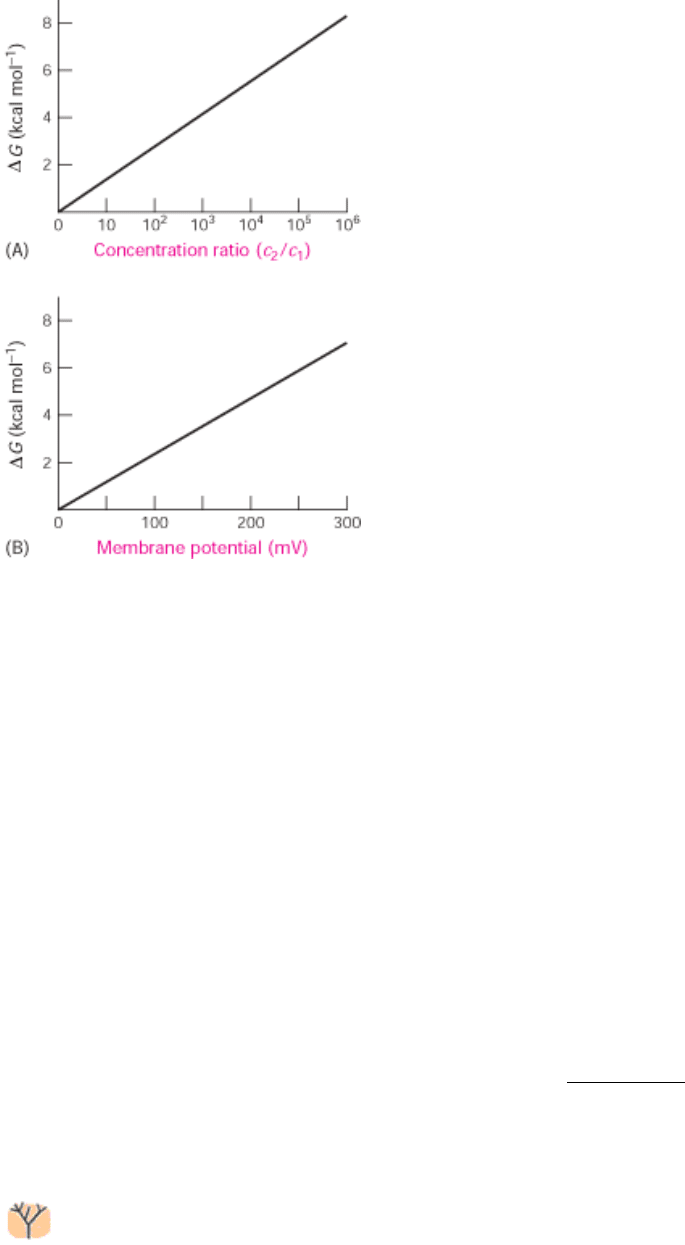
Figure 13.2. Free Energy and Transport. The free-energy change in transporting (A) an uncharged solute from a
compartment at concentration c
1
to one at c
2
and (B) a singly charged species across a membrane to the side having the
same charge as that of the transported ion. Note that the free-energy change imposed by a membrane potential of 59 mV
is equivalent to that imposed by a concentration ratio of 10 for a singly charged ion at 25°C.
I. The Molecular Design of Life 13. Membrane Channels and Pumps
13.2. A Family of Membrane Proteins Uses ATP Hydrolysis to Pump Ions Across
Membranes
The extracellular fluid of animal cells has a salt concentration similar to that of sea water. However, cells must control
their intracellular salt concentrations to prevent unfavorable interactions with high concentrations of ions such as calcium
and to facilitate specific processes. For instance, most animal cells contain a high concentration of K
+
and a low
concentration of Na
+
relative to the external medium. These ionic gradients are generated by a specific transport system,
an enzyme that is called the Na
+
-K
+
pump or the Na
+
-K
+
ATPase. The hydrolysis of ATP by the pump provides the
energy needed for the active transport of Na
+
out of the cell and K
+
into the cell, generating the gradient. The pump is
called the Na
+
-K
+
ATPase because the hydrolysis of ATP occurs only when Na
+
and K
+
are bound to the pump.
Moreover, this ATPase, like all such enzymes, requires Mg
2+
(Section 9.4.2). The active transport of Na
+
and K
+
is of
great physiological significance. Indeed, more than a third of the ATP consumed by a resting animal is used to pump
these ions. The Na
+
-K
+
gradient in animal cells controls cell volume, renders neurons and muscle cells electrically
excitable, and drives the active transport of sugars and amino acids.
The subsequent purification of other ion pumps has revealed a large family of evolutionarily related ion pumps
including proteins from bacteria, archaea, and all eukaryotes. These pumps are specific for an array of ions. Of
particular interest are the Ca
2+
ATPase, the enzyme that transports Ca
2+
out of the cytoplasm and into the sarcoplasmic
reticulum of muscle cells, and the gastric H
+
-K
+
ATPase, the enzyme responsible for pumping sufficient protons into the
stomach to lower the pH below 1.0. These enzymes and the hundreds of known homologs, including the Na
+
-K
+
ATPase, are referred to as P-type ATPases because they form a key phosphorylated intermediate. In the formation of this
intermediate, a phosphoryl group obtained from the hydrolysis of ATP is linked to the side chain of a specific conserved

aspartate residue in the ATPase (Figure 13.3).
13.2.1. The Sarcoplasmic Reticulum Ca
2+
ATPase Is an Integral Membrane Protein
We will consider the structural and mechanistic features of these enzymes by examining the Ca
2+
ATPase found in the
sarcoplasmic reticulum (SR Ca
2+
ATPase) of muscle cells. This enzyme, which constitutes 80% of the sarcoplasmic
reticulum membrane protein, plays an important role in muscle contraction, which is triggered by an abrupt rise in the
cytosolic calcium level. Muscle relaxation depends on the rapid removal of Ca
2+
from the cytosol into the sarcoplasmic
reticulum, a specialized compartment for calcium storage, by the SR Ca
2+
ATPase. This pump maintains a Ca
2+
concentration of approximately 0.1 µM in the cytosol compared with 1.5 mM in the sarcoplasmic reticulum.
The SR Ca
2+
ATPase is a single 110-kd polypeptide with a transmembrane domain consisting of 10 α helices. A large
cytoplasmic head piece constitutes nearly half the molecular weight of the protein and consists of three distinct domains
(Figure 13.4). The three cytoplasmic domains of the SR Ca
2+
ATPase have distinct functions. One domain (N) binds the
ATP nucleotide, another (P) accepts the phosphoryl group on its conserved aspartate residue, and the third (A) may serve
as an actuator for the N domain. The relation between these three domains changes significantly on ATP hydrolysis. The
crystal structure in the absence of ATP shows the likely nucleotide-binding site separated by more than 25 Å from the
phosphorylation site, suggesting that the N and P domains move toward one another during the catalytic cycle. This
closure is facilitated by ATP binding and by the binding of Ca
2+
to the membrane-spanning helices.
The results of mechanistic studies of the SR Ca
2+
ATPase and other P-type ATPases have revealed two common
features. First, as we have seen, each protein can be phosphorylated on a specific aspartate residue. For the SR Ca
2+
ATPase, this reaction takes place at Asp 351 only in the presence of relatively high cytosolic concentrations of Ca
2+
.
Second, each pump can interconvert between at least two different conformations, denoted by E
1
and E
2
. Thus, at least
four conformational states
E
1
, E
1
-P, E
2
-P, and E
2
participate in the transport process. From these four states, it is
possible to construct a plausible mechanism of action for these enzymes, although further studies are necessary to
confirm the mechanism and provide more details (Figure 13.5):
1. The postulated reaction cycle begins with the binding of ATP and two Ca
2+
ions to the E
1
state.
2. The enzyme cleaves ATP, transferring the γ-phosphoryl group to the key aspartate residue. Calcium must be bound to
the enzyme for the phosphorylation to take place. Phosphorylation shifts the conformational equilibrium of the ATPase
toward E
2
.
3. The transition from the E
1
to the E
2
state causes the ion-binding sites to "evert" so that the ions can dissociate only to
the luminal side of the membrane.
4. In the E
2
-P state, the enzyme has low affinity for the Ca
2+
ions, so they are released.
5. With the release of Ca
2+
, the phosphoaspartate residue is hydrolyzed, and the phosphate group is released.
6. The enzyme, devoid of a covalently attached phosphoryl group, is not stable in the E
2
form. It everts back to the E
1
form, completing the cycle.
Essentially the same mechanism is employed by the Na
+
-K
+
ATPase. The E
1
state binds three Na
+
ions and transports
them across the membrane and out of the cell as a result of the protein's phosphorylation and transition to the E
2
state.
The three Na
+
ions are released into the extracellular medium. The E
2
state of this enzyme also binds ions namely, two
K
+
ions. These K
+
ions are carried across the membrane in the opposite direction by eversion driven by the hydrolysis of
the phosphoaspartate residue and are released into the cytosol.

The change in free energy accompanying the transport of Na
+
and K
+
can be calculated (Section 13.1.1). Suppose that
the concentration of Na
+
outside and inside the cell is 143 and 14 mM, respectively, and that of K
+
is 4 and 157 mM. At
a membrane potential of -50 mV and a temperature of 37 °C, the free-energy change in transporting 3 moles of Na
+
out
of and 2 moles of K
+
into the cell is +10.0 kcal (+41.8 kJ mol
-1
). The hydrolysis of a single ATP per transport cycle
provides sufficient free energy, about -12 kcal mol
-1
(-50 kJ mol
-1
) under cellular conditions, to drive the uphill transport
of these ions.
13.2.2. P-Type ATPases Are Evolutionarily Conserved and Play a Wide Range of Roles
Analysis of the complete yeast genome revealed the presence of 16 proteins that clearly belong to the P-type
ATPase family. More detailed sequence analysis suggests that 2 of these proteins transport H
+
ions, 2 transport Ca
2
+
, 3 transport Na
+
, and 2 transport metals such as Cu
2+
. In addition, 5 members of this family appear to participate in the
transport of phospholipids with amino acid head groups. These latter proteins assist in the maintenance of membrane
asymmetry by transporting lipids such as phosphatidyl serine from the outer to the inner leaflet of the bilayer membrane
(Figure 13.6). Such enzymes have been termed "flippases."
All members of this protein family employ the same fundamental mechanism. The free energy of ATP hydrolysis drives
membrane transport by effecting conformational changes associated with the addition and removal of a phosphoryl
group at an analogous aspartate site in each protein.
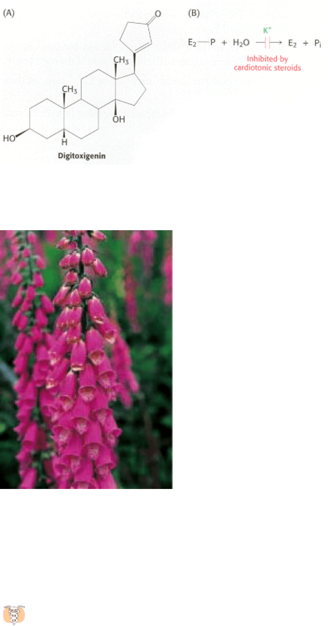
13.2.3. Digitalis Specifically Inhibits the Na
+
-K
+
Pump by Blocking Its
Dephosphorylation
Certain steroids derived from plants are potent inhibitors (K
i
10 nM) of the Na
+
-K
+
pump. Digitoxigenin and ouabain
are members of this class of inhibitors, which are known as cardiotonic steroids because of their strong effects on the
heart (Figure 13.7). These compounds inhibit the dephosphorylation of the E
2
-P form of the ATPase when applied on the
extracellular face of the membrane.
Digitalis, a mixture of cardiotonic steroids derived from the dried leaf of the foxglove plant (Digitalis purpurea),
is of great clinical significance. Digitalis increases the force of contraction of heart muscle, which makes it a
choice drug in the treatment of congestive heart failure. Inhibition of the Na
+
-K
+
pump by digitalis leads to a higher
level of Na
+
inside the cell. The diminished Na
+
gradient results in slower extrusion of Ca
2+
by the sodium calcium
exchanger (Section 13.4). The subsequent increase in the intracellular level of Ca
2+
enhances the contractility of cardiac
muscle. It is interesting to note that digitalis was effectively used long before the discovery of the Na
+
-K
+
ATPase. In
1785, William Withering, a British physician, heard tales of an elderly woman, known as "the old woman of
Shropshire," who cured people of "dropsy" (which today would be recognized as congestive heart failure) with an extract
of foxglove. Withering conducted the first scientific study of the effects of foxglove on congestive heart failure and
documented its effectiveness.
I. The Molecular Design of Life 13. Membrane Channels and Pumps 13.2. A Family of Membrane Proteins Uses ATP Hydrolysis to Pump Ions Across Membranes
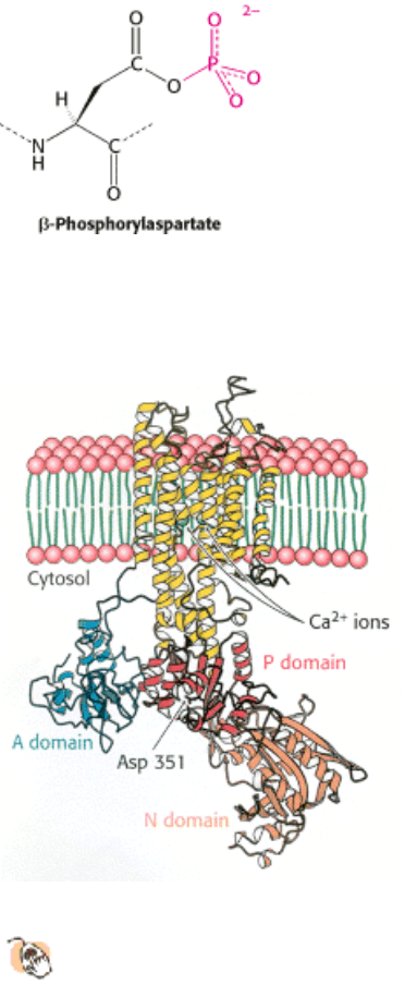
Figure 13.3. Phosphoaspartate. Phosphoaspartate (also referred to as β-aspartyl phosphate) is a key intermediate in the
reaction cycles of P-type ATPases.
I. The Molecular Design of Life 13. Membrane Channels and Pumps 13.2. A Family of Membrane Proteins Uses ATP Hydrolysis to Pump Ions Across Membranes
Figure 13.4. Structure of SR CA
2+
ATPase. This enzyme, the calcium pump of the sarcoplasmic reticulum, comprises
a membrane-spanning domain of 10 α helices and a cytoplasmic headpiece consisting of three domains (N, P, and
A). Two calcium ions (green) bind within the membrane-spanning region. The aspartate residue characteristic of
this protein family is indicated.
I. The Molecular Design of Life 13. Membrane Channels and Pumps 13.2. A Family of Membrane Proteins Uses ATP Hydrolysis to Pump Ions Across Membranes
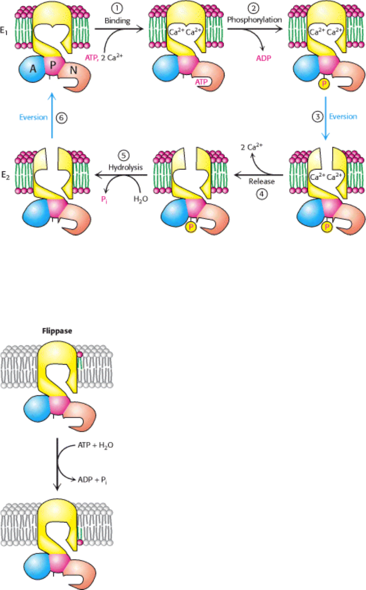
Figure 13.5. Mechanism of P-Type ATPase Action. The binding of Ca
2+
and the phosphorylation of the ATPase (steps
1 and 2), illustrated here for the Ca
2+
ATPase, lead to the eversion of the binding sites (step 3) and the release of Ca
2+
to
the luminal side of the membrane (step 4). Hydrolysis of phosphoaspartate (step 5) and eversion (step 6) reset the
enzyme to its initial state.
I. The Molecular Design of Life 13. Membrane Channels and Pumps 13.2. A Family of Membrane Proteins Uses ATP Hydrolysis to Pump Ions Across Membranes
Figure 13.6. P-Type ATPases Can Transport Lipids. Flippases are enzymes that maintain membrane asymmetry by
"flipping" phospholipids (displayed with a red head group) from the outer to the inner layer of the membrane.
I. The Molecular Design of Life 13. Membrane Channels and Pumps 13.2. A Family of Membrane Proteins Uses ATP Hydrolysis to Pump Ions Across Membranes
Figure 13.7. Digitoxigenin. Cardiotonic steroids such as digitoxigenin inhibit the Na
+
-K
+
pump by blocking the
dephosphorylation of E
2
-P.
I. The Molecular Design of Life 13. Membrane Channels and Pumps 13.2. A Family of Membrane Proteins Uses ATP Hydrolysis to Pump Ions Across Membranes
Foxglove (Digitalis purpurea) is the source of digitalis, one of the most widely used drugs. [Inga Spence/Visuals
Unlimited.]
I. The Molecular Design of Life 13. Membrane Channels and Pumps
13.3. Multidrug Resistance and Cystic Fibrosis Highlight a Family of Membrane
Proteins with ATP-Binding Cassette Domains
Tumor cells in culture often become resistant to drugs that were initially quite toxic to the cells. Remarkably, the
development of resistance to one drug also makes the cells less sensitive to a range of other compounds. This
phenomenon is known as multidrug resistance. In a significant discovery, the onset of multidrug resistance was found to
correlate with the expression and activity of a membrane protein with an apparent molecular mass of 170 kd. This
protein acts as an ATP-dependent pump that extrudes a wide range of small molecules from cells that express it. The
protein is called the multidrug resistance protein (MDR) or P-glycoprotein ("glyco" because it includes a carbohydrate
moiety). Thus, when cells are exposed to a drug, the MDR pumps the drug out of the cell before the drug can exert its
effects. A related protein was discovered through genetic studies of the hereditary disease cystic fibrosis (Section 1.1.4).
In one of the first studies leading to the identification of a specific genetic change causing human disease, investigators
performed a comparative genetic analysis of many people having this disease and family members who did not have the
disease. The gene found to be mutated in the affected persons encodes a protein, now called cystic fibrosis
transmembrane conductance regulator (CFTR). CFTR acts as an ATP-regulated chloride channel in the plasma
membranes of epithelial cells. As mentioned in Chapter 1, cystic fibrosis results from a decrease in fluid and salt
secretion by CFTR. As a consequence of this defect, secretion from the pancreas is blocked and heavy, dehydrated
mucus accumulates in the lungs, leading to chronic lung infections.
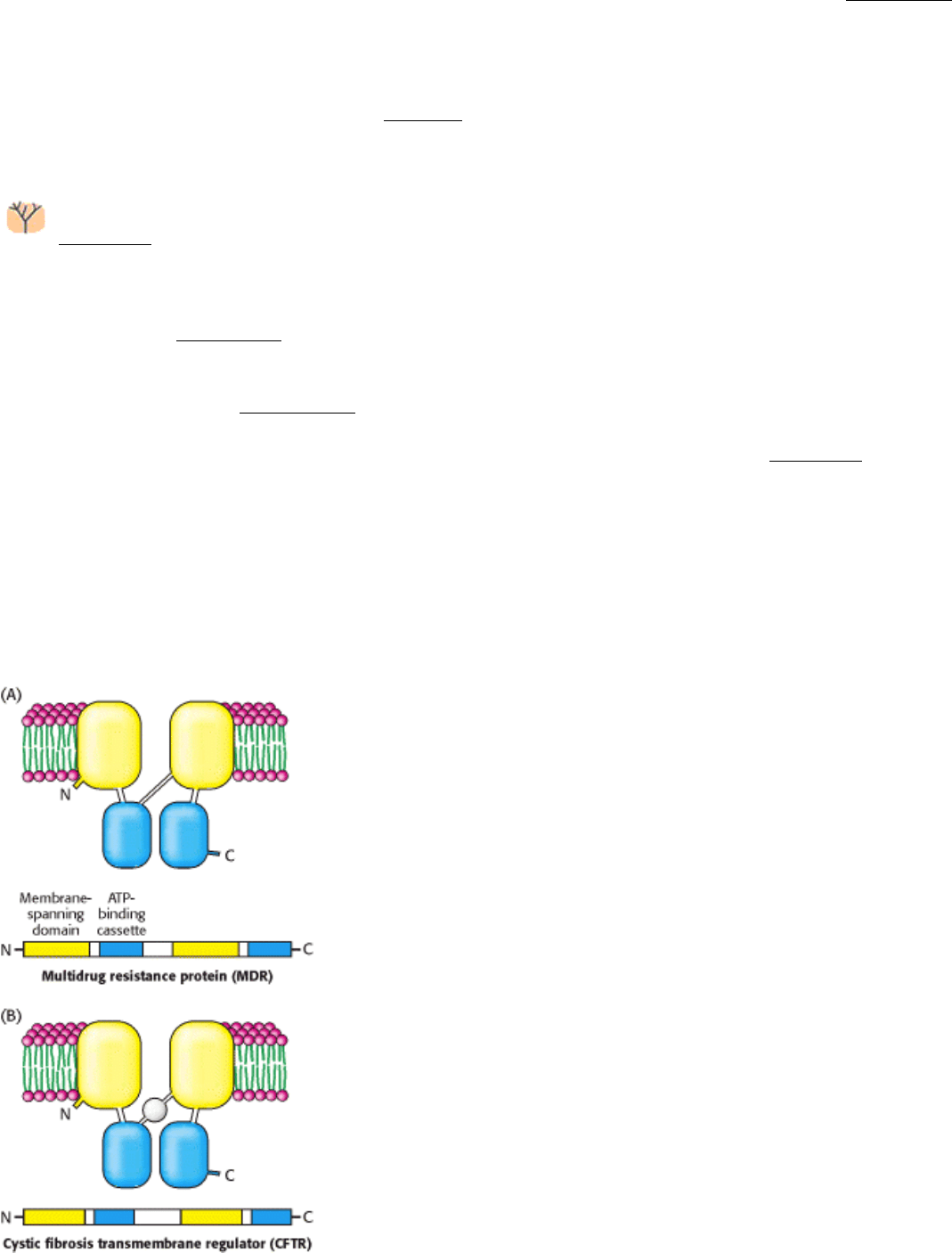
Analysis of the amino acid sequences of MDR, CFTR, and homo logous proteins revealed a common architecture
(Figure 13.8). Each protein comprises four domains: two membrane-binding domains of unknown structure and
two ATP-binding domains. The ATP-binding domains of these proteins are called ATP-binding cassettes (or ABCs) and
are homologous to domains in a large family of transport proteins of bacteria and archaea. Indeed, with 79 members, the
ABC proteins are the largest single family identified in the E. coli genome. The ABC proteins are members of the P-loop
NTPase superfamily (Section 9.4.4). Some ABC proteins, particularly those of prokaryotes, are multisubunit proteins
constructed such that the membrane-spanning domains and the ABC domains are present on separate polypeptide chains.
The consolidation of enzymatic activities of several polypeptide chains in prokaryotes to a single chain in eukaryotes is a
theme that we will see again (Section 22.4.4). For example, the histidine permease of Salmonella typhimurium, which
transports the amino acid histidine into the bacterium, consists of (1) two different protein subunits with membrane-
spanning domains (HisQ and HisM) and (2) a homodimeric protein (HisP) with ABC domains (Figure 13.9). A soluble,
histidine-binding protein (HisJ) binds histidine after the amino acid enters the cell.
Like other members of the P-loop NTPase superfamily, proteins with ABC domains undergo conformational changes on
ATP binding and hydrolysis. These structural changes are coupled within each dimeric transporter unit in a manner that
allows these membrane proteins to drive the uptake or efflux of specific compounds or to act as gates for open
membrane channels.
I. The Molecular Design of Life 13. Membrane Channels and Pumps 13.3. Multidrug Resistance and Cystic Fibrosis Highlight a Family of Membrane Proteins with ATP-Binding Cassette Domains
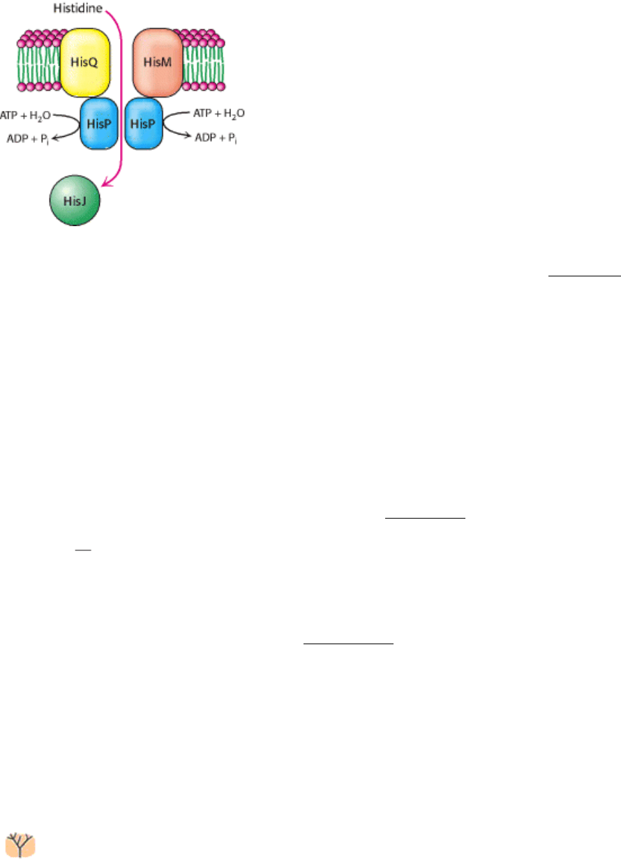
Figure 13.8. ABC Transporters. The multidrug resistance protein (MDR) and the cystic fibrosis transmembrane
regulator (CFTR) are homologous proteins composed of two transmembrane domains and two ATP-binding domains,
called ATP-binding cassettes (ABCs).
I. The Molecular Design of Life 13. Membrane Channels and Pumps 13.3. Multidrug Resistance and Cystic Fibrosis Highlight a Family of Membrane Proteins with ATP-Binding Cassette Domains
Figure 13.9. Histidine Permease. In the histidine permease of S. typhimurium, the membrane-spanning regions (yellow
and orange) and ABC regions (blue) are on separate polypeptide chains (compare with Figure 13.8). ATP hydrolysis
drives the transport of histidine into the cell.
I. The Molecular Design of Life 13. Membrane Channels and Pumps
13.4. Secondary Transporters Use One Concentration Gradient to Power the
Formation of Another
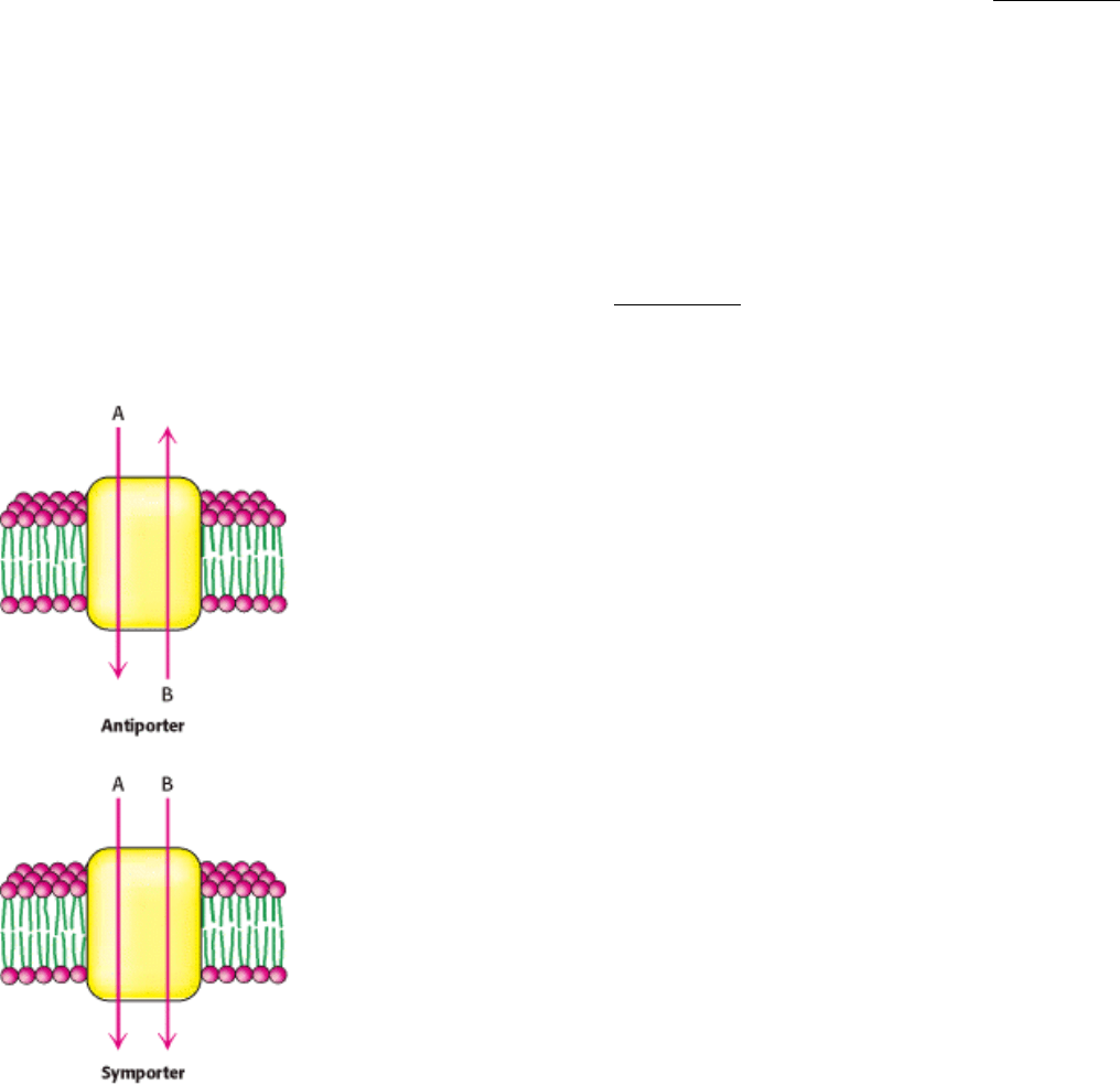
Many active-transport processes are not directly driven by the hydrolysis of ATP. Instead, the thermodynamically uphill
flow of one species of ion or molecule is coupled to the downhill flow of a different species. Membrane proteins that
pump ions or molecules uphill by this means are termed secondary transporters or cotransporters. These proteins can be
classified as either antiporters or symporters. Antiporters couple the downhill flow of one species to the uphill flow of
another in the opposite direction across the membrane; symporters use the flow of one species to drive the flow of a
different species in the same direction across the membrane (Figure 13.10).
The sodium
calcium exchanger in the plasma membrane of an animal cell is an antiporter that uses the electrochemical
gradient of Na
+
to pump Ca
2+
out of the cell. Three Na
+
ions enter the cell for each Ca
2+
ion that is extruded. The cost of
transport by this exchanger is paid by the Na
+
-K
+
- ATPase pump, which generates the requisite sodium gradient.
Because Ca
2+
is a vital messenger inside the cell, its concentration must be tightly controlled. The exchanger has lower
affinity for Ca
2
+
than does the Ca
2
+
ATPase (Section 13.2.1), but its capacity to extrude Ca
2
+
is greater. The
exchanger can lower the cytosolic Ca
2+
level to several micromolar; submicromolar Ca
2+
levels are attained by the
subsequent action of the Ca
2+
ATPase. The exchanger can extrude about 2000 Ca
2+
ions per second, compared with only
30 ions per second for the Ca
2+
-ATPase pump.
Glucose is pumped into some animal cells by a symporter powered by the simultaneous entry of Na
+
. The entry of Na
+
provides a free-energy input of 2.2 kcal mol
-1
(9.2 kJ mol
-1
) under typical cellular conditions (external [Na
+
] = 143 mM,
internal [Na
+
] = 14 mM, and membrane potential = -50 mV). This free-energy input is sufficient to generate a 66-fold
concentration gradient of an uncharged molecule such as glucose.
Secondary transporters are ancient molecular machines, common today in bacteria and archaea as well as in
eukaryotes. For example, approximately 160 (of approximately 4000) proteins encoded by the E. coli genome
appear to be secondary transporters. Sequence comparison and hydropathy analysis suggest that members of the largest
family have 12 transmembrane helices that appear to have arisen by duplication and fusion of a membrane protein with 6
transmembrane helices. Included in this family is the lactose permease of E. coli. This symporter uses the H
+
gradient
across the E. coli membrane generated by the oxidation of fuel molecules to drive the uptake of lactose and other sugars
against a concentration gradient. The permease has a proton-binding site and a lactose-binding site (Figure 13.11). A
proton and a lactose molecule bind to sites facing the outside of the cell. The permease, with both binding sites full,
everts, releasing first the proton and then the lactose inside the bacterium. Another eversion places the empty sites on the
outside. Thus, the energetically uphill transfer of one lactose molecule is coupled to the downhill transport of one proton.
Further analysis of the three-dimensional structures is underway and should provide more information about their
mechanisms of action as well as the evolutionary relationships within this large group of ancient proteins.
These observations reveal how different energy currencies are interconverted. A single energy currency, ATP, is used by
P-type ATPases to generate gradients of a small number of types of ions, particularly Na
+
and H
+
, across membranes.
These gradients then serve as an energy source for the large number of secondary transporters, which allow many
different molecules to be taken up or transported out of cells (Figure 13.12).
I. The Molecular Design of Life 13. Membrane Channels and Pumps 13.4. Secondary Transporters Use One Concentration Gradient to Power the Formation of Another
Figure 13.10. Secondary Transporters. These transporters employ the downhill flow of one gradient to power the
formation of another gradient. In antiporters, the chemical species move in opposite directions. In symporters, the two
species move in the same direction.
I. The Molecular Design of Life 13. Membrane Channels and Pumps 13.4. Secondary Transporters Use One Concentration Gradient to Power the Formation of Another
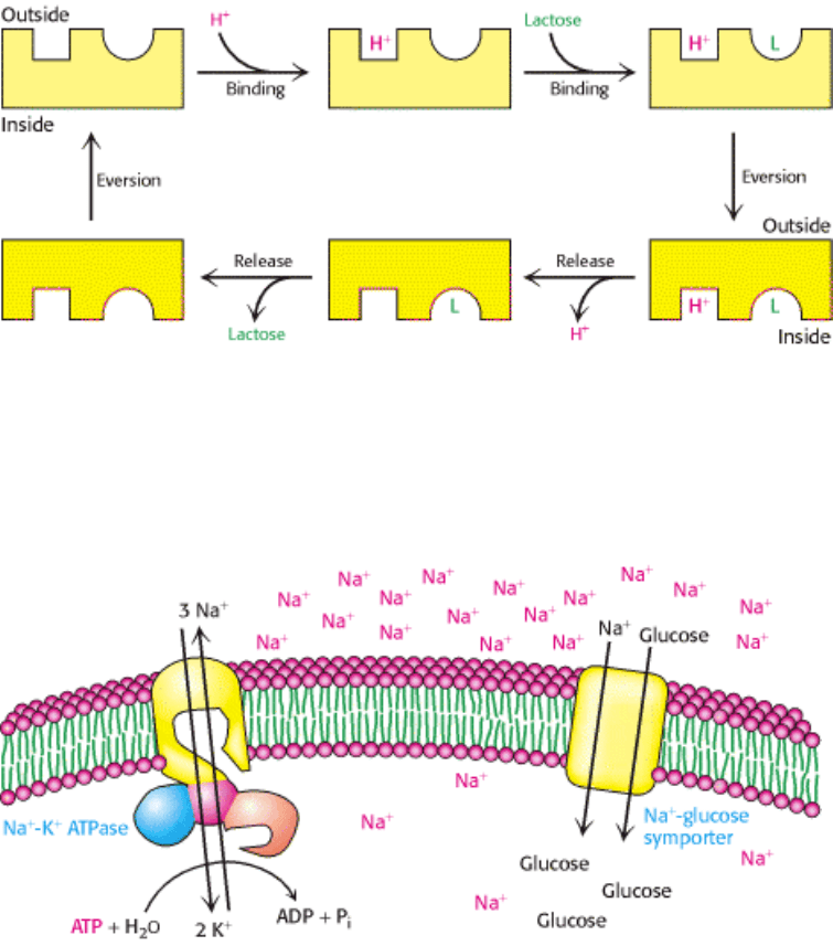
Figure 13.11. Action of Lactose Permease. Lactose permease pumps lactose into bacterial cells by drawing on the
proton-motive force. The binding sites evert when a lactose molecule (L) and a proton (H
+
) are bound to external sites.
After these species are released inside the cell, the binding sites again evert to complete the transport cycle. Lactose
permease is an example of a symporter.
I. The Molecular Design of Life 13. Membrane Channels and Pumps 13.4. Secondary Transporters Use One Concentration Gradient to Power the Formation of Another
Figure 13.12. Energy Transduction by Membrane Proteins. The Na
+
-K
+
ATPase converts the free energy of
phosphoryl transfer into the free energy of a Na
+
ion gradient. The ion gradient can then be used to pump materials into
the cell, through the action of a secondary transporter such as the Na
+
-glucose symporter.
I. The Molecular Design of Life 13. Membrane Channels and Pumps
13.5. Specific Channels Can Rapidly Transport Ions Across Membranes
Pumps and secondary transporters can transport ions at rates approaching several thousand ions per second. Other
membrane proteins, ion channels, which are passive transport systems, are capable of ion-transport rates that are more
than 1000 times as high. These rates of transport through ion channels are close to rates expected for ions diffusing freely
through aqueous solution. Yet, ion channels are not simply tubes that span membranes through which ions can rapidly
flow. Instead, they are highly sophisticated molecular machines that respond to chemical and physical changes in their
environments and undergo precisely timed conformational changes that facilitate their roles as essential components of
the nervous and other systems.
Several key properties characterize ion channels:
1. Ion channels can be highly selective for particular ions. For example, some channels allow the flow of K
+
very
effectively but do not allow appreciable levels of Na
+
to cross the membrane. Other channels transport positively
charged ions (cations), but block the flow of negatively charged ions (anions). The selectivities of some ion-channel