Berg J.M., Tymoczko J.L., Stryer L. Biochemistry
Подождите немного. Документ загружается.

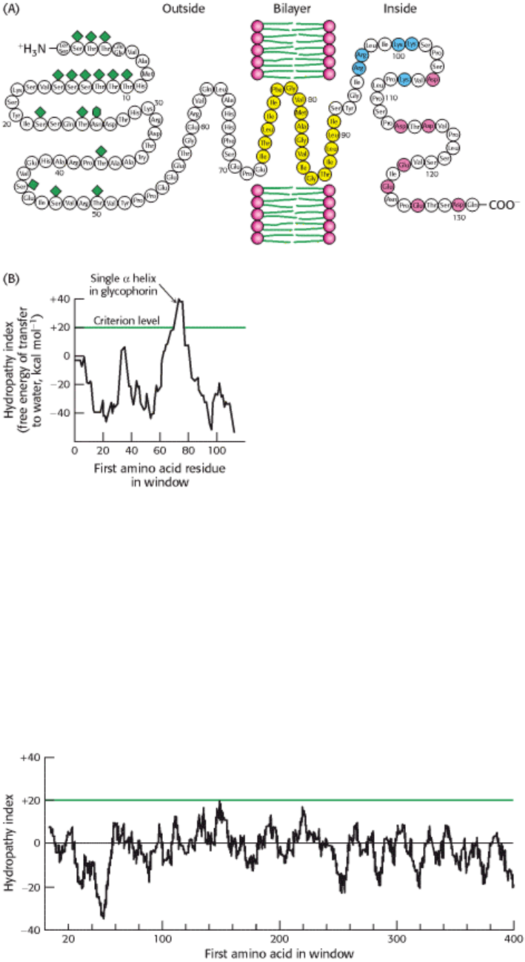
Figure 12.27. Locating the Membrane-Spanning Helix of Glycophorin. (A) Amino acid sequence and
transmembrane disposition of glycophorin A from the red-cell membrane. Fifteen O-linked carbohydrate units are shown
as diamond shapes, and an N-linked unit is shown as a lozenge shape. The hydrophobic residues (yellow) buried in the
bilayer form a transmembrane α helix. The carboxyl-terminal part of the molecule, located on the cytosolic side of the
membrane, is rich in negatively charged (red) and positively charged (blue) residues. (B) Hydropathy plot for
glycophorin. The free energy for transfering a helix of 20 residues from the membrane to water is plotted as a function of
the position of the first residue of the helix in the sequence of the protein. Peaks of greater than +20 kcal mol
-1
in
hydropathy plots are indicative of potential transmembrane helices. [(A) Courtesy of Dr. Vincent Marchesi; (B) after D.
M. Engelman, T. A. Steitz, and A. Goldman. Identifying nonpolar transbilayer helices in amino acid sequences of
membrane proteins. Annu. Rev. Biophys. Biophys. Chem. 15(1986):343. Copyright © 1986 by Annual Reviews, Inc. All
rights reserved.]
I. The Molecular Design of Life 12. Lipids and Cell Membranes 12.5. Proteins Carry Out Most Membrane Processes
Figure 12.28. Hydropathy Plot for Porin. No strong peaks are observed for this intrinsic membrane protein because it
is constructed from membrane-spanning β strands rather than α helices.
I. The Molecular Design of Life 12. Lipids and Cell Membranes
12.6. Lipids and Many Membrane Proteins Diffuse Rapidly in the Plane of the
Membrane
Biological membranes are not rigid, static structures. On the contrary, lipids and many membrane proteins are constantly
in lateral motion, a process called lateral diffusion. The rapid lateral movement of membrane proteins has been
visualized by means of fluorescence microscopy through the use of the technique of fluorescence recovery after
photobleaching (FRAP; Figure 12.29). First, a cell-surface component is specifically labeled with a fluorescent
chromophore. A small region of the cell surface (~3 µm
2
) is viewed through a fluorescence microscope. The fluorescent
molecules in this region are then destroyed (bleached) by a very intense light pulse from a laser. The fluorescence of this
region is subsequently monitored as a function of time by using a light level sufficiently low to prevent further
bleaching. If the labeled component is mobile, bleached molecules leave and unbleached molecules enter the illuminated
region, which results in an increase in the fluorescence intensity. The rate of recovery of fluorescence depends on the
lateral mobility of the fluorescence-labeled component, which can be expressed in terms of a diffusion coefficient, D.
The average distance s traversed in time t depends on D according to the expression
The diffusion coefficient of lipids in a variety of membranes is about 1 µ m
2
s
-1
. Thus, a phospholipid molecule diffuses
an average distance of 2 µ m in 1 s. This rate means that a lipid molecule can travel from one end of a bacterium to the
other in a second. The magnitude of the observed diffusion coefficient indicates that the viscosity of the membrane is
about 100 times that of water, rather like that of olive oil.
In contrast, proteins vary markedly in their lateral mobility. Some proteins are nearly as mobile as lipids, whereas others
are virtually immobile. For example, the photoreceptor protein rhodopsin (Section 32.3.1), a very mobile protein, has a
diffusion coefficient of 0.4 µm
2
s
-1
. The rapid movement of rhodopsin is essential for fast signaling. At the other
extreme is fibronectin, a peripheral glycoprotein that interacts with the extracellular matrix. For fibronectin, D is less
than 10
-4
µm
2
s
-1
. Fibronectin has a very low mobility because it is anchored to actin filaments on the inside of the
plasma membrane through integrin, a transmembrane protein that links the extracellular matrix to the cytoskeleton.
12.6.1. The Fluid Mosaic Model Allows Lateral Movement but Not Rotation Through
the Membrane
On the basis of the dynamic properties of proteins in membranes, S. Jonathan Singer and Garth Nicolson proposed the
concept of a fluid mosaic model for the overall organization of biological membranes in 1972 (Figure 12.30). The
essence of their model is that membranes are two-dimensional solutions of oriented lipids and globular proteins. The
lipid bilayer has a dual role: it is both a solvent for integral membrane proteins and a permeability barrier. Membrane
proteins are free to diffuse laterally in the lipid matrix unless restricted by special interactions.
Although the lateral diffusion of membrane components can be rapid, the spontaneous rotation of lipids from one face of
a membrane to the other is a very slow process. The transition of a molecule from one membrane surface to the other is
called transverse diffusion or flip-flop (Figure 12.31) The flip-flop of phospholipid molecules in phosphatidyl choline
vesicles has been directly measured by electron spin resonance techniques, which show that a phospholipid molecule flip-
flops once in several hours. Thus, a phospholipid molecule takes about 10
9
times as long to flip-flop across a membrane
as it takes to diffuse a distance of 50 Å in the lateral direction. The free-energy barriers to flip-flopping are even larger
for protein molecules than for lipids because proteins have more extensive polar regions. In fact, the flip-flop of a protein
molecule has not been observed. Hence, membrane asymmetry can be preserved for long periods.
12.6.2. Membrane Fluidity Is Controlled by Fatty Acid Composition and Cholesterol

Content
Many membrane processes, such as transport or signal transduction, depend on the fluidity of the membrane lipids,
which in turn depends on the properties of fatty acid chains, which can exist in an ordered, rigid state or in a relatively
disordered, fluid state. The transition from the rigid to the fluid state occurs rather abruptly as the temperature is raised
above T
m
, the melting temperature (Figure 12.32). This transition temperature depends on the length of the fatty acyl
chains and on their degree of unsaturation (Table 12.3). The presence of saturated fatty acyl residues favors the rigid
state because their straight hydrocarbon chains interact very favorably with each other. On the other hand, a cis double
bond produces a bend in the hydrocarbon chain. This bend interferes with a highly ordered packing of fatty acyl chains,
and so T
m
is lowered (Figure 12.33). The length of the fatty acyl chain also affects the transition temperature. Long
hydrocarbon chains interact more strongly than do short ones. Specifically, each additional -CH
2
- group makes a
favorable contribution of about -0.5 kcal mol
-1
(-2.1 kJ mol
-1
) to the free energy of interaction of two adjacent
hydrocarbon chains.
Bacteria regulate the fluidity of their membranes by varying the number of double bonds and the length of their fatty
acyl chains. For example, the ratio of saturated to unsaturated fatty acyl chains in the E. coli membrane decreases from
1.6 to 1.0 as the growth temperature is lowered from 42°C to 27°C. This decrease in the proportion of saturated residues
prevents the membrane from becoming too rigid at the lower temperature.
In animals, cholesterol is the key regulator of membrane fluidity. Cholesterol contains a bulky steroid nucleus with a
hydroxyl group at one end and a flexible hydrocarbon tail at the other end. Cholesterol inserts into bilayers with its long
axis perpendicular to the plane of the membrane. The hydroxyl group of cholesterol forms a hydrogen bond with a
carbonyl oxygen atom of a phospholipid head group, whereas the hydrocarbon tail of cholesterol is located in the
nonpolar core of the bilayer. The different shape of cholesterol compared with phospholipids disrupts the regular
interactions between fatty acyl chains. In addition, cholesterol appears to form specific complexes with some
phospholipids. Such complexes may concentrate in specific regions within membranes. One result of these interactions is
the moderation of membrane fluidity, making membranes less fluid but at the same time less subject to phase transitions.
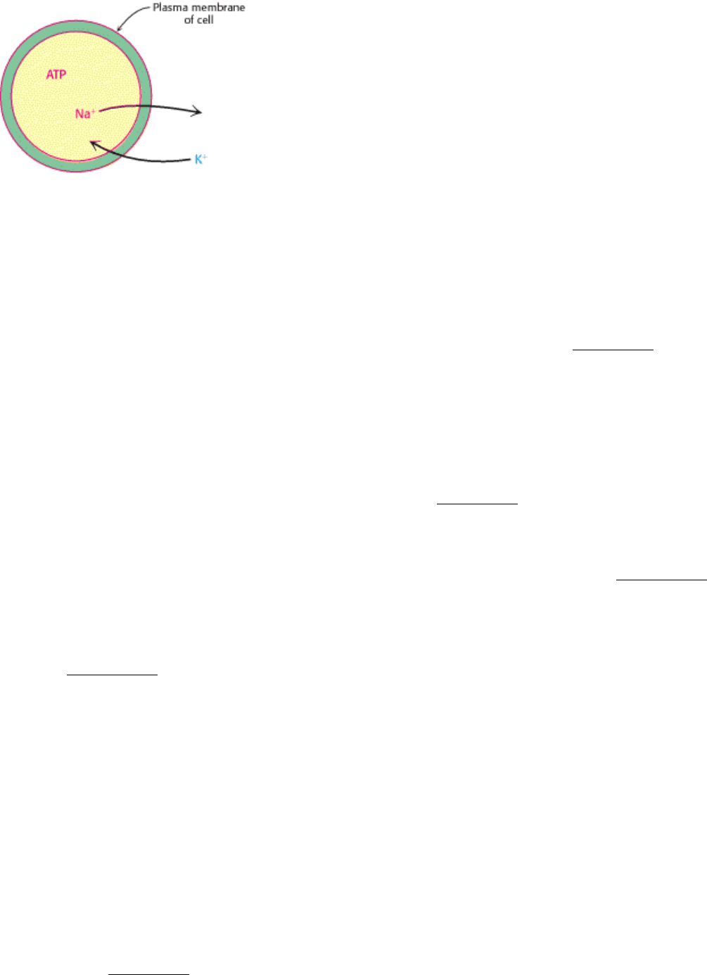
12.6.3. All Biological Membranes Are Asymmetric
Membranes are structurally and functionally asymmetric. The outer and inner surfaces of all known biological
membranes have different components and different enzymatic activities. A clear-cut example is the pump that regulates
the concentration of Na
+
and K
+
ions in cells (Figure 12.34). This transport protein is located in the plasma membrane of
nearly all cells in higher organisms. The Na
+
-K
+
pump is oriented so that it pumps Na
+
out of the cell and K
+
into it.
Furthermore, ATP must be on the inside of the cell to drive the pump. Ouabain, a specific inhibitor of the pump, is
effective only if it is located outside.
Membrane proteins have a unique orientation because they are synthesized and inserted into the membrane in an
asymmetric manner. This absolute asymmetry is preserved because membrane proteins do not rotate from one side of the
membrane to the other and because membranes are always synthesized by the growth of preexisting membranes. Lipids,
too, are asymmetrically distributed as a consequence of their mode of biosynthesis, but this asymmetry is usually not
absolute, except for glycolipids. In the red-blood-cell membrane, sphingomyelin and phosphatidyl choline are
preferentially located in the outer leaflet of the bilayer, whereas phosphatidyl ethanolamine and phosphatidyl serine are
located mainly in the inner leaflet. Large amounts of cholesterol are present in both leaflets.
I. The Molecular Design of Life 12. Lipids and Cell Membranes 12.6. Lipids and Many Membrane Proteins Diffuse Rapidly in the Plane of the Membrane
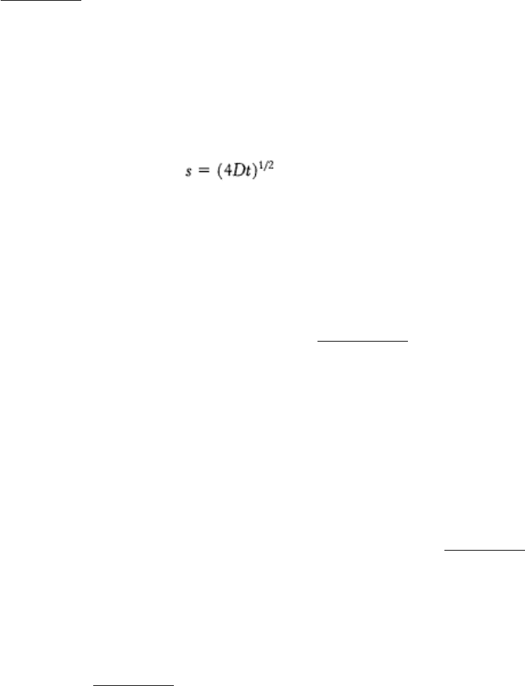
Figure 12.29. Fluorescence Recovery After Photobleaching (FRAP) Technique. (A) The cell-surface fluoresces
because of a labeled surface component. (B) The fluorescent molecules of a small part of the surface are bleached by an
intense light pulse. (C) The fluorescence intensity recovers as bleached molecules diffuse out of the region and
unbleached molecules diffuse into it. (D) The rate of recovery depends on the diffusion coefficient.
I. The Molecular Design of Life 12. Lipids and Cell Membranes 12.6. Lipids and Many Membrane Proteins Diffuse Rapidly in the Plane of the Membrane
Figure 12.30. Fluid Mosaic Model. [After S. J. Singer and G. L. Nicolson. Science 175(1972):723.]
I. The Molecular Design of Life 12. Lipids and Cell Membranes 12.6. Lipids and Many Membrane Proteins Diffuse Rapidly in the Plane of the Membrane
Figure 12.31. Lipid Movement in Membranes. Lateral diffusion of lipids is much more rapid than transverse diffusion
(flip-flop).

I. The Molecular Design of Life 12. Lipids and Cell Membranes 12.6. Lipids and Many Membrane Proteins Diffuse Rapidly in the Plane of the Membrane
Figure 12.32. The Phase-Transition, or Melting, Temperature (Tm) for a Phospholipid Membrane. As the
temperature is raised, the phospholipid membrane changes from a packed, ordered state to a more random one.
I. The Molecular Design of Life 12. Lipids and Cell Membranes 12.6. Lipids and Many Membrane Proteins Diffuse Rapidly in the Plane of the Membrane
Table 12.3. The melting temperature of phosphatidyl choline containing different pairs of identical
fatty acid chains
Number of carbons Number of double bonds Fatty acid Tm (°C)
Common name Systematic name
22 0 Behenate n-Docosanote 75
18 0 Stearate n-Octadecanoate 58
16 0 Palmitate n-Hexadecanoate 41
14 0 Myristate n-Tetradecanoate 24
18 1 Oleate
cis-∆
9
-Octadecenoate
- 22
I. The Molecular Design of Life 12. Lipids and Cell Membranes 12.6. Lipids and Many Membrane Proteins Diffuse Rapidly in the Plane of the Membrane
Figure 12.33. Packing of Fatty Acid Chains in a Membrane. The highly ordered packing of fatty acid chains is
disrupted by the presence of cis double bonds. The space-filling models show the packing of (A) three molecules of
stearate (C
18
, saturated) and (B) a molecule of oleate (C
18
, unsaturated) between two molecules of stearate.
I. The Molecular Design of Life 12. Lipids and Cell Membranes 12.6. Lipids and Many Membrane Proteins Diffuse Rapidly in the Plane of the Membrane
Figure 12.34. Asymmetry of the Na
+
-K
+
transport system in plasma membranes. The Na
+
-K
+
transport system
pumps Na
+
out of the cell and K
+
into the cell.
I. The Molecular Design of Life 12. Lipids and Cell Membranes
12.7. Eukaryotic Cells Contain Compartments Bounded by Internal Membranes
Thus far we have considered only the plasma membrane of cells. Many bacteria such as E. coli have two membranes
separated by a cell wall (made of proteins, peptides, and carbohydrates) lying in between (Figure 12.35). The inner
membrane acts as the permeability barrier, and the outer membrane and the cell wall provide additional protection. The
outer membrane is quite permeable to small molecules owing to the presence of porins. The region between the two
membranes containing the cell wall is called the periplasm. Other bacteria and archaea have only a single membrane
surrounded by a cell wall.
Eukaryotic cells, with the exception of plant cells, do not have cell walls, and their cell membranes consist of a single
lipid bilayer. In plant cells, the cell wall is on the outside of the plasma membrane. Eukaryotic cells are distinguished by
the use of membranes inside the cell to form internal compartments (Figure 12.36). For example, peroxisomes,
organelles that play a major role in the oxidation of fatty acids for energy conversion, are defined by a single membrane.
Mitochondria, the organelles in which ATP is synthesized, are surrounded by two membranes. Much like the case for a
bacterium, the outer membrane is quite permeable to small molecules, whereas the inner membrane is not. Indeed,
considerable evidence now indicates that mitochondria evolved from bacteria by endosymbiosis (Section 18.1.2). A
double membrane also surrounds the nucleus. However, the nuclear envelope is not continuous but, instead, consists of a
set of closed membranes that come together at structures called nuclear pores. These pores regulate transport into and
out of the nucleus. The nuclear membranes are linked to another membrane-defined structure, the endoplasmic
reticulum, which plays a host of cellular roles, including drug detoxification and the modification of proteins for
secretion (Section 11.3.4). Thus, a eukaryotic cell comprises interacting compartments, and transport into and out of
these compartments is essential to many biochemical processes.
12.7.1. Proteins Are Targeted to Specific Compartments by Signal Sequences
The compartmentalization of eukaryotic cells makes possible many processes that must be separated from the remainder
of the cellular environment to function properly. Specific proteins are found in peroxisomes, others in mitochondria, and
still others in the nucleus. How do proteins end up in the proper compartment? Even for bacteria, some targeting of
proteins is required: some proteins are secreted from the cell, whereas others remain in the cytosol.
Proteins include specific sequences that serve as address labels to direct the molecules to the proper location. For
example, most peroxisomal proteins end with a sequence, Ser-Lys-Leu-COO
-
, that acts as an autonomous targeting
signal. The removal of this sequence from a protein that normally resides in peroxisomes blocks its import into that
organelle, whereas the addition of this sequence to a protein that normally resides in the cytosol can direct that protein to
peroxisomes. A protein destined to pass through both mitochondrial membranes usually has a targeting sequence at its
amino terminus (Figure 12.37). Unlike the peroxisomal targeting sequence, these amino-terminal sequences are highly
variable; no clear consensus exists. They are typically from 15 to 35 residues long and rich in positively charged residues

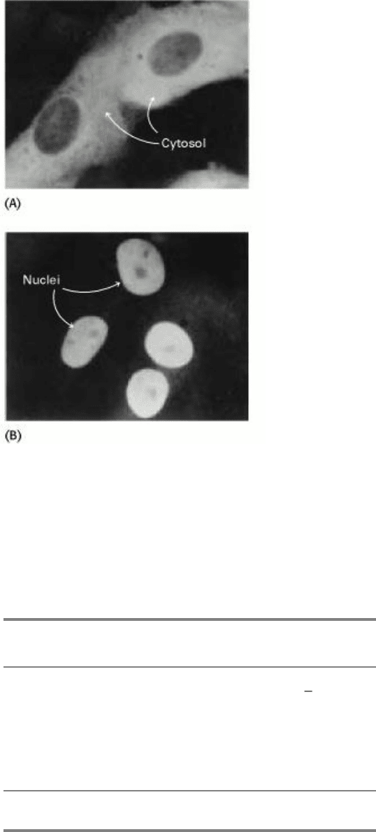
and in serines and threonines. Proteins destined for the nucleus have internal targeting sequences. A typical nuclear
localization signal contains five consecutive positively charged residues such as Lys-Lys-Lys-Arg-Lys. The addition of
such a sequence to a protein not found in the nucleus can direct it to the nucleus (Figure 12.38). Other sequences can
direct proteins out of the nucleus. The known targeting sequences are given in Table 12.4.
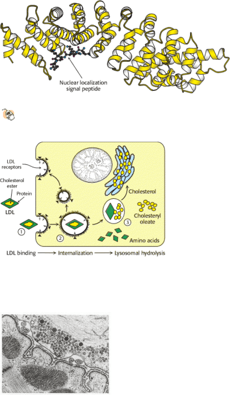
Targeting sequences act by binding to specific proteins associated with each organelle. The determination of the
structure of a protein, α-karyopherin, that binds to the nuclear localization signal reveals how the protein recognizes
such a targeting sequence (Figure 12.39). A peptide containing the appropriate sequence binds to a specific site on the
protein. The target peptide is held in an extended conformation through interactions between the target peptide backbone
and asparagine side chains of the α-karyopherin while each of the basic residues lies in a deep pocket near the bottom,
lined with negatively charged residues. Proteins that bind to the other targeting signal sequences presumably also have
structures that allow recognition of the required features. Note that we have considered only how proteins are marked for
different compartments. Later, we will consider the mechanisms by which proteins actually cross membranes (Section
11.3.2).
12.7.2. Membrane Budding and Fusion Underlie Several Important Biological
Processes
Membranes must be able to separate or join together to take up, transport, and release molecules. Many take up
molecules through the process of receptor- mediated endocytosis (Figure 12.40). Here, a protein or larger complex
initially binds to a receptor on the cell surface. After the protein is bound, specialized proteins act to cause the membrane
in the vicinity of the bound protein to invaginate. The invaginated membrane eventually breaks off and fuses to form a
vesicle.
Receptor-mediated endocytosis plays a key role in cholesterol metabolism (Section 26.3.3). Some cholesterol in the
blood is in the form of a lipid-protein complex called low-density lipoprotein (LDL). Low density lipoprotein binds to an
LDL receptor, an integral membrane protein. The segment of the plasma membrane containing the LDL-LDL receptor
complex then invaginates and buds off from the membrane. The LDL separates from the receptor, which is recycled
back to the membrane in a separate vesicle. The vesicle containing the LDL fuses with a lysosome, an organelle
containing an array of digestive enzymes. The cholesterol is released into the cell for storage or use in membrane
biosynthesis, and the remaining protein components are degraded. Various hormones, transport proteins, and antibodies
employ receptormediated endocytosis to gain entry into a cell. A less advantageous consequence is that this pathway is
available to viruses and toxins as a means of entry into cells. The reverse process
the fusion of a vesicle to a
membrane is a key step in the release of neurotransmitters from a neuron into the synaptic cleft (Figure 12.41).
Although the processes of budding and fusion appear deceptively simple, the structures of the intermediates in the
budding and fusing processes and the detailed mechanisms remain active areas of investigation.
I. The Molecular Design of Life 12. Lipids and Cell Membranes 12.7. Eukaryotic Cells Contain Compartments Bounded by Internal Membranes
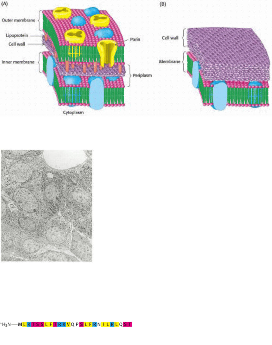
Figure 12.35. Cell Membranes of Prokaryotes. A schematic view of the membrane in bacterial cells surrounded by
(A) two membranes or (B) one membrane.
I. The Molecular Design of Life 12. Lipids and Cell Membranes 12.7. Eukaryotic Cells Contain Compartments Bounded by Internal Membranes
Figure 12.36. Internal Membranes of Eukaryotes. Electron micrograph of a thin section of a hormone-secreting cell
for the rat pituitary, showing the presence of internal structures bounded by membranes. [Biophoto Associates/Photo
Researchers.]
I. The Molecular Design of Life 12. Lipids and Cell Membranes 12.7. Eukaryotic Cells Contain Compartments Bounded by Internal Membranes
Figure 12.37. A Mitochondrial Targeting Sequence. This sequence is recognized by receptors on the external face of
the outer mitochondrial membrane. A protein bearing the sequence will be imported into the mitochondrion.
Hydrophobic residues are shown in yellow, basic ones in blue, and serine and threonine in red.
I. The Molecular Design of Life 12. Lipids and Cell Membranes 12.7. Eukaryotic Cells Contain Compartments Bounded by Internal Membranes
Figure 12.38. Movement of a Protein Into the Nucleus. Localization of (A) unmodified pyruvate kinase, and (B)
pyruvate kinase containing a nuclear localization signal sequence attached to its amino terminus. The protein was
visualized by fluorescence microscopy after staining with a specific antibody. [From W. D. Richardson, B. L. Roberts,
and A. E. Smith. Cell 44(1986):79.]
I. The Molecular Design of Life 12. Lipids and Cell Membranes 12.7. Eukaryotic Cells Contain Compartments Bounded by Internal Membranes
Table 12.4. Targeting sequences
Target Signal
Nucleus -KKXK or -(K/R)
2
-X
10 12
-(K/R)*
Peroxisome
-SKL-COO
-
Mitochondrion N-terminal amphipathic helix
Endoplasmic reticulum
-KDEL-COO
-
(ER retention)
*
The "/" means that either K or R is required.
I. The Molecular Design of Life 12. Lipids and Cell Membranes 12.7. Eukaryotic Cells Contain Compartments Bounded by Internal Membranes
Figure 12.39. Protein Targeting Signal Recognition.
The structure of the nuclear localization signal-binding protein α-
karyopherin (also known as α-importin) with a nuclear localization signal peptide bound to its major recognition
site.
I. The Molecular Design of Life 12. Lipids and Cell Membranes 12.7. Eukaryotic Cells Contain Compartments Bounded by Internal Membranes
Figure 12.40. Receptor-Mediated Endocytosis. The process of receptor-mediated endocytosis is illustrated for the
cholesterol-carrying complex, low-density lipoprotein (LDL): (1) LDL binds to a specific receptor, the LDL receptor; (2)
this complex invaginates to form an internal vesicle; (3) after separation from its receptor, the LDL-containing vesicle
fuses with a lysosome, leading to degradation of the LDL and release of the cholesterol.
I. The Molecular Design of Life 12. Lipids and Cell Membranes 12.7. Eukaryotic Cells Contain Compartments Bounded by Internal Membranes
Figure 12.41. Neurotransmitter Release. Neurotransmitter-containing synaptic vesicles are arrayed near the plasma
membrane of a nerve cell. Synaptic vesicles fuse with the plasma membrane, releasing the neurotransmitter into the
synaptic cleft. [T. Reese/Don Fawcett/ Photo Researchers.]