Berg J.M., Tymoczko J.L., Stryer L. Biochemistry
Подождите немного. Документ загружается.


We previously encountered the cAMP-dependent protein kinase in Section 10.4.2. This protein kinase and others are the
link that transduces changes in the concentrations of free second messengers into changes in the covalent structures of
proteins. Although these changes are less transient than the changes in secondary-messenger concentrations, protein
phosphorylation is not irreversible. Indeed, protein phosphatases are enzymes that hydrolytically remove specific
phosphoryl groups from modified proteins.
4. The signal is terminated. Protein phosphatases are one mechanism for the termination of a signaling process. After a
signaling process has been initiated and the information has been transduced to affect other cellular processes, the
signaling processes must be terminated. Without such termination, cells lose their responsiveness to new signals.
Moreover, signaling processes that fail to be terminated properly may lead to uncontrolled cell growth and the possibility
of cancer.
Essentially every biochemical process presented in the remainder of this book either is a component of a signal-
transduction pathway or can be affected by one. As we shall see, the use of protein modules in various combinations is a
clear, even dominant, theme in the construction of signal-transduction proteins. Signal-transduction proteins have
evolved by the addition of such ancillary modules to core domains to facilitate interactions with other proteins or cell
membranes. By controlling which proteins interact with one another, these modules play important roles in determining
the wiring diagrams for signal-transduction circuits.
We begin by considering the largest and one of the most important classes of receptor, the seven-transmembrane-helix
receptors.
II. Transducing and Storing Energy 15. Signal-Transduction Pathways: An Introduction to Information Metabolism
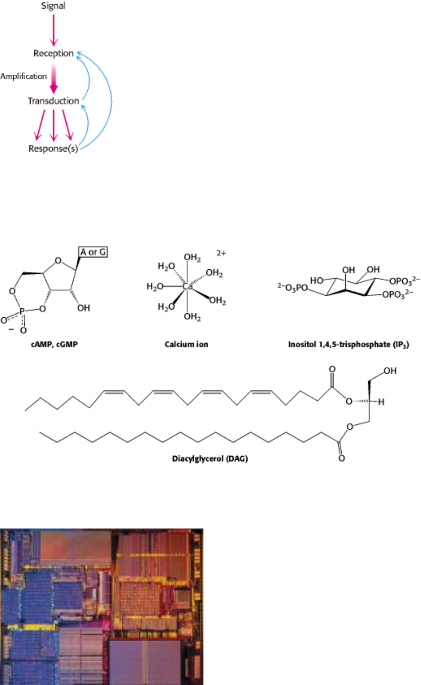
Figure 15.1. Principles of Signal Transduction. An environmental signal, such as a hormone, is first received by
interaction with a cellular component, most often a cell-surface receptor. The information that the signal has arrived is
then converted into other chemical forms, or transduced. The signal is often amplified before evoking a response.
Feedback pathways regulate the entire signaling process.
II. Transducing and Storing Energy 15. Signal-Transduction Pathways: An Introduction to Information Metabolism
Figure 15.2. Common Second Messengers. Second messengers are intracellular molecules that change in concentration
in response to environmental signals. That change in concentration conveys information inside the cell.
II. Transducing and Storing Energy 15. Signal-Transduction Pathways: An Introduction to Information Metabolism
Molecular switches. Signal transduction circuits in biological systems have molecular on/off switches that, like those in
a computer chip (above), transmit information when "on." Common among these are G proteins (right), which transmit a
signal when bound to GTP and are silent when bound to GDP. [(Left) Courtesy of Intel.]
II. Transducing and Storing Energy 15. Signal-Transduction Pathways: An Introduction to Information Metabolism
15.1. Seven-Transmembrane-Helix Receptors Change Conformation in Response to
Ligand Binding and Activate G Proteins
Conceptual Insights, Signaling Pathways: Response and Recovery.
Animations in this media module show how 7TM receptors and G proteins are
employed in two sensory-system signaling pathways.
The seven-transmembrane-helix (7TM) receptors are responsible for transmitting information initiated by signals as
diverse as photons, odorants, tastants, hormones, and neurotransmitters (Table 15.1). Several thousand such receptors are
known, and the list continues to grow. As the name indicates, these receptors contain seven helices that span the
membrane bilayer. The receptors are sometimes referred to as serpentine receptors because the single polypeptide chain
"snakes" through the membrane seven times (Figure 15.3A). A well-characterized member of this family is rhodopsin.
The "ligand" for this protein, which plays an essential role in vision, is a photon (Section 32.3.1). An example of a
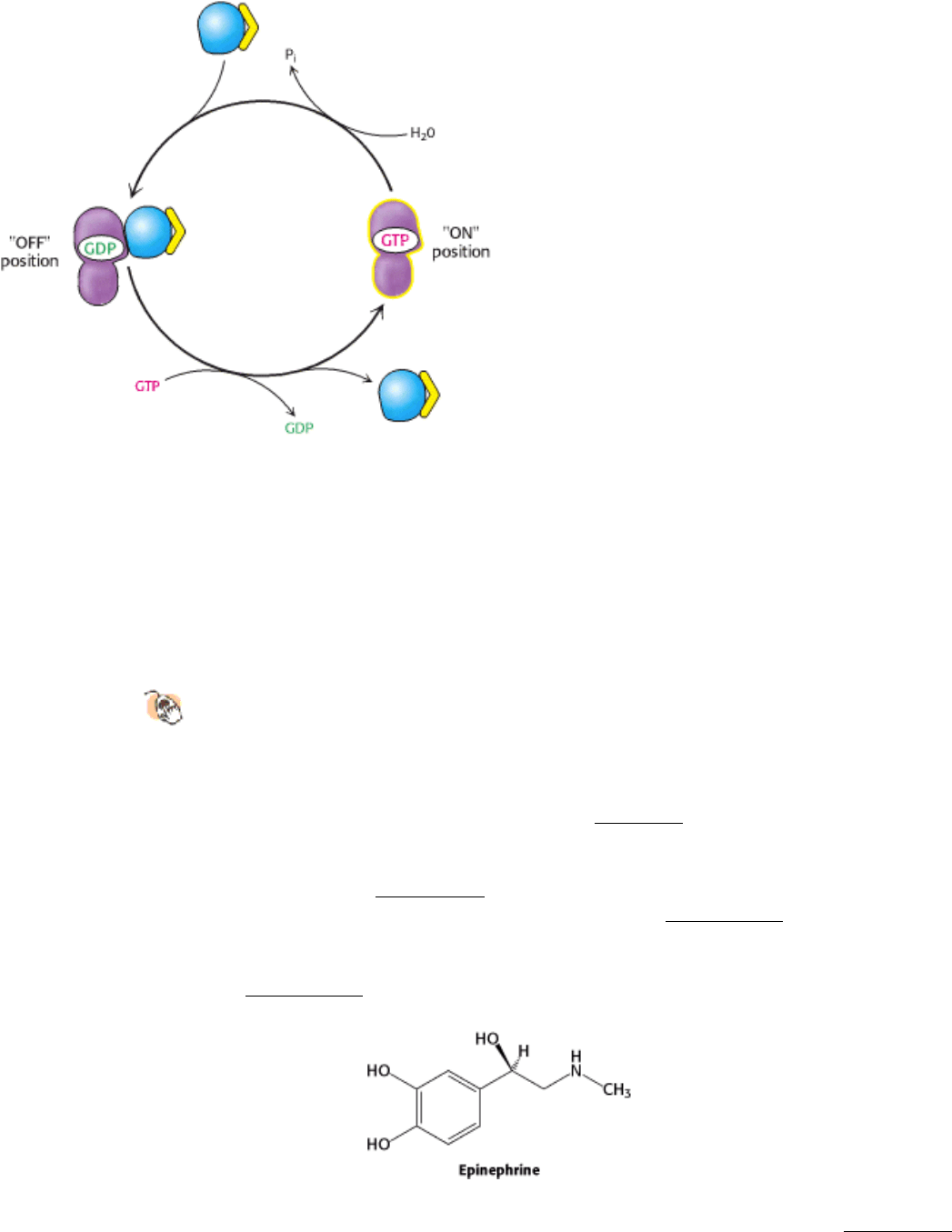
receptor that responds to chemical signals is the β-adrenergic receptor. This protein binds epinephrine (also called
adrenaline), a hormone responsible for the "fight or flight" response. We will address the biochemical roles of this
hormone in more detail later (Section 21.3.1).
Recently, the three-dimensional structure of bovine rhodopsin was determined in its unactivated form (Figure 15.3B). A
variety of evidence reveals that the 7TM receptors, particularly their cytoplasmic loops and their carboxyl termini,
change conformation in response to ligand binding, although the details of these conformational changes remain to be
established. Thus, the binding of a ligand from outside the cell induces a conformational change in the 7TM receptor
that can be detected inside the cell. Even though vision and response to hormones would seem to have little in common,

a comparison of the amino acid sequences of rhodopsin and the β-adrenergic receptor clearly reveals homology. On the
basis of this sequence comparison, the β-adrenergic receptor is expected to have a structure quite similar to that of
rhodopsin. As we shall see, these receptors also have in common the next step in their signaling transduction cascades.
15.1.1. Ligand Binding to 7TM Receptors Leads to the Activation of G Proteins
What is the next step in the pathway after the binding of epinephrine by the β-adrenergic receptor? An important clue
was Martin Rodbell's finding that GTP in addition to hormone is essential for signal transduction to proceed. Equally
revealing was the observation that hormone binding stimulates GTP hydrolysis. These findings led to the discovery by
Alfred Gilman that a guanyl nucleotide-binding protein is an intermediary in signal transduction from the 7TM
receptors. This signal-coupling protein is termed a G protein (G for guanyl nucleotide). The activated G protein
stimulates the activity of adenylate cyclase, an enzyme that increases the concentration of cAMP by forming it from
ATP (Figure 15.4).
15.1.2. G Proteins Cycle Between GDP- and GTP-Bound Forms
How do these G proteins operate? In the unactivated state, the guanyl nucleotide bound to the G protein is GDP. In this
form, the G protein exists as a heterotrimer consisting of α, β, and γ subunits; the α subunit (referred to as G
α
) binds
the nucleotide (Figure 15.5). The α subunit is a member of the P-loop NTPases family (Section 9.4.1) and the P-loop that
participates in nucleotide binding. The β subunit contains a seven-bladed propeller structure, and the γ subunit comprises
a pair of α helices that wrap around the β subunit (Figure 15.6). The α and γ subunits are usually anchored to the
membrane by covalently attached fatty acids. The role of the hormone-bound receptor is to catalyze the exchange of
GTP for bound GDP. The hormone-receptor complex interacts with the heterotrimeric G protein and opens the
nucleotide-binding site so that GDP can depart and GTP from solution can bind. The α subunit simultaneously
dissociates from the β γ dimer (G
β
γ
). The structure of the G
α
subunit conforms tightly to the GTP molecule; in
particular, three stretches of polypeptide (termed switch I, switch II, and switch III) interact either directly or indirectly
with the γ phosphate of GTP (Figure 15.7). These structural changes are responsible for the reduced affinity of G
α
for G
β
γ
. The dissociation of the G-protein heterotrimer into G
α
and G
β
γ
units transmits the signal that the receptor has
bound its ligand. Moreover, the surfaces of G
α
and G
β
γ
that had formed the trimer interface are now exposed to interact
with other proteins.
A single hormone-receptor complex can stimulate nucleotide exchange in many G-protein heterotrimers. Thus, hundreds
of G
α
molecules are converted from their GDP into their GTP forms for each bound molecule of hormone, giving an
amplified response. All 7TM receptors appear to be coupled to G proteins, and so the 7TM receptors are sometimes
referred to as G-protein-coupled receptors or GPCRs. Do all of the signals that function by means of 7TM receptors
funnel through the same G protein? Indeed not. Different G proteins exist, and they can affect downstream targets in
different ways when activated. For example, in regard to the G protein coupled to the β-adrenergic receptor, the α
subunit binds to adenylate cyclase and stimulates its enzymatic activity. This subunit is referred to as G
α
s
, the s in the
subscript indicating the subunit's stimulatory role. The human genome contains more than 15 genes encoding the α
subunits, 5 encoding the β subunits, and 10 encoding the γ subunits. Thus, in principle, there could be more than a
thousand heterotrimeric G proteins; however, the number of combinations that actually exists is not known. Selected
members of this family are shown in Table 15.2. Only a small subset of these proteins is expressed in a particular cell.
15.1.3. Activated G Proteins Transmit Signals by Binding to Other Proteins
The adenylate cyclase enzyme that is activated by the epinephrine-β-adrenergic receptor complex is a membrane protein
that contains 12 presumed membrane-spanning helices. The enzymatically active part of the protein is formed from two
large intracellular domains: one is located between transmembrane helices 6 and 7 and the other after the last

transmembrane helix. The structure was determined for a complex formed between G
α
s
bound to a GTP analog and
protein fragments corresponding to the active adenylate cyclase (Figure 15.8). As expected, the G protein binds to
adenylate cyclase through the surface that had bound the β γ dimer when the G protein was in its GDP form. The
activation of the G protein exposes this surface and subtly changes it so that it now binds the surface of adenylate cyclase
in preference to G
β
γ
. The interaction of G
α
s
with adenylate cyclase favors a more catalytically active conformation of
the enzyme, thus stimulating cAMP production. The net result is that the binding of epinephrine to the receptor on the
cell surface increases the rate of cAMP production inside the cell.
15.1.4. G Proteins Spontaneously Reset Themselves Through GTP Hydrolysis
The ability to shut down signal-transduction pathways is as critical as the ability to turn them on. How is the signal
initiated by activated 7TM receptors switched off? G
α
subunits have intrinsic GTPase activity, hydrolyzing bound GTP
to GDP and P
i
. This hydrolysis reaction is slow, however, requiring from seconds to minutes and thus allowing the GTP
form of G
α
to activate downstream components of the signal-transduction pathway before GTP hydrolysis deactivates
the subunit. In essence, the bound GTP acts as a built-in clock that spontaneously resets the G
α
subunit after a short
time period. After GTP hydrolysis and the release of P
i
, the GDP-bound form of G
α
then reassociates with G
β
γ
to
reform the heterotrimeric protein (Figure 15.9).
The hormone-bound activated receptor must be reset as well to prevent the continuous activation of G proteins. This
resetting is accomplished by two processes (Figure 15.10). First, the hormone dissociates, returning the receptor to its
initial, unactivated state. The likelihood that the receptor remains in its unbound state depends on the concentration of
hormone. Second, the hormone-receptor complex is deactivated by the phosphorylation of serine and threonine residues
in the carboxyl-terminal tail. In the example under consideration, β-adrenergic receptor kinase phosphorylates the
carboxyl-terminal tail of the hormone-receptor complex but not the unoccupied receptor. Finally, the binding of β -
arrestin, binds to the phosphorylated receptor and further diminishes its G-protein-activating ability. Phosphorylation
and the binding of β-arrestin account for the desensitization (adaptation) of the receptor subsequent to prolonged
exposure to epinephrine. The epinephrine-initiated cascade, like many other signal-transduction processes, has evolved
to respond to changes in the strength of stimuli rather than to their absolute level. Adaptation is advantageous because it
enables receptors to respond to changes in the level of stimuli over a wide range of background levels.
15.1.5. Cyclic AMP Stimulates the Phosphorylation of Many Target Proteins by
Activating Protein Kinase A
Let us continue to follow the information flow down this signal-transduction pathway. The increased concentration of
cAMP can affect a wide range of cellular processes. For example, it enhances the degradation of storage fuels, increases
the secretion of acid by the gastric mucosa, leads to the dispersion of melanin pigment granules, diminishes the
aggregation of blood platelets, and induces the opening of chloride channels. How does cAMP influence so many
cellular processes? Is there a common denominator for its diverse effects? Indeed there is. Most effects of cyclic AMP in
eukaryotic cells are mediated by activation of a single protein kinase. This key enzyme is called protein kinase A (PKA).
As discussed in Section 10.4.2, PKA consists of two regulatory (R) chains and two catalytic (C) chains. In the absence of
cAMP, the R
2
C
2
complex is catalytically inactive. The binding of cAMP to the regulatory chains releases the catalytic
chains, which are enzymatically active on their own. Activated PKA then phosphorylates specific serine and threonine
residues in many targets to alter their activity. The significance and far reach of the adenylate cyclase cascade are seen in
the following examples:
1. In glycogen metabolism (Section 21.5), PKA phosphorylates two enzymes that lead to the breakdown of this
polymeric store of glucose and the inhibition of further glycogen synthesis.
2. PKA stimulates the expression of specific genes by phosphorylating a transcriptional activator called the cAMP-
response element binding (CREB) protein (Section 31.3.6). This activity of PKA illustrates that signal-transduction
pathways can extend into the nucleus to alter gene expression.
3. Synaptic transmission between pairs of neurons in Aplysia (a marine snail) is enhanced by serotonin, a
neurotransmitter that is released by adjacent interneurons. Serotonin binds to a 7TM receptor to trigger an adenylate
cyclase cascade. The rise in cAMP level activates PKA, which facilitates the closing of potassium channels by
phosphorylating them. Closure of potassium channels increases the excitability of the target cell.
Thus, signal-transduction pathways that include 7TM receptors, the activation of adenylate cyclase, and the activation of
PKA can modulate enzyme activities, gene-expression patterns, and membrane excitability.
II. Transducing and Storing Energy 15. Signal-Transduction Pathways: An Introduction to Information Metabolism 15.1. Seven-Transmembrane-Helix Receptors Change Conformation in Response to Ligand Binding and Activate G Proteins
Table 15.1. Biological functions mediated by 7TM receptors
Smell
Taste
Vision
Neurotransmission
Hormone secretion
Chemotaxis
Exocytosis
Control of blood pressure
Embryogenesis
Cell growth and differentiation
Development
Viral infection
Carcinogenesis
Source: After J. S. Gutkind, J. Biol. Chem. 273(1998):1839.
II. Transducing and Storing Energy 15. Signal-Transduction Pathways: An Introduction to Information Metabolism 15.1. Seven-Transmembrane-Helix Receptors Change Conformation in Response to Ligand Binding and Activate G Proteins
Figure 15.3. A 7TM Receptor.
(A) Schematic representation of a 7TM receptor showing how it passes through the
membrane seven times. (B) Three-dimensional structure of rhodopsin, a 7TM receptor taking part in visual signal
transduction. As the first 7TM receptor whose structure has been determined, its structure provides a framework
for understanding other 7TM receptors. A linked photoreceptor molecule, retinal, is present in the position where, in at
least other 7TM receptors, ligands likely bind.
II. Transducing and Storing Energy 15. Signal-Transduction Pathways: An Introduction to Information Metabolism 15.1. Seven-Transmembrane-Helix Receptors Change Conformation in Response to Ligand Binding and Activate G Proteins
Figure 15.4. The β -Adrenergic Receptor Signal-Transduction Pathway. On binding of ligand, the receptor activates
a G protein that in turn activates the enzyme adenylate cyclase. Adenylate cyclase generates the second messenger
cAMP. The increase in cAMP results in a biochemical response to the initial signal.

II. Transducing and Storing Energy 15. Signal-Transduction Pathways: An Introduction to Information Metabolism 15.1. Seven-Transmembrane-Helix Receptors Change Conformation in Response to Ligand Binding and Activate G Proteins
Figure 15.5. A Heterotrimeric G Protein.
(A) A ribbon diagram shows the relation between the three subunits. In this
complex, the α subunit (gray and purple) is bound to GDP. (B) A schematic representation of the heterotrimeric G
protein.
II. Transducing and Storing Energy 15. Signal-Transduction Pathways: An Introduction to Information Metabolism 15.1. Seven-Transmembrane-Helix Receptors Change Conformation in Response to Ligand Binding and Activate G Proteins
Figure 15.6. The β γ Subunits of the Heterotrimeric G Protein.
Two views illustrate the interaction between the β and
the γ subunits. The helices of the γ subunit (yellow) wrap around the β subunit (blue). The seven-bladed propeller
structure of the β subunit is readily apparent in the representation on the right.

II. Transducing and Storing Energy 15. Signal-Transduction Pathways: An Introduction to Information Metabolism 15.1. Seven-Transmembrane-Helix Receptors Change Conformation in Response to Ligand Binding and Activate G Proteins
Figure 15.7. Conformational Changes in G
α
On Nucleotide Exchange. (Left) Prior to activation, Gα binds GDP.
(Right) On GTP for GDP exchanges, the three switch regions (shown in blue) close upon the nucleoside
triphosphate, generating the active conformation.
II. Transducing and Storing Energy 15. Signal-Transduction Pathways: An Introduction to Information Metabolism 15.1. Seven-Transmembrane-Helix Receptors Change Conformation in Response to Ligand Binding and Activate G Proteins
Table 15.2. G-protein families and their functions
G
α
class Initiating signal Downstream signal
G
α
s
β-Adrenergic amines, glucagon, parathyroid hormone, many others
Stimulates adenylate cyclase
G
α
i
Acetylcholine, α-adrenergic amines, many neurotransmitters
Inhibits adenylate cyclase
G
α
t
Photons Stimulates cGMP phosphodiesterase
G
α
q
Acetylcholine, α-adrenergic amines, many neurotransmitters
Increases IP
3
and intracellular calcium
G
α
13
Thrombin, other agonists
Stimulates Na
+
and H
+
exchange
Source: Z. Farfel, H. R. Bourne, and T. Iiri. N. Engl. J. Med. 340(1999):1012.
II. Transducing and Storing Energy 15. Signal-Transduction Pathways: An Introduction to Information Metabolism 15.1. Seven-Transmembrane-Helix Receptors Change Conformation in Response to Ligand Binding and Activate G Proteins

Figure 15.8. Adenylate Cyclase Is Activated by G
α
s
. (A) Adenylate cyclase is an integral membrane protein with two
large cytoplasmic domains that form the catalytic structure. (B) G
α
s
bound to GTP binds to the catalytic part of
the cyclase, inducing a structural change that stimulates enzyme activity. The surface of G
α
s
that interacts with
adenylate cyclase is the one that is exposed on release of G
β
γ
.
II. Transducing and Storing Energy 15. Signal-Transduction Pathways: An Introduction to Information Metabolism 15.1. Seven-Transmembrane-Helix Receptors Change Conformation in Response to Ligand Binding and Activate G Proteins
Figure 15.9. Resetting G
α
. On hydrolysis of the bound GTP by the intrinsic GTPase activity of G
α
, G
α
reassociates
with the β γ subunits to form the heterotrimeric G protein, thereby terminating the activation of adenyl cyclase.
II. Transducing and Storing Energy 15. Signal-Transduction Pathways: An Introduction to Information Metabolism 15.1. Seven-Transmembrane-Helix Receptors Change Conformation in Response to Ligand Binding and Activate G Proteins
Figure 15.10. Signal Termination. Signal transduction by the 7TM receptor is halted (1) by dissociation of the signal
molecule from the receptor and (2) by phosphorylation of the cytoplasmic C-terminal tail of the receptor and the