Berg J.M., Tymoczko J.L., Stryer L. Biochemistry
Подождите немного. Документ загружается.


II. Transducing and Storing Energy 15. Signal-Transduction Pathways: An Introduction to Information Metabolism 15.3. Calcium Ion Is a Ubiquitous Cytosolic Messenger
Figure 15.20. Calcium Imaging. A series of images shows Ca
2+
spreading across a cell. These images were obtained
through the use of a fluorescent calcium-binding dye. The images are false colored: red represents high Ca
2+
concentrations and blue low Ca
2+
concentrations. [Courtesy of Dr. Masashi Isshiki, Dept. of Nephrology, University of
Tokyo, and Dr. G. W. Anderson, Dept. of Cell Biology, University of Texas Southwestern Medical School.]
II. Transducing and Storing Energy 15. Signal-Transduction Pathways: An Introduction to Information Metabolism 15.3. Calcium Ion Is a Ubiquitous Cytosolic Messenger
Figure 15.21. EF Hand.
Formed by a helix-loop-helix unit, an EF hand is a binding site for calcium in many calcium
sensing proteins. Here, the E helix is yellow, the F helix is blue, and calcium is represented by the green sphere.
II. Transducing and Storing Energy 15. Signal-Transduction Pathways: An Introduction to Information Metabolism 15.3. Calcium Ion Is a Ubiquitous Cytosolic Messenger
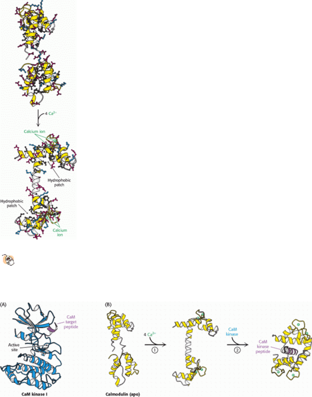
Figure 15.22. Conformational Changes in Calmodulin on Calcium Binding.
In the absence of calcium (top), the EF
hands have hydrophobic cores. On binding of a calcium ion (green sphere) to each EF hand, structural changes
expose hydrophobic patches on the calmodulin surface. These patches serve as docking regions for target proteins.
Acidic residues are shown in red, basic residues in blue, and hydrophobic residues in black. The central helix in
calmodulin remains somewhat flexible, even in the calcium-bound state.
II. Transducing and Storing Energy 15. Signal-Transduction Pathways: An Introduction to Information Metabolism 15.3. Calcium Ion Is a Ubiquitous Cytosolic Messenger
Figure 15.23. Calmodulin Binds to Amphipathic α Helices. (A) An amphipatic α helix (purple) in CaM kinase I is a
target for calmodulin. (B) After calcium binding (1), the two halves of calmodulin clamp down around the target
helix (2), binding it through hydrophobic and ionic interactions. In CaM kinase I, this interaction extracts a C-
terminal α helix, allowing the enzyme to adopt an active conformation.
II. Transducing and Storing Energy 15. Signal-Transduction Pathways: An Introduction to Information Metabolism
15.4. Some Receptors Dimerize in Response to Ligand Binding and Signal by Cross-
phosphorylation
The 7TM receptors initiate signal-transduction pathways through changes in tertiary structure that are induced by ligand
binding. A fundamentally different mechanism is utilized by a number of other classes of receptors. For these receptors,
ligand binding leads to changes in quaternary structures
specifically, the formation of receptor dimers. As we shall see,
receptor dimerization is crucial because protein kinase domains associated with the intracellular domains of the receptors
are brought together in such a way that they can phosphorylate one another. Such cross-phosphorylation initiates further
signaling.
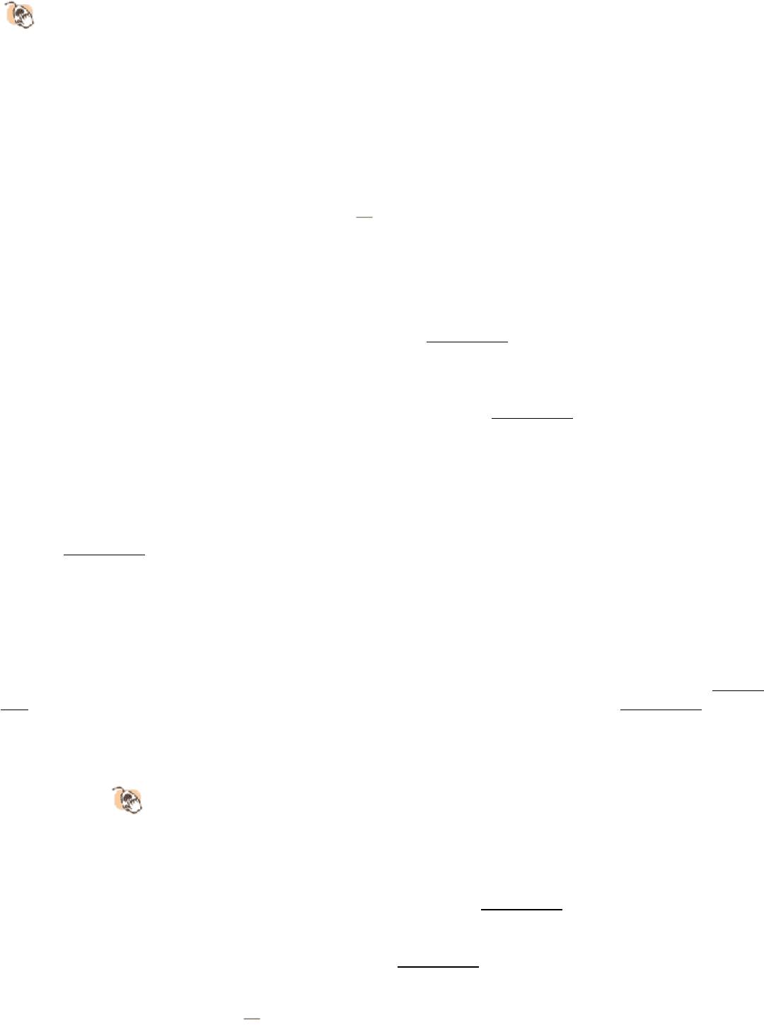
We consider human growth hormone and its receptor as our first example. Growth hormone is a monomeric protein of
217 amino acids that forms a compact four-helix bundle structure (Figure 15.24). The growth-hormone receptor
comprises 638 amino acids, divided into an extracellular domain of 250 amino acids, a single membrane-spanning helix,
and an intracellular domain of 350 amino acids. In the absence of bound hormone, the receptor is present as a monomer.
Growth hormone binds to the extracellular domain of the receptor. Remarkably, each monomeric hormone binds to two
receptor molecules, thus promoting the formation of a dimer of the receptor (Figure 15.25). The binding between the
hormone and the receptors is highly cooperative; once a hormone has bound to a single receptor molecule, the binding of
the second receptor is highly favored.
This process takes place outside the cell. Dimerization of the extracellular domains of the receptor brings together the
intracellular domains as well. Associated with each intracellular domain is a molecule of a protein kinase termed Janus
kinase 2 (JAK2) in an unactivated form. Janus kinases have modular structures consisting of four previously described
domains (Figure 15.26).
The carboxyl terminus is a protein kinase domain; its amino acid sequence and known biochemical properties suggest
that this domain functions as a tyrosine kinase. Adjacent to this domain is a second region that is clearly homologous to a
protein kinase, although several key residues have been changed and the biochemical activity of this domain is not well
established. Indeed, this pair of kinase-like domains accounts for the name of these proteins; Janus is the two-headed
Roman god of gateways. At the amino terminus is a 300-amino-acid domain, called the ERM domain, that helps anchor
JAK2 to membranes. In between this domain and the kinase domains is an SH2 domain (SH for Src homology; Section
15.4). SH2 domains are 100-amino-acid domains that bind peptides containing phosphotyrosine (Figure 15.27). The
domains bind phosphotyrosine through interactions with conserved arginine residues among other residues.
Structural Insights, SH2 Domains: An Example of Modular Regulatory
Domains, takes a close look at phosphotyrosine-SH2 domain interactions and
the diverse ways they can affect protein function.
Dimerization of the growth-hormone receptors brings together the JAK2 proteins associated with each intracellular
domain, apparently delivering a key loop (termed the activation loop) of one kinase domain into the active site of the
kinase bound to the other receptor, which results in cross-phosphorylation (Figure 15.28). How phosphorylation of JAK2
leads to its activation has not been directly established. However, the results of studies of other kinases reveal that the
activation loop is in a conformation unsuitable for catalysis in the unphosphorylated form but changes to an active
conformation when phosphoryl groups are added to key sites (Figure 15.29).
When activated by cross-phosphorylation, JAK2 can phosphorylate other substrates. In the present case, at least two
important proteins are phosphorylated
a regulator of gene expression called STAT5 (STAT for signal transducers and

activators of transcription) and the growth-hormone receptor itself. What are the consequences of the phosphorylation of
these two proteins? STAT5 is phosphorylated on a tyrosine residue near the carboxyl terminus of the protein. The
phosphotyrosine residue binds to an SH2 domain of another STAT5 molecule. Reciprocal interactions lead to the
formation of a stable STAT dimer (Figure 15.30). The dimerized STAT protein, which has a much greater affinity for
specific binding sites on DNA than does a monomeric protein, moves to the nucleus, where it binds to the DNA binding
sites to regulate gene expression. The phosphorylation of the growth-hormone receptor may have several consequences.
First, the phosphorylated receptor may serve as a docking site for JAK2 through its SH2 domain. Second, other proteins
may associate with the phosphorylated receptor, participating in other signaling pathways.
15.4.1. Some Receptors Contain Tyrosine Kinase Domains Within Their Covalent
Structures
Growth factors such as insulin, epidermal growth factor (EGF), and platelet-derived growth factor bind to the
extracellular domains of transmembrane receptors that have tyrosine kinase domains present within their intracellular
domains. For these proteins, which are found in multicellular organisms but not in yeast, genes encoding extracellular
domains and the signaling kinases fused in the course of evolution. These receptor tyrosine kinases (RTKs) signal by
mechanisms quite similar to those discussed for the pathway initiated by the growth-hormone receptor.
Consider, for example, epidermal growth factor, a 6-kd polypeptide that stimulates the growth of epidermal and
epithelial cells (Figure 15.31). This 53-residue growth factor is produced by the cleavage of an EGF precursor, a large
transmembrane protein. Such processing, which is common for growth factors and hormones, is reminiscent of the
processing of zymogens into active enzymes (Section 10.5). The first step in the signal-transduction pathway is the
binding of EGF to the epidermal growth factor receptor, a single polypeptide chain consisting of 1186 residues. The
receptor tyrosine kinase is monomeric and enzymatically inactive in the absence of the growth factor. The binding of
EGF to the extracellular domain causes the receptor to dimerize and undergo cross-phosphorylation and activation.
The insulin receptor is a disulfide-bonded dimer of α β pairs even when insulin is not bound. Nevertheless, insulin is still
required for the activation of the kinase, demonstrating that dimerization is necessary but not sufficient for activation.
The binding of the growth factor must convert the subunits of the dimer into a conformation that brings appropriate
tyrosine residues from one chain into the active site of the other chain so that cross-phosphorylation can take place.
An elegant experiment demonstrated the commonality of the receptor tyrosine kinase signaling mechanism. The EGF
receptor and the insulin receptor both contain intrinsic tyrosine kinases. Do these receptors transfer information across
the membrane in the same way? This question was answered by synthesizing a gene that encoded a chimeric receptor
the extracellular part came from the insulin receptor, and the membrane-spanning and cytosolic parts came from the
EGF receptor. The striking result was that the binding of insulin induced tyrosine kinase activity, as evidenced by rapid
autophosphorylation. Hence, the insulin receptor and the EGF receptor employ a common mechanism of signal
transmission across the plasma membrane.
How is the signal transferred beyond the receptor tyrosine kinase? We have seen that activated tyrosine kinases can
phosphorylate other proteins and that phosphotyrosines on the phosphorylated receptors can act as docking sites for SH2
domains on other proteins. A key adaptor protein links the phosphorylation of the EGF receptor to the stimulation of cell
growth through a chain of protein phosphorylations (Figure 15.32). On phosphorylation of the receptor, the SH2 domain
of the adaptor protein Grb-2 binds to the phosphotyrosine residues of the receptor tyrosine kinase. Grb-2 then recruits a
protein called Sos, which interacts with Grb-2 through two SH3 domains, domains that bind proline-rich stretches of
polypeptide and, like SH2 domains, are recurring domains that mediate protein-protein interactions. Sos, in turn, binds to
and activates Ras, a very prominent signal-transduction component that we will consider in Section 15.4.2. Finally, Ras,
in its activated form, binds to other components of the molecular circuitry leading to the activation of the specific serine-
threonine protein kinases that phosphorylate specific targets that promote cell growth. We see here another example of
how a signal-transduction pathway is constructed. Specific protein-protein interactions (through SH2, SH3, and other
domains not considered here) link the original ligand-binding event to the final result stimulation of cell growth.

15.4.2. Ras, Another Class of Signaling G Protein
We now turn our attention to another important family of signal proteins, the small G proteins, or small GTPases. This
large superfamily of proteins
grouped into subfamilies called Ras, Rho, Arf, Rab, and Ran plays a major role in a
host of cell functions including growth, differentiation, cell motility, cytokinesis, and transport of materials throughout
the cell (Table 15.3). Like their relatives the heterotrimeric G proteins (Section 15.1.2), the small G proteins cycle
between an active GTP-bound form and an inactive GDP-bound form. They differ from the heterotrimeric G proteins in
being smaller (20 25 kd versus 30 35 kd) and monomeric. Nonetheless, the two families are related by divergent
evolution, and small G proteins have many key mechanistic and structural motifs in common with the G
α
subunit of the
heterotrimeric G proteins.
In their activated GTP-bound form, small G proteins such as Ras stimulate cell growth and differentiation. Recall that
Sos is the immediate upstream link to Ras in the circuit conveying the EGF signal. How does Sos activate Ras? Sos
binds to Ras, reaches into the nucleotide-binding pocket, and opens it up, allowing GDP to escape and GTP to enter in its
place (Figure 15.33). This process is presumably analogous to the stimulation of nucleotide exchange in heterotrimeric G
proteins by activated 7TM receptors, a process for which structural details are not yet available. Sos is referred to as a
guaninenucleotide exchange factor (GEF). Thus, the binding of EGF to the EGF receptor leads to the conversion of Ras
into its GTP form through the intermediacy of Grb-2 and Sos (Figure 15.34).
Like the G
α
protein, Ras possesses an intrinsic GTPase activity, which serves to terminate the signal and return the
system to the inactive state. This activity is slow but is augmented by helper proteins termed GTPase-activating proteins
(GAPs). The guanine-nucleotide exchange factors and the GTPase-activating proteins allow the G-protein cycle to
proceed with rates appropriate for a balanced level of downstream signaling.
II. Transducing and Storing Energy 15. Signal-Transduction Pathways: An Introduction to Information Metabolism 15.4. Some Receptors Dimerize in Response to Ligand Binding and Signal by Cross-phosphorylation
Figure 15.24. Human Growth Hormone Structure.
Human growth hormone forms a four-helix bundle.
II. Transducing and Storing Energy 15. Signal-Transduction Pathways: An Introduction to Information Metabolism 15.4. Some Receptors Dimerize in Response to Ligand Binding and Signal by Cross-phosphorylation
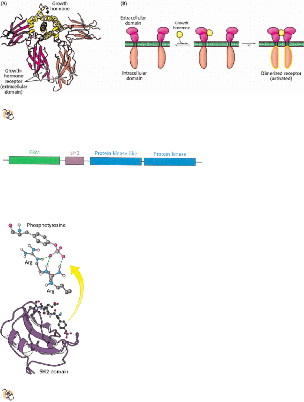
Figure 15.25. Binding of Growth Hormone Leads to Receptor Dimerization.
(A) A single growth-hormone molecule
(yellow) interacts with the extracellular domain of two receptors (red and orange). (B) The binding of one
hormone molecule to two receptors leads to the formation of a receptor dimer. Dimerization is a key step in this
signal-transduction pathway.
II. Transducing and Storing Energy 15. Signal-Transduction Pathways: An Introduction to Information Metabolism 15.4. Some Receptors Dimerize in Response to Ligand Binding and Signal by Cross-phosphorylation
Figure 15.26. Janus Kinase Domain Structure. A Janus kinase (JAK) includes four recognized domains: an ERM
domain that favors interactions with membranes, an SH2 domain that binds phosphotyrosine-containing peptides, and
two domains homologous to protein kinases. Only the second protein kinase domain appears to be enzymatically
functional.
II. Transducing and Storing Energy 15. Signal-Transduction Pathways: An Introduction to Information Metabolism 15.4. Some Receptors Dimerize in Response to Ligand Binding and Signal by Cross-phosphorylation
Figure 15.27. Recognition of Phosphotyrosine by SH2 Domains.
The structure of an SH2 domain (purple) bound to a
phosphotyrosine-containing peptide. The hydrogen-bonding interactions between the phosphotyrosine residue and
two arginine residues are shown; interactions with other residues are omitted for clarity.

II. Transducing and Storing Energy 15. Signal-Transduction Pathways: An Introduction to Information Metabolism 15.4. Some Receptors Dimerize in Response to Ligand Binding and Signal by Cross-phosphorylation
Figure 15.28. Cross-Phosphorylation of Jaks Induced by Receptor Dimerization. The binding of growth hormone
leads to receptor dimerization, which brings two JAKs together in such a way that each phosphorylates key residues on
the other. The activated JAKs remain bound to the receptor.
II. Transducing and Storing Energy 15. Signal-Transduction Pathways: An Introduction to Information Metabolism 15.4. Some Receptors Dimerize in Response to Ligand Binding and Signal by Cross-phosphorylation
Figure 15.29. Activation of a Protein Kinase by Phosphorylation.
In the unphosphorylated state, a key loop is in a
conformation unsuitable for catalysis. Phosphorylation (at two sites in the case shown) stabilizes an active
conformation.
II. Transducing and Storing Energy 15. Signal-Transduction Pathways: An Introduction to Information Metabolism 15.4. Some Receptors Dimerize in Response to Ligand Binding and Signal by Cross-phosphorylation

Figure 15.30. Phosphorylation-Induced Dimerization of STAT Proteins. The phosphorylation of a key tyrosine
residue on each STAT protein leads to an interaction between the phosphotyrosine and an SH2 domain on another STAT
monomer. The STAT dimer produced by these reciprocal interactions has a high affinity for specific DNA sequences
and is able to alter gene expression after binding to DNA.
II. Transducing and Storing Energy 15. Signal-Transduction Pathways: An Introduction to Information Metabolism 15.4. Some Receptors Dimerize in Response to Ligand Binding and Signal by Cross-phosphorylation
Figure 15.31. Structure of Epidermal Growth Factor.
This protein growth factor is stabilized by three disulfide
bonds.
II. Transducing and Storing Energy 15. Signal-Transduction Pathways: An Introduction to Information Metabolism 15.4. Some Receptors Dimerize in Response to Ligand Binding and Signal by Cross-phosphorylation
Figure 15.32. Structure of Grb-2, an Adaptor Protein.
Grb-2 consists of two SH3 domains and a central SH2 domain.
The SH2 domain binds to phosphotyrosine resides on an activated receptor while the SH3 domains bind proline-
rich regions on other proteins such as Sos.
II. Transducing and Storing Energy 15. Signal-Transduction Pathways: An Introduction to Information Metabolism 15.4. Some Receptors Dimerize in Response to Ligand Binding and Signal by Cross-phosphorylation
Table 15.3. Ras superfamily of GTPases
Subfamily Function
Ras Regulates cell growth through serine-threonine protein kinases
Rho Reorganizes cytoskeleton through serine-threonine protein kinases
Arf Activates the ADP-ribosyltransferase of the cholera toxin A subunit; regulates vesicular trafficking
pathways; activates phospholipase D
Rab Plays a key role in secretory and endocytotic pathways
Ran Functions in the transport of RNA and protein into and out of the nucleus
II. Transducing and Storing Energy 15. Signal-Transduction Pathways: An Introduction to Information Metabolism 15.4. Some Receptors Dimerize in Response to Ligand Binding and Signal by Cross-phosphorylation
Figure 15.33. Structure of Sos, a Guanine-Nucleotide Exchange Factor.
Sos (yellow) binds to Ras and opens up its
nucleotide-binding site, allowing GDP to escape and GTP to bind. In the GTP-bound form, Ras can bind to and
activate other proteins, including protein kinases.
II. Transducing and Storing Energy 15. Signal-Transduction Pathways: An Introduction to Information Metabolism 15.4. Some Receptors Dimerize in Response to Ligand Binding and Signal by Cross-phosphorylation

Figure 15.34. Egf Signaling Pathway. The binding of epidermal growth factor (EGF) to its receptor leads to cross-
phosphorylation of the receptor. The phosphorylated receptor binds Grb-2, which, in turn, binds Sos. Sos stimulates the
exchange of GTP for GDP in Ras. Activated Ras binds to and stimulates protein kinases (not shown).
II. Transducing and Storing Energy 15. Signal-Transduction Pathways: An Introduction to Information Metabolism
15.5. Defects in Signaling Pathways Can Lead to Cancer and Other Diseases
In light of their complexity, it comes as no surprise that signal-transduction pathways occasionally fail, leading to
pathological or disease states. Cancer, a set of diseases characterized by uncontrolled or inappropriate cell growth, is
strongly associated with defects in signal-transduction proteins. Indeed, the study of cancer, particularly cancer caused
by certain viruses, has contributed greatly to our understanding of signal-transduction proteins and pathways.
For example, Rous sarcoma virus is a retrovirus that causes sarcoma (a cancer of tissues of mesodermal origin such as
muscle or connective tissue) in chickens. In addition to the genes necessary for viral replication, this virus carries a gene
termed v-src. The v-src gene is an oncogene; it leads to the transformation of susceptible cell types. The protein encoded
by v-src is a protein tyrosine kinase that includes SH2 and SH3 domains (Figure 15.35). Indeed, the names of these
domains derive from the fact that they are Src homology domains. The v-Src protein is similar in amino acid sequence to
a protein normally found in chicken muscle cells referred to as c-Src (for cellular Src). The c-src gene does not induce
cell transformation and is termed a proto-oncogene. The protein that it encodes is a signal-transduction protein that
regulates cell growth. As we shall see, small differences in the amino acid sequences between the proteins encoded by
the proto-oncogene and the oncogene are responsible for the oncogene product being trapped in the "on" position.
An examination of the structure of c-Src in an inactive conformation reveals an intricate relation between the three major
domains. The SH3 domain lies nearest the amino terminus, followed by the SH2 domain and then the kinase domain.
There is also an extended carboxyl-terminal stretch that includes a phosphotyrosine residue. The phosphotyrosine residue
is bound within the SH2 domain, whereas the linker between the SH2 domain and the kinase domain is bound by the
SH3 domain. These interactions hold the kinase domain in an inactive conformation. The Src protein in this form can be
activated by three distinct processes (Figure 15.36): (1) the phosphotyrosine residue bound in the SH2 pocket can be
displaced by another phosphotyrosine-containing polypeptide with a higher affinity for this SH2 domain, (2) the
phosphoryl group on the tyrosine residue can be removed by a phosphatase, and (3) the linker can be displaced from the
SH3 domain by a polypeptide with a higher affinity for this SH3 domain. Thus, Src responds to the presence of one of a
set of distinct signals. The amino acid sequence of the viral oncogene is more than 90% identical with its cellular
counterpart. Why does it have such a different biological activity? The C-terminal 19 amino acids of c-Src are replaced
by a completely different stretch of 11 amino acids, and this region lacks the key tyrosine residue that is phosphorylated
in inactive c-Src. Since the discovery of Src, many other mutated protein kinases have been identified as oncogenes.
How did the Rous sarcoma virus acquire the mutated version of src? In an infection, a viral genome may pick up a
gene from its host in such a way that the region encoding the last few amino acids is missing. Such a modified
gene may have given the Rous sarcoma virus a selective advantage because it will have favored viral growth when
introduced with the virus into a host cell.
Impaired GTPase activity in a regulatory protein also can lead to cancer. Indeed, ras is one of the genes most commonly
mutated in human tumors. Mammalian cells contain three 21-kd Ras proteins (H-, K-, and N-Ras) that cycle between
GTP and GDP forms. The most common mutations in tumors lead to a loss of the ability to hydrolyze GTP. Thus, the
Ras protein is trapped in the "on" position and continues to stimulate cell growth.
15.5.1. Protein Kinase Inhibitors May Be Effective Anticancer Drugs
The widespread occurrence of over active protein kinases in cancer cells suggests that molecules that inhibit these
enzymes might act as antitumor agents. Recent results have dramatically supported this concept. More than 90%
of patients with chronic myologenous leukemia (CML) show a specific chromosomal defect in affected cells (Figure
15.37). The translocation of genetic material between chromosomes 9 and 22 causes the c-abl gene, which encodes a
tyrosine kinase, to be inserted into the bcr gene on chromosome 22. The result is the production of a fusion protein called